. 2016 Feb;107(2):133-9.
doi: 10.1111/cas.12848. Epub 2015 Dec 23.

Role for Daple in non-canonical Wnt signaling during gastric cancer invasion and metastasis

Affiliations

Role for Daple in non-canonical Wnt signaling during gastric cancer invasion and metastasis

Hosne Ara et al. Cancer Sci. 2016 Feb.

Abstract

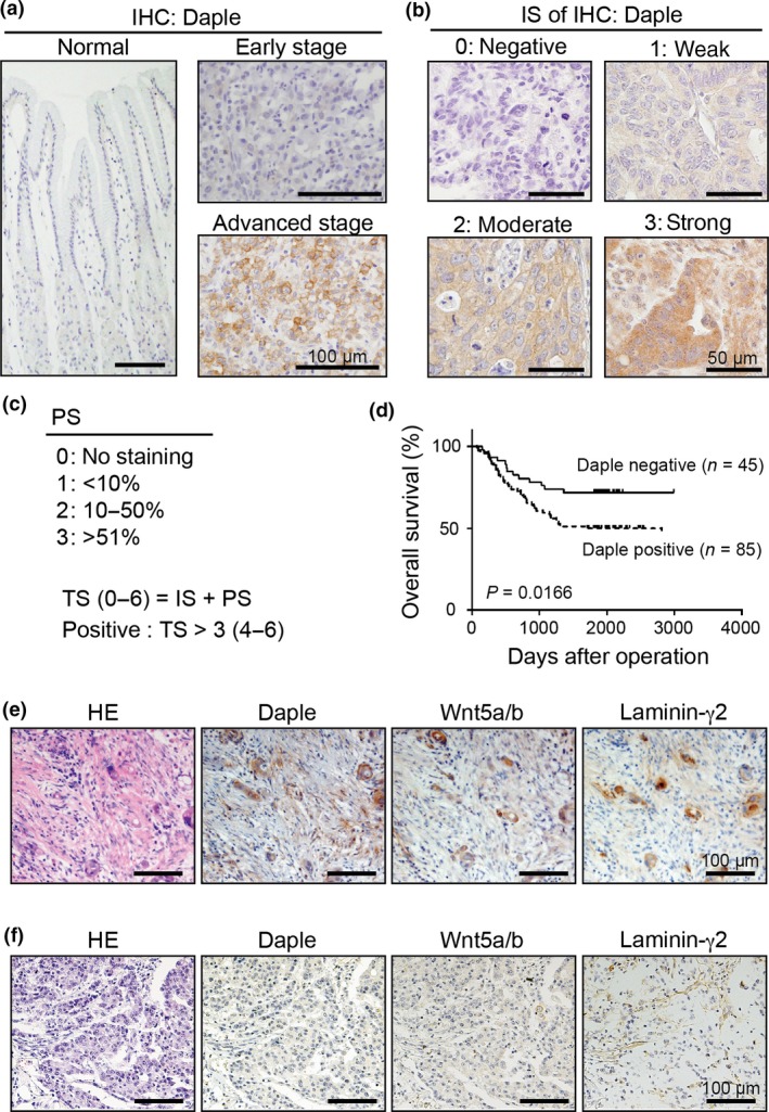
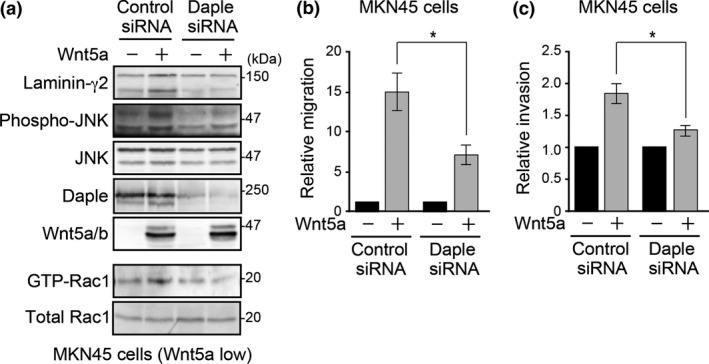
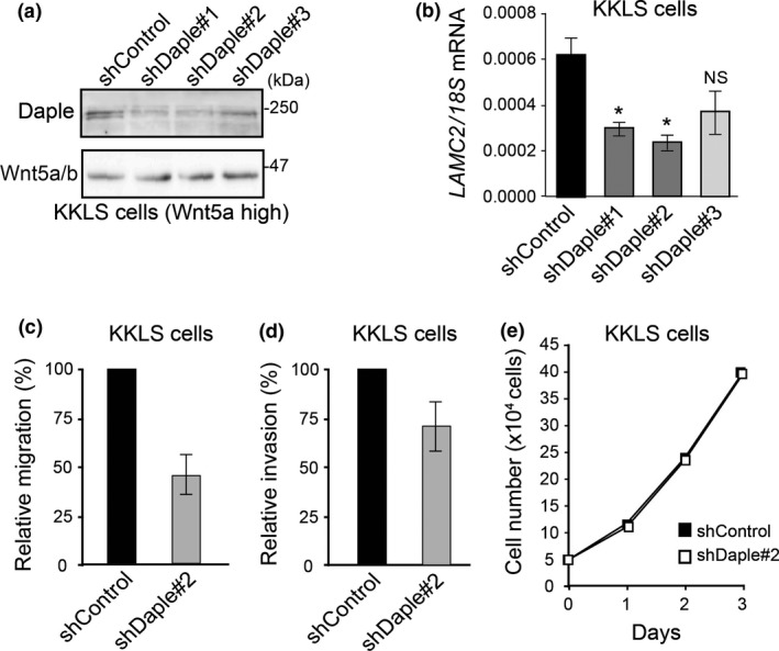
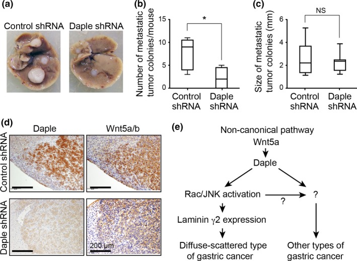
In gastric cancer, the non-canonical Wnt signaling pathway is activated by Wnt5a, which has a critical role in disease outcome. Previous studies have shown that Wnt5a mediates the expression of the extracellular matrix protein laminin γ2 through Rac and JNK activation to promote gastric cancer progression. However, the mechanism of this regulatory pathway has not been completely addressed. The scaffold protein Dvl is a major component of the Wnt signaling pathway. Here, we show that Dvl-associating protein with a high frequency of leucine residues (Daple) mediates Wnt5a-induced laminin γ2 expression. Immunohistochemical analysis showed marked expression of Daple in advanced clinical stages of gastric cancer, where it highly correlated with Wnt5a/b and laminin γ2 expression, the depth of wall invasion, and the frequency of lymph node metastasis. In cultured cancer cells, Daple depletion led to the suppression of Wnt5a-induced Rac and JNK activation, laminin γ2 expression, and cell migration and invasion. Accordingly, Daple depletion also suppressed liver metastasis in a mouse xenograft model of gastric cancer. These results suggest that the non-canonical Wnt signaling pathway contributes to gastric cancer progression at least in part via Daple, which provides a new therapeutic opportunity for the treatment of the disease.

Keywords: Daple; Wnt signaling; gastric cancer; invasion; metastasis.


Figures

Figure 1
Figure 1
Expression of Daple in gastric cancer. (a) Representative images of immunohistochemical (IHC) staining for Daple. Sections as indicated including an invasive region of the advanced stage of gastric cancer (bottom right) were stained with anti‐Daple antibody. Scale bars, 100 μm. (b) Representative images for representative Daple staining intensity for each intensity score (IS) (0–3). Scale bars, 50 μm. (c) Frequency and distribution of Daple expression was judged with the proportion score (PS) as indicated in the panel. The sum of IS and PS was used as a total score (TS) for the determination of Daple positivity (box). TS > 3 was judged as positive. (d) Kaplan–Meier survival curves of patients with gastric cancer segregated by Daple expression status. (e,f) Representative images for Daple, Wnt5a/b and laminin γ2 expression in diffuse‐scattered (e) or diffuse‐adherent (f) types of gastric cancer. Scale bars, 100 μm.
Figure 2
Figure 2
Daple regulates Wnt5a‐induced Rac/JNK activation and laminin γ2 expression in gastric cancer cells. (a) Daple knockdown inhibited Wnt5a‐induced laminin γ2 expression, Rac activation and JNK phosphorylation. Western blot analysis of total cell lysates from MKN45 cells transfected with the indicated combinations of plasmids (control or Wnt5a) and siRNA (control or Daple siRNA). For Rac activation analysis (lower two panels), GTP‐bound Rac1 was pulled down with GSTPBD and precipitated samples were probed with Rac1 antibody. (b,c) Wnt5a‐induced migration or invasion was attenuated by Daple knockdown. Transwell migration (b) or invasion (c) assays of MKN45 cells transfected with the indicated combinations of plasmids. Migrated cell numbers were expressed as the relative migration divided by that of Wnt5a (−) cells. The results represent the means ± SE. *P < 0.05.
Figure 3
Figure 3
Daple knockdown attenuates migration and invasion of high‐Wnt5a‐expressing KKLS cells. (a) Western blot of total cell lysates of control and Daple knockdown KKLS cells using three different shRNA (shDaple#1‐3). (b) LAMC2 expression in control and Daple knockdown KKLS cells quantified by real‐time RTPCR, normalized to 18S ribosomal RNA (18S) expression. The results represent the means ± SE. *P < 0.05 compared with control shRNA. NS, not significant. (c,d) Migration (c) and invasion (d) assays of control shRNA or Daple shRNA#2‐expressing KKLS cells. The number of migrated cells was expressed as relative migration (%) divided by that of control cells. The results represent the means ± SE. (e) Daple knockdown had no effect on KKLS cell proliferation. 1 × 10⁵ KKLS cells stably expressing control (closed square) or Daple (open square) shRNA were cultured for 3 days; cell numbers were counted each day.
Figure 4
Figure 4
Daple knockdown attenuates gastric cancer cell metastasis. (a) Representative images of metastatic tumors that developed in the liver of nude mice at 5 weeks after intrasplenic injection of control (left) or Daple knockdown (right) KKLS cells. (b) Total numbers of metastatic hepatic nodules per mouse (n = 6) were counted and quantified. The box plot shows median (horizontal line), 25th to 75th percentile (box), and total range (bars). *P < 0.05 (Mann–Whitney test). (c) Metastatic nodule size (n = 12) was measured in each group. NS, not significant (Mann–Whitney test). (d) Representative images of anti‐Daple and anti‐Wnt5a/b antibody‐stained metastatic tumor tissues. Scale bars, 100 μm. (e) Proposed model for Daple function in the non‐canonical Wnt signaling pathway. In particular gastric cancers including the diffuse‐scattered type, Daple induces laminin γ2 expression through Rac and JNK activation downstream of Wnt5a stimulation (left). The current results also suggest the existence of laminin γ2‐independent roles in other types of gastric cancer.

References

    1. Ferlay J, Soerjomataram I, Dikshit R et al Cancer incidence and mortality worldwide: sources, methods and major patterns in GLOBOCAN 2012. Int J Cancer 2015; 136: E359–86. - PubMed
    1. Deng N, Goh LK, Wang H et al A comprehensive survey of genomic alterations in gastric cancer reveals systematic patterns of molecular exclusivity and co‐occurrence among distinct therapeutic targets. Gut 2012; 61: 673–84. - PMC - PubMed
    1. Yang W, Raufi A, Klempner SJ. Targeted therapy for gastric cancer: molecular pathways and ongoing investigations. Biochim Biophys Acta 2014; 1846: 232–7. - PubMed
    1. El‐Rifai W, Powell SM. Molecular biology of gastric cancer. Semin Radiat Oncol 2002; 12: 128–40. - PubMed
    1. Gotoda T, Yanagisawa A, Sasako M et al Incidence of lymph node metastasis from early gastric cancer: estimation with a large number of cases at two large centers. Gastric Cancer 2000; 3: 219–25. - PubMed

Publication types

Substances