

An ultrastructural study of Trichophyton rubrum induced onychomycosis
- PMID: 26578095
- PMCID: PMC4650305
- DOI: 10.1186/s12879-015-1240-1
An ultrastructural study of Trichophyton rubrum induced onychomycosis
Abstract
Background: Trichophyton rubrum (T.rubrum) caused onychomycosis is the most common nail fungal disease. The common diagnostic methods are direct microscopic examination and fungal culture. In this study we used scanning electron microscopy (SEM) and transmission electron microscopy (TEM) to study the subungual ultrastructural changes in T. rubrum induced onychomycosis.
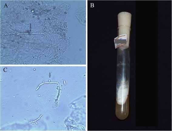
Methods: Six outpatients with onychomycosis were recruited and T.rubrum infection was confirmed by fungal culture. Six toenail samples were collected and prepared for SEM characterization. The cultured fugal colonies were prepared for SEM and TEM characterization.
Results: 1) SEM showed significant structural damages and the formation of a thin layer or a single layer of keratinocytes in all infected nail plates. Hyphae (piercing or penetrating keratinocytes layers), arthrospores and local bacterial aggregation were observed on the ventral surface of the nail plates. 2) SEM of the cultured fungal colony showed relatively straight, highly branched hyphae and microconidias; TEM showed branching hyphae that were composed of double-layer cell walls. Hyphae had nucleus, mitochondria, liposomes, lysosomes, scattered rough endoplasmic reticulum, myeloid bodies and aggregated ribosomes. There were high-density particles outside the hyphae.
Conclusion: SEM showed a large number of hyphae penetrated the keratinocytes layer, suggesting that T. rubrum can cause severe damage to the stratum corneum. TEM showed the ultrastructural features of T. rubrum-induced infection before treatment.
Figures


References
-
- Aljabre SH, Richardson MD, Scott EM, Rashid A, Shankland GS. Adherence of arthroconidia and germlings of anthropophilic and zoophilic varieties of Trichophyton mentagrophytes to human corneocytes as an early event in the pathogenesis of dermatophytosis. Clin Exp Dermatol. 1993;18(3):231–235. doi: 10.1111/j.1365-2230.1993.tb02176.x. - DOI - PubMed
-
- Samdani AJ, Dykes PJ, Marks R. The proteolytic activity of strains of T. mentagrophytes and T. rubrum isolated from tinea pedis and tinea unguium infections. Journal of medical and veterinary mycology : bi-monthly publication of the International Society for Human and Animal Mycology. 1995;33(3):167–170. doi: 10.1080/02681219580000351. - DOI - PubMed
-
- Xiaofang L, Weida L, Hui C, Xianjin C, Yongnian S, Guixia L. Determination of keratin degradation by fungi isolated from onychomycosis. Chin J Derm Venereol. 2007;21(8):455–457.
MeSH terms
LinkOut - more resources
Full Text Sources
Other Literature Sources
