. 2016 Jan 1;291(1):413-24.
doi: 10.1074/jbc.M115.686709. Epub 2015 Nov 17.

Hypoxia Potentiates Palmitate-induced Pro-inflammatory Activation of Primary Human Macrophages

Affiliations

Hypoxia Potentiates Palmitate-induced Pro-inflammatory Activation of Primary Human Macrophages

Ryan G Snodgrass et al. J Biol Chem. .

Abstract

Pro-inflammatory cytokines secreted by adipose tissue macrophages (ATMs) contribute to chronic low-grade inflammation and obesity-induced insulin resistance. Recent studies have shown that adipose tissue hypoxia promotes an inflammatory phenotype in ATMs. However, our understanding of how hypoxia modulates the response of ATMs to free fatty acids within obese adipose tissue is limited. We examined the effects of hypoxia (1% O2) on the pro-inflammatory responses of human monocyte-derived macrophages to the saturated fatty acid palmitate. Compared with normoxia, hypoxia significantly increased palmitate-induced mRNA expression and protein secretion of IL-6 and IL-1β. Although palmitate-induced endoplasmic reticulum stress and nuclear factor κB pathway activation were not enhanced by hypoxia, hypoxia increased the activation of JNK and p38 mitogen-activated protein kinase signaling in palmitate-treated cells. Inhibition of JNK blocked the hypoxic induction of pro-inflammatory cytokine expression, whereas knockdown of hypoxia-induced transcription factors HIF-1α and HIF-2α alone or in combination failed to reduce IL-6 and only modestly reduced IL-1β gene expression in palmitate-treated hypoxic macrophages. Enhanced pro-inflammatory cytokine production and JNK activity under hypoxia were prevented by inhibiting reactive oxygen species generation. In addition, silencing of dual-specificity phosphatase 16 increased normoxic levels of IL-6 and IL-1β and reduced the hypoxic potentiation in palmitate-treated macrophages. The secretome of hypoxic palmitate-treated macrophages promoted IL-6 and macrophage chemoattractant protein 1 expression in primary human adipocytes, which was sensitive to macrophage JNK inhibition. Our results reveal that the coexistence of hypoxia along with free fatty acids exacerbates macrophage-mediated inflammation.

Keywords: cytokine; fatty acid; hypoxia; inflammation; macrophage.


Figures

FIGURE 1.
FIGURE 1.
Hypoxia potentiates palmitate-induced, pro-inflammatory cytokine expression and secretion in primary human macrophages. A–E, mRNA expression of TNF-α (A), IL-6 (B), IL-1β (C), IL-8 (D), and IL-10 (E) in primary human macrophages treated with 500 μm palmitate (C16:0) or BSA alone for 9 h under normoxia (20% O2) or hypoxia (1% O2). F–J, protein concentrations of TNF-α (F), IL-6 (G), IL-1β (H), IL-8 (I), and IL-10 (J) in cell culture supernatants after 24-h treatment with C16:0 or BSA alone under normoxia or hypoxia. K–O, mRNA expression of Tnfa (K), Il6 (L), Il1b (M), Nos2 (N), and Il10 (O) in murine SVFs treated with 500 μm C16:0 or BSA alone for 9 h under normoxia (20% O2) or hypoxia (1% O2). Results are presented as mean ± S.E. of at least four independent experiments. *, p < 0.05; **, p < 0.01; ***, p < 0.001; ns, not significant.
FIGURE 2.
FIGURE 2.
Reduced oxygen levels dose-dependently potentiate palmitate-induced, pro-inflammatory cytokine expression. A–D, mRNA expression of TNF-α (A), IL-6 (B), IL-1β (C), and IL-8 (D) in primary human macrophages treated with 500 μm palmitate (C16:0) or BSA alone for 9 h under normoxia (20% O2) or moderate hypoxia (5% and 2.5% O2). E–H, mRNA expression of TNF-α (E), IL-6 (F), IL-1β (G), and IL-8 (H) in primary human macrophages treated with 500 μm myristate (C14:0), 500 μm stearate (C18:0), or BSA alone for 9 h under normoxia (20% O2) or hypoxia (1% O2). Results are presented as mean ± S.E. of at least four independent experiments. *, p < 0.05; **, p < 0.01; ***, p < 0.001.
FIGURE 3.
FIGURE 3.
Hypoxia does not induce or potentiate palmitate-induced markers of ER stress. A and B, mRNA expression of GRP78 (A) and CHOP (B) at 9 h in macrophages treated with 500 μm palmitate (C16:0) under normoxia (20% O2) or hypoxia (1% O2). C, phosphorylation of IRE1 9 h after treatment with C16:0 or BSA alone under normoxia or hypoxia. Results are presented as mean ± S.E. of at least four independent experiments. *, p < 0.05; **, p < 0.01; ***, p < 0.001.
FIGURE 4.
FIGURE 4.
Enhanced palmitate-induced, pro-inflammatory cytokine production under hypoxia is HIF-1α- and HIF-2α-independent. A–F, mRNA expression levels of HIF-1α (A), HIF-2α (B), the HIF target gene GLUT1 (C), IL-6 (D), IL-1β (E), and IL-8 (F) in macrophages transfected with control, HIF-1α, HIF-2α, or HIF-1α/HIF-2α siRNA and treated with 500 μm palmitate (C16:0) for 9 h under normoxia (20% O2) or hypoxia (1% O2). G–I, protein concentrations of IL-6 (G), IL-1β (H), and IL-8 (I) in macrophages transfected with control, HIF-1α, HIF-2α, or HIF-1α/HIF-2α siRNA and treated with 500 μm C16:0 for 24 h under normoxia (20% O2) or hypoxia (1% O2). Results are presented as mean ± S.E. of at least three independent experiments. C–F, the expression in hypoxic C16:0-treated control siRNA macrophages was set to 1. *, p < 0.05.
FIGURE 5.
FIGURE 5.
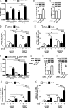
Hypoxia enhances palmitate-induced cytokine production through MAPK. A–C, phosphorylation of p38, c-Jun, and ERK 1/2 1, 3, and 9 h after treatment with 500 μm palmitate (C16:0) in normoxic (20% O2) and hypoxic (1% O2) macrophages. D, phosphorylation of p38 and c-Jun following treatment with 500 μm myristate (C14:0), 500 μm stearate (C18:0), or BSA alone for 3 h under normoxia or hypoxia. E–G, mRNA expression of IL-6 (E), IL-1β (F), and IL-8 (G) in macrophages pretreated with JNK inhibitor (SP600125, 10 μm) or p38 inhibitor (SB203580, 10 μm) and then co-incubated for 9 h with 500 μm C16:0. Results are presented as mean ± S.E. of at least three independent experiments. *, p < 0.05.
FIGURE 6.
FIGURE 6.
Mitochondrial ROS in palmitate-induced inflammation under hypoxia. A, mitochondrial ROS measured by MitoSOX fluorescence in primary human macrophages treated with 500 μm palmitate (C16:0) or BSA alone for 1 h under normoxia (20% O2) or hypoxia (1% O2). B–D, mRNA expression of IL-6 (B) IL-1β (C), and IL-8 (D) in normoxic and hypoxic macrophages pretreated with 5 mm NAC or 1 μm MitoTEMPO (MT) and then co-incubated for 9 h with 500 μm C16:0. E–H, phosphorylation of c-Jun (E and G) and p38 (F and H) in hypoxic macrophages pretreated with NAC or MitoTEMPO and then co-incubated with 500 μm C16:0 for 3 h. Results are presented as mean ± S.E. of at least three independent experiments. MitoSOX fluorescence in normoxic C16:0-treated macrophages was set to 1. *, p < 0.05; **, p < 0.01; ***, p < 0.001.
FIGURE 7.
FIGURE 7.
Attenuating DUSP expression increases c-Jun phosphorylation and potentiates palmitate-induced, pro-inflammatory cytokine expression. A, mRNA expression of DUSP16 in macrophages transfected with DUSP16 siRNA and then treated with 500 μm palmitate (C16:0) for 9 h under normoxia and hypoxia. B and C, phosphorylation of c-Jun (B) and p38 (C) following transfection with DUSP16 siRNA. D and E, mRNA expression of IL-6 (D) and IL-1β (E) in macrophages transfected with DUSP16 siRNA and then treated with 500 μm C16:0 for 9 h under normoxia and hypoxia. F and G, mRNA expression (F) and protein (G) of DUSP1 in macrophages transfected with DUSP1 siRNA and then treated with 500 μm C16:0 for 9 h under normoxia and hypoxia. H and I, phosphorylation of c-Jun (H) and p38 (I) following transfection with DUSP1 siRNA. J and K, mRNA expression of IL-6 (J) and IL-1β (K) in macrophages transfected with DUSP1 siRNA and then treated with 500 μm C16:0 for 9 h under normoxia and hypoxia. Results are presented as mean ± S.E. of at least three independent experiments. D, E, J, and K, the expression in normoxic C16:0-treated control siRNA macrophages was set to 1. *, p < 0.05; **, p < 0.01; ***, p < 0.001; ns, not significant.
FIGURE 8.
FIGURE 8.
Conditioned media from hypoxic, palmitate-treated human macrophages induce an inflammatory response in primary human adipocytes. A–C, mRNA expression levels of IL-6 (A), MCP-1 (B), and Adiponectin (C) in normoxic adipocytes following treatment with macrophage-conditioned media. D, mRNA expression levels of IL-6 and MCP-1 in normoxic adipocytes following treatment with conditioned media prepared from hypoxic, palmitate-treated macrophages preincubated with 10 μm SP600125 or 10 μm SB203580. Expression in adipocytes incubated with conditioned media from C16:0-, vehicle-treated hypoxic macrophages was set to 1. E, proposed model by which decreased oxygen tension potentiates palmitate-induced, pro-inflammatory cytokine production in macrophages through enhanced JNK activity. Results are presented as mean ± S.E. of at least five independent experiments. *, p < 0.05.

Similar articles

Cited by

References

    1. Chawla A., Nguyen K. D., and Goh Y. P. (2011) Macrophage-mediated inflammation in metabolic disease. Nat. Rev. Immunol. 11, 738–749 - PMC - PubMed
    1. McNelis J. C., and Olefsky J. M. (2014) Macrophages, immunity, and metabolic disease. Immunity 41, 36–48 - PubMed
    1. Han J. M., and Levings M. K. (2013) Immune regulation in obesity-associated adipose inflammation. J. Immunol. 191, 527–532 - PubMed
    1. Sun K., Kusminski C. M., and Scherer P. E. (2011) Adipose tissue remodeling and obesity. J. Clin. Invest. 121, 2094–2101 - PMC - PubMed
    1. Amano S. U., Cohen J. L., Vangala P., Tencerova M., Nicoloro S. M., Yawe J. C., Shen Y., Czech M. P., and Aouadi M. (2014) Local proliferation of macrophages contributes to obesity-associated adipose tissue inflammation. Cell Metab. 19, 162–171 - PMC - PubMed

Publication types

MeSH terms