Clinical Trial
. 2015 Nov 17;112(46):14331-6.
doi: 10.1073/pnas.1514748112. Epub 2015 Nov 2.

Role of HIV-1 matrix protein p17 variants in lymphoma pathogenesis

Affiliations
Clinical Trial

Role of HIV-1 matrix protein p17 variants in lymphoma pathogenesis

Riccardo Dolcetti et al. Proc Natl Acad Sci U S A. .

Erratum in

Abstract

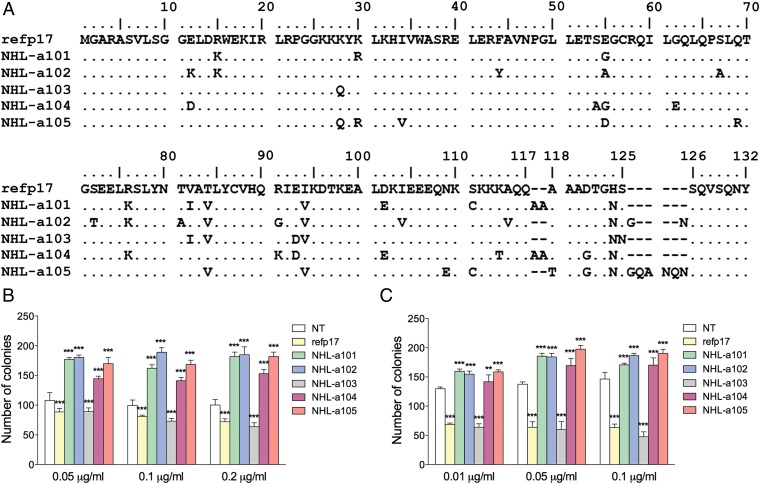
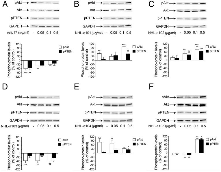
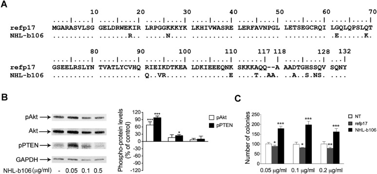
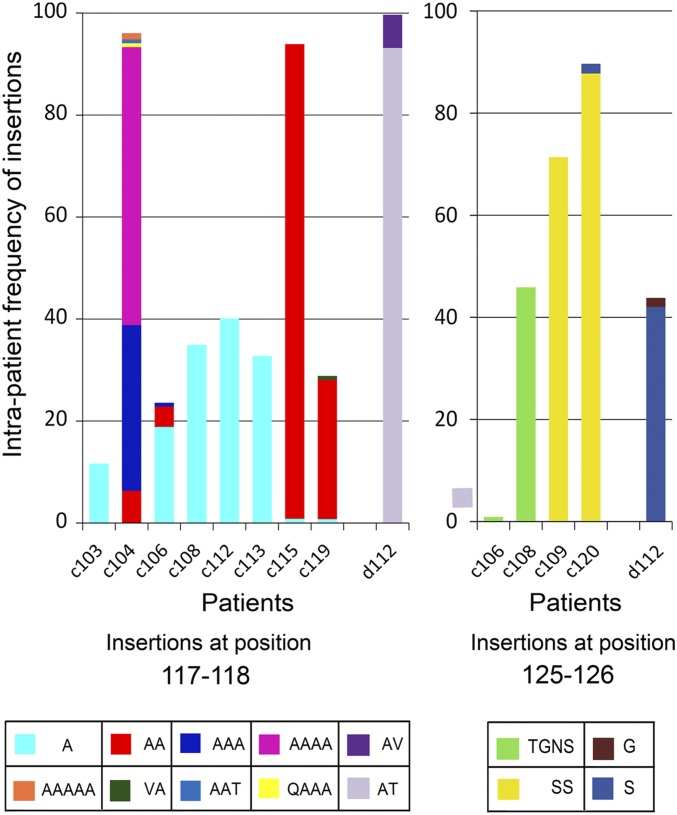
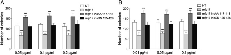
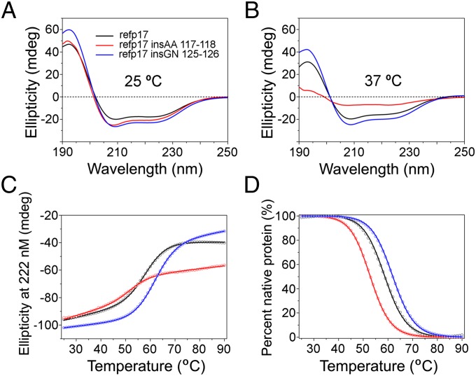
Although in decline after successful anti-HIV therapy, B-cell lymphomas are still elevated in HIV-1-seropositive (HIV+) persons, and the mechanisms are obscure. The HIV-1 matrix protein p17 persists in germinal centers long after HIV-1 drug suppression, and some p17 variants (vp17s) activate Akt signaling and promote growth of transformed B cells. Here we show that vp17s derived from four of five non-Hodgkin lymphoma (NHL) tissues from HIV+ subjects display potent B-cell growth-promoting activity. They are characterized by amino acid insertions at position 117-118 (Ala-Ala) or 125-126 (Gly-Asn or Gly-Gln-Ala-Asn-Gln-Asn) among some other mutations throughout the sequence. Identical dominant vp17s are found in both tumor and plasma. Three of seven plasma samples from an independent set of NHL cases manifested multiple Ala insertions at position 117-118, and one with the Ala-Ala profile also promoted B-cell growth and activated Akt signaling. Ultradeep pyrosequencing showed that vp17s with C-terminal insertions are more frequently detected in plasma of HIV+ subjects with than without NHL. Insertion of Ala-Ala at position 117-118 into reference p17 (refp17) was sufficient to confer B-cell growth-promoting activity. In contrast, refp17 bearing the Gly-Asn insertion at position 125-126 did not, suggesting that mutations not restricted to the C terminus can also account for this activity. Biophysical analysis revealed that the Ala-Ala insertion mutant is destabilized compared with refp17, whereas the Gly-Asn form is stabilized. This finding provides an avenue for further exploration of structure function relationships and new treatment strategies in combating HIV-1-related NHL.

Keywords: AIDS; B-cell clonogenicity; HIV-1 matrix protein p17; non-Hodgkin lymphoma; p17 variants.


Conflict of interest statement

The authors declare no conflict of interest.

Figures

Fig. 1.
Fig. 1.
Alignment and comparison among amino acid sequences of refp17 and vp17s isolated from HIV–NHL patients and effects of different recombinant p17s on B-cell clonogenicity. (A) Sequences are represented by the single-letter amino acid code. Amino acid positions are referred to the prototype genotype B strain BH10 (UniProtKB P04585; refp17), adopted as reference for this analysis. Each amino acid residue of NHL-derived vp17s not differing from the refp17 sequence is represented by a dot. (B) Raji and (C) BJAB cells were plated in 12-well plates, and after 4 d, medium was replaced by fresh medium with various concentrations, 0.01, 0.05, 0.1, and 0.2 μg/mL of refp17 and lymphoma-associated variants, as indicated. Cells not treated (NT) were used as negative control. The cell growth was analyzed by using 3-[4, 5-Dimethylthiazol-2-y1]-2, 5-diphenyltetrazolium bromide (MTT). Data represent the average number of colonies ± SD from three independent experiments performed in triplicate. The statistical significance between control and treated cultures was calculated using one-way ANOVA performed separately for each concentration of p17 variants, across the three groups. Bonferroni’s posttest was used to compare data. *P < 0.05, **P < 0.01, ***P < 0.001.
Fig. S1.
Fig. S1.
Alignment and comparison among amino acid sequences of refp17 and p17s isolated from HIV+ non-NHL patients and effects of different recombinant p17s on B-cell clonogenicity. (A) Sequences are represented by the single-letter amino acid code. Amino acid positions are referred to the prototype genotype B strain BH10 (UniProtKB P04585; refp17), adopted as the reference for this analysis. Each amino acid residue of p17s not differing from the refp17 sequence is represented by a dot. (B) Raji cells were plated in 12-well plates, and after 4 d, medium was replaced by fresh medium with various concentrations, 0.05, 0.1, and 0.2 μg/mL, of refp17 and p17 derived from blood samples of HIV+ non-NHL patients, namely d205, d206, d210, d212, and d215, as indicated. Cells not treated (NT) were used as negative control, whereas cells treated with vp17 NHL-a101 and NHL-a102 were used as the positive clonogenic control. The cell growth was analyzed by using MTT. Data represent the average number of colonies ± SD from three independent experiments performed in triplicate. The statistical significance between control and treated cultures was calculated using one-way ANOVA performed separately for each concentration of p17 across the three groups. Bonferroni’s posttest was used to compare data. *P < 0.05, **P < 0.01, ***P < 0.001.
Fig. 2.
Fig. 2.
Effects of refp17 and lymphoma-associated vp17 stimulation on Akt and PTEN activity in Raji cells. Cells were treated for 5 min with 0.05, 0.1, and 0.5 μg/mL of refp17 (A) and lymphoma-associated p17 variants NHL-a101 (B), NHL-a102 (C), NHL-a103 (D), NHL-a104 (E), and NHL-a105 (F). Untreated cells were used as control (lane 1). Western blot analysis of Raji lysates shows that refp17 and NHL-a103 inhibit the activation of Akt and maintain PTEN in an active state (A), as shown by the respective phosphorylation state, verified by densitometric analysis and plotting of the pAkt/Akt and pPTEN/GAPDH. On the contrary, NHL-a101, NHL-a102, NHL-a104, and NHLa105 induce the activation of Akt and maintain PTEN in an inactive state, as shown by the increased phosphorylation, verified by densitometric analysis and plotting of the pAkt/Akt and pPTEN/GAPDH. In the Upper panels, blots from one representative experiment of three with similar results are shown. In the Lower panels, values reported for phosphorylation of Akt and PTEN are the mean ± SD of three independent experiments. Statistical analysis was performed by one-way ANOVA, and the Bonferroni’s posttest was used to compare data. *P < 0.05, **P < 0.01, ***P < 0.001.
Fig. S2.
Fig. S2.
Effect of NHL-b106 stimulation on the PTEN/Akt signaling pathway and on B-cell clonogenicity. (A) Alignment and comparison among p17 and NHL-b106 amino acid sequences. Sequences are represented by the single-letter amino acid code. Each amino acid residue of NHL-b106 not differing from refp17 is represented by a dot. (B) Raji cells were treated for 5 min with 0.05, 0.1, and 0.5 μg/mL of NHL-b106. Cells not treated (NT) were used as the control. Western blot analysis of lysates shows that NHL-b106 induces the activation of Akt and maintains PTEN in an inactive phosphorylated state, verified by densitometric analysis and plotting of pAkt/Akt and pPTEN/GAPDH. In the Left panels, blots from one representative experiment of three with similar results are shown. In the Right panels, values reported for pAkt and pPTEN are the mean ± SD of three independent experiments. Statistical analysis was performed by one-way ANOVA, and the Bonferroni.s posttest was used to compare data. *P < 0.05, ***P < 0.001. (C) Raji were plated in 12-well plates, and after 4 d, medium was replaced by fresh medium with various concentrations, 0.05, 0.1, and 0.2 μg/mL, of refp17 and NHL-b106. Cells not treated (NT) were used as the negative control. The cell growth was analyzed by using MTT. Data represent the average number of colonies ± SD from three independent experiments performed in triplicate. The statistical significance between control and treated cultures was calculated using one-way ANOVA performed separately for each concentration of p17 variant across the three groups. Bonferroni’s posttest was used to compare data. *P < 0.05, **P < 0.01, ***P < 0.001.
Fig. 3.
Fig. 3.
Frequency of amino acid insertions at positions 117–118 and 125–126 in vp17 of HIV+ patients with or without NHL. Bars represent the frequency of amino acid insertions detected at positions 117–118 (Left panel) and 125–126 (Right panel) in patients belonging to groups C and D. Each insertion is identified by a color code (see table at the bottom of the histogram). *Amino acid positions are referred to the prototype genotype B strain BH10 (UniProtKB P04585; refp17), adopted as the reference for this analysis.
Fig. 4.
Fig. 4.
Effect of Ala–Ala or Gly–Asn insertion at position 117–118 and 125–126, respectively, of the refp17 backbone on B-cell clonogenicity. Raji (A) and BJAB (B) were plated in 12-well plates, and after 4 d, medium was replaced by fresh medium with various concentrations, 0.01, 0.05, 0.1, and 0.2 μg/mL of refp17, refp17 insAA 117–118, and refp17 insGN 125–126, as indicated. Cells not treated (NT) were used as the negative control. The cell growth was analyzed by using MTT. Data represent the average number of colonies SD from three independent experiments performed in triplicate. The statistical significance between control and treated cultures was calculated using one-way ANOVA performed separately for each concentration of p17 variants, across the three groups. Bonferroni’s posttest was used to compare data. ***P < 0.001.
Fig. 5.
Fig. 5.
Effects of the Ala–Ala and Gly–Asn insertions in the C terminus of refp17 on protein conformation and stability. (A) CD spectra of refp17, refp17 insAA 117–118, and refp17 insGN 125–126, each at 2.5 µM, collected at 25 °C in 10 mM phosphate buffer, pH 7.4. (B) CD spectra of the three recombinant p17 proteins at 37 °C under otherwise identical conditions. (C) Thermal denaturation of refp17, refp17 insAA 117–118, and refp17 insGN 125–126, each at 10 µM in PBS, pH 7.4, as monitored at 222 nm by CD spectroscopy. (D) The experimental data normalized according to a two-state protein denaturation model from which Tm values were derived.

References

    1. Shiels MS, et al. Cancer burden in the HIV-infected population in the United States. J Natl Cancer Inst. 2011;103(9):753–762. - PMC - PubMed
    1. Vandenhende MA, et al. ANRS EN20 Mortalité 2010 study group Cancer-related causes of death among HIV-infected patients in France in 2010: Evolution since 2000. PLoS One. 2015;10(6):e0129550. - PMC - PubMed
    1. Coté TR, et al. AIDS/Cancer Study Group Non-Hodgkin’s lymphoma among people with AIDS: Incidence, presentation and public health burden. Int J Cancer. 1997;73(5):645–650. - PubMed
    1. Gloghini A, Dolcetti R, Carbone A. Lymphomas occurring specifically in HIV-infected patients: From pathogenesis to pathology. Semin Cancer Biol. 2013;23(6):457–467. - PubMed
    1. Knowles DM. Etiology and pathogenesis of AIDS-related non-Hodgkin’s lymphoma. Hematol Oncol Clin North Am. 2003;17(3):785–820. - PubMed

Publication types

MeSH terms

Substances

LinkOut - more resources