

LIM homeobox protein 5 (Lhx5) is essential for mamillary body development
- PMID: 26578897
- PMCID: PMC4621302
- DOI: 10.3389/fnana.2015.00136
LIM homeobox protein 5 (Lhx5) is essential for mamillary body development
Abstract
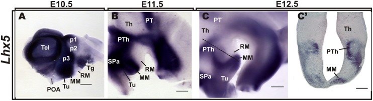
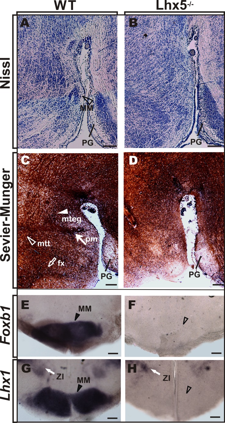
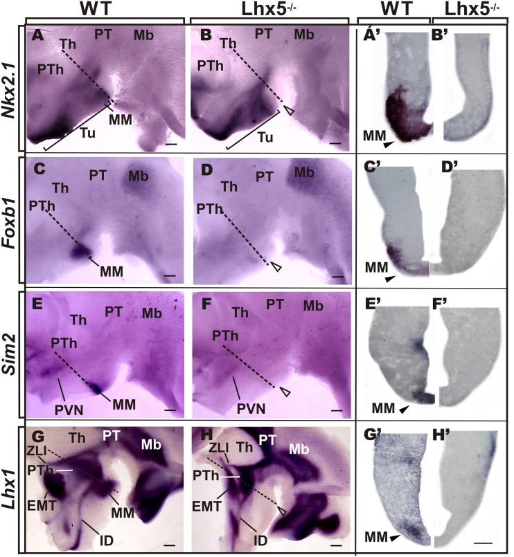
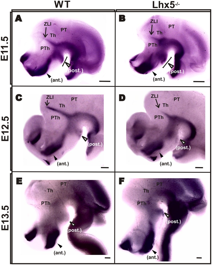
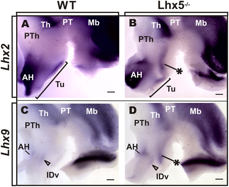
The mamillary body (MM) is a group of hypothalamic nuclei related to memory and spatial navigation that interconnects hippocampal, thalamic, and tegmental regions. Here we demonstrate that Lhx5, a LIM-HD domain transcription factor expressed early in the developing posterior hypothalamus, is required for the generation of the MM and its derived tracts. The MM markers Foxb1, Sim2, and Lhx1 are absent in Lhx5 knock-out mice from early embryonic stages, suggesting abnormal specification of this region. This was supported by the absence of Nkx2.1 and expansion of Shh in the prospective mamillary area. Interestingly, we also found an ectopic domain expressing Lhx2 and Lhx9 along the anterio-posterior hypothalamic axis. Our results suggest that Lhx5 controls early aspects of hypothalamic development by regulating gene expression and cellular specification in the prospective MM.
Keywords: diencephalon; embryonic development; hypothalamus; mouse; transcription factor.
Figures
Similar articles
-
Lhx5 controls mamillary differentiation in the developing hypothalamus of the mouse.Front Neuroanat. 2015 Aug 14;9:113. doi: 10.3389/fnana.2015.00113. eCollection 2015. Front Neuroanat. 2015. PMID: 26321924 Free PMC article.
-
LIM-homeodomain genes as territory markers in the brainstem of adult and developing Xenopus laevis.J Comp Neurol. 2005 May 9;485(3):240-54. doi: 10.1002/cne.20498. J Comp Neurol. 2005. PMID: 15791640
-
LIM-homeodomain genes as developmental and adult genetic markers of Xenopus forebrain functional subdivisions.J Comp Neurol. 2004 Apr 19;472(1):52-72. doi: 10.1002/cne.20046. J Comp Neurol. 2004. PMID: 15024752
-
The olfactory amygdala in amniotes: an evo-devo approach.Anat Rec (Hoboken). 2013 Sep;296(9):1317-32. doi: 10.1002/ar.22744. Epub 2013 Jul 31. Anat Rec (Hoboken). 2013. PMID: 23904411 Review.
-
Sonic hedgehog signaling in the development of the mouse hypothalamus.Front Neuroanat. 2015 Jan 6;8:156. doi: 10.3389/fnana.2014.00156. eCollection 2014. Front Neuroanat. 2015. PMID: 25610374 Free PMC article. Review.
Cited by
-
Multiple allelic configurations govern long-range Shh enhancer-promoter communication in the embryonic forebrain.Mol Cell. 2024 Dec 19;84(24):4698-4710.e6. doi: 10.1016/j.molcel.2024.10.042. Epub 2024 Nov 22. Mol Cell. 2024. PMID: 39579767
-
Generation of ventralized human thalamic organoids with thalamic reticular nucleus.Cell Stem Cell. 2023 May 4;30(5):677-688.e5. doi: 10.1016/j.stem.2023.03.007. Epub 2023 Apr 4. Cell Stem Cell. 2023. PMID: 37019105 Free PMC article.
-
Neighbor-specific gene expression revealed from physically interacting cells during mouse embryonic development.Proc Natl Acad Sci U S A. 2023 Jan 10;120(2):e2205371120. doi: 10.1073/pnas.2205371120. Epub 2023 Jan 3. Proc Natl Acad Sci U S A. 2023. PMID: 36595695 Free PMC article.
-
Cellular taxonomy and spatial organization of the murine ventral posterior hypothalamus.Elife. 2020 Oct 29;9:e58901. doi: 10.7554/eLife.58901. Elife. 2020. PMID: 33119507 Free PMC article.
-
Genome-Wide DNA Methylation and Gene Expression Profiles in Cows Subjected to Different Stress Level as Assessed by Cortisol in Milk.Genes (Basel). 2020 Jul 25;11(8):850. doi: 10.3390/genes11080850. Genes (Basel). 2020. PMID: 32722461 Free PMC article.
References
-
- Alvarez-Bolado G., Zhou X., Voss A. K., Thomas T., Gruss P. (2000). Winged helix transcription factor Foxb1 is essential for access of mammillothalamic axons to the thalamus. Development 127 1029–1038. - PubMed
LinkOut - more resources
Full Text Sources
Other Literature Sources
Research Materials