Review
. 2015 Oct 30:9:145.
doi: 10.3389/fnsys.2015.00145. eCollection 2015.

Prefrontal-amygdala fear networks come into focus

Affiliations
Review

Prefrontal-amygdala fear networks come into focus

Maithe Arruda-Carvalho et al. Front Syst Neurosci. .

Abstract

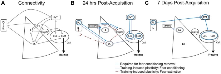
The ability to form associations between aversive threats and their predictors is fundamental to survival. However, fear and anxiety in excess are detrimental and are a hallmark of psychiatric diseases such as post-traumatic stress disorder (PTSD). PTSD symptomatology includes persistent and intrusive thoughts of an experienced trauma, suggesting an inability to downregulate fear when a corresponding threat has subsided. Convergent evidence from human and rodent studies supports a role for the medial prefrontal cortex (mPFC)-amygdala network in both PTSD and the regulation of fear memory expression. In particular, current models stipulate that the prelimbic (PL) and infralimbic (IL) subdivisions of the rodent mPFC bidirectionally regulate fear expression via differential recruitment of amygdala neuronal subpopulations. However, an array of recent studies that employ new technical approaches has fundamentally challenged this interpretation. Here we explore how a new emphasis on the contribution of inhibitory neuronal populations, subcortical structures and the passage of time is reshaping our understanding of mPFC-amygdala circuits and their control over fear.

Keywords: basal amygdala; central amygdala; consolidation; infralimbic; intercalated; optogenetics; paraventricular thalamus; prelimbic.


Figures

Figure 1
Figure 1
Medial prefrontal cortex (mPFC)-amygdala contributions to conditioned fear and extinction. (A) Established monosynaptic connections between mPFC and amygdala subregions. Double arrows indicate bidirectional projections. (B) Model depicting brain regions (circles) or projection pathways (arrows) implicated in fear conditioning or extinction, as expressed at 24 h after fear or extinction training. *IL-BLA projections display plasticity at 24 h after extinction, and have been shown to be necessary for extinction acquisition, but are not required for extinction retrieval at 24 h after acquisition (Cho et al., ; Do-Monte et al., 2015a). (C) Model depicting brain regions (circles) or projection pathways (arrows) implicated in fear conditioning, as expressed at 7 days after fear conditioning. Designation of regions and pathways required for retrieval is based on the results of in vivo cell- or projection-specific optogenetic or chemogenetic manipulations. Designation of pathways displaying training-associated synaptic plasticity is based on ex vivo brain slice recordings obtained at 24 h or 7 days after fear conditioning or extinction. BA, basal amygdala; CeA, central amygdala; CeL, lateral division of the central amygdala; CeM, medial division of the central amygdala; IL, infralimbic cortex; LA, lateral amygdala; mpITC, medial paracapsular intercalated cells; PL, prelimbic cortex; PVT, paraventricular thalamus. Additional references used to compile these models, but not discussed within the main text, include (Amano et al., ; Clem and Huganir, ; Nabavi et al., 2014).

References

    1. Amano T., Unal C. T., Paré D. (2010). Synaptic correlates of fear extinction in the amygdala. Nat. Neurosci. 13, 489–494. 10.1038/nn.2499 - DOI - PMC - PubMed
    1. Amir A., Amano T., Pare D. (2011). Physiological identification and infralimbic responsiveness of rat intercalated amygdala neurons. J. Neurophysiol. 105, 3054–3066. 10.1152/jn.00136.2011 - DOI - PMC - PubMed
    1. Arruda-Carvalho M., Clem R. L. (2014). Pathway-selective adjustment of prefrontal-amygdala transmission during fear encoding. J. Neurosci. 34, 15601–15609. 10.1523/jneurosci.2664-14.2014 - DOI - PMC - PubMed
    1. Asede D., Bosch D., Lüthi A., Ferraguti F., Ehrlich I. (2015). Sensory inputs to intercalated cells provide fear-learning modulated inhibition to the basolateral amygdala. Neuron 86, 541–554. 10.1016/j.neuron.2015.03.008 - DOI - PubMed
    1. Barrett D., Shumake J., Jones D., Gonzalez-Lima F. (2003). Metabolic mapping of mouse brain activity after extinction of a conditioned emotional response. J. Neurosci. 23, 5740–5749. - PMC - PubMed

LinkOut - more resources