Potential effects of UV radiation on photosynthetic structures of the bloom-forming cyanobacterium Cylindrospermopsis raciborskii CYRF-01
- PMID: 26579108
- PMCID: PMC4627488
- DOI: 10.3389/fmicb.2015.01202
Potential effects of UV radiation on photosynthetic structures of the bloom-forming cyanobacterium Cylindrospermopsis raciborskii CYRF-01
Abstract
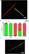
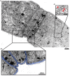
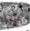
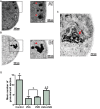
Cyanobacteria are aquatic photosynthetic microorganisms. While of enormous ecological importance, they have also been linked to human and animal illnesses around the world as a consequence of toxin production by some species. Cylindrospermopsis raciborskii, a filamentous nitrogen-fixing cyanobacterium, has attracted considerable attention due to its potential toxicity and ecophysiological adaptability. We investigated whether C. raciborskii could be affected by ultraviolet (UV) radiation. Non-axenic cultures of C. raciborskii were exposed to three UV treatments (UVA, UVB, or UVA + UVB) over a 6 h period, during which cell concentration, viability and ultrastructure were analyzed. UVA and UVA + UVB treatments showed significant negative effects on cell concentration (decreases of 56.4 and 64.3%, respectively). This decrease was directly associated with cell death as revealed by a cell viability fluorescent probe. Over 90% of UVA + UVB- and UVA-treated cells died. UVB did not alter cell concentration, but reduced cell viability in almost 50% of organisms. Transmission electron microscopy (TEM) revealed a drastic loss of thylakoids, membranes in which cyanobacteria photosystems are localized, after all treatments. Moreover, other photosynthetic- and metabolic-related structures, such as accessory pigments and polyphosphate granules, were damaged. Quantitative TEM analyses revealed a 95.8% reduction in cell area occupied by thylakoids after UVA treatment, and reduction of 77.6 and 81.3% after UVB and UVA + UVB treatments, respectively. Results demonstrated clear alterations in viability and photosynthetic structures of C. raciborskii induced by various UV radiation fractions. This study facilitates our understanding of the subcellular organization of this cyanobacterium species, identifies specific intracellular targets of UVA and UVB radiation and reinforces the importance of UV radiation as an environmental stressor.
Keywords: cell death; cell viability; cyanobacteria; thylakoid membranes; transmission electron microscopy; ultraviolet radiation.
Figures


Similar articles
-
The Cyanobacterium Cylindrospermopsis raciborskii (CYRF-01) Responds to Environmental Stresses with Increased Vesiculation Detected at Single-Cell Resolution.Front Microbiol. 2018 Feb 21;9:272. doi: 10.3389/fmicb.2018.00272. eCollection 2018. Front Microbiol. 2018. PMID: 29515552 Free PMC article.
-
Increasing Temperature Counteracts the Negative Effect of UV Radiation on Growth and Photosynthetic Efficiency of Microcystis aeruginosa and Raphidiopsis raciborskii.Photochem Photobiol. 2021 Jul;97(4):753-762. doi: 10.1111/php.13377. Epub 2021 Jan 18. Photochem Photobiol. 2021. PMID: 33394510
-
Effects of ultraviolet radiation (UVA+UVB) on young gametophytes of Gelidium floridanum: growth rate, photosynthetic pigments, carotenoids, photosynthetic performance, and ultrastructure.Photochem Photobiol. 2014 Sep-Oct;90(5):1050-60. doi: 10.1111/php.12296. Epub 2014 Jun 23. Photochem Photobiol. 2014. PMID: 24893751
-
Mechanisms of ultraviolet (UV) B and UVA phototherapy.J Investig Dermatol Symp Proc. 1999 Sep;4(1):70-2. doi: 10.1038/sj.jidsp.5640185. J Investig Dermatol Symp Proc. 1999. PMID: 10537012 Review.
-
Broad-spectrum sunscreens provide better protection from solar ultraviolet-simulated radiation and natural sunlight-induced immunosuppression in human beings.J Am Acad Dermatol. 2008 May;58(5 Suppl 2):S149-54. doi: 10.1016/j.jaad.2007.04.035. J Am Acad Dermatol. 2008. PMID: 18410801 Review.
Cited by
-
Limiting factors in the operation of photosystems I and II in cyanobacteria.Microb Biotechnol. 2024 Aug;17(8):e14519. doi: 10.1111/1751-7915.14519. Microb Biotechnol. 2024. PMID: 39101352 Free PMC article. Review.
-
Cytotoxic effects of zinc oxide nanoparticles on cyanobacterium Spirulina (Arthrospira) platensis.PeerJ. 2018 Jun 1;6:e4682. doi: 10.7717/peerj.4682. eCollection 2018. PeerJ. 2018. PMID: 29876145 Free PMC article.
-
Nanoparticles for Mitigation of Harmful Cyanobacterial Blooms.Toxins (Basel). 2024 Jan 12;16(1):41. doi: 10.3390/toxins16010041. Toxins (Basel). 2024. PMID: 38251256 Free PMC article. Review.
-
Inferred ancestry of scytonemin biosynthesis proteins in cyanobacteria indicates a response to Paleoproterozoic oxygenation.Geobiology. 2022 Nov;20(6):764-775. doi: 10.1111/gbi.12514. Epub 2022 Jul 18. Geobiology. 2022. PMID: 35851984 Free PMC article.
-
Biologically-Based and Physiochemical Life Support and In Situ Resource Utilization for Exploration of the Solar System-Reviewing the Current State and Defining Future Development Needs.Life (Basel). 2021 Aug 18;11(8):844. doi: 10.3390/life11080844. Life (Basel). 2021. PMID: 34440588 Free PMC article. Review.
References
-
- Adams D. G. (2002). “Symbiotic Interactions,” in The Ecology of Cyanobacteria, eds Whitton B. A., Potts M. (Berlin: Springer; ), 523–561.
-
- Agusti S., Alou E. V. A., Hoyer M. V., Frazer T. K., Canfield D. E. (2006). Cell death in lake phytoplankton communities. Freshwater Biol. 51 1496–1506. 10.1111/j.1365-2427.2006.01584.x - DOI
LinkOut - more resources
Full Text Sources
Other Literature Sources

