. 2015 Oct 29:6:553.
doi: 10.3389/fimmu.2015.00553. eCollection 2015.

Effector and Central Memory Poly-Functional CD4(+) and CD8(+) T Cells are Boosted upon ZOSTAVAX(®) Vaccination

Affiliations

Effector and Central Memory Poly-Functional CD4(+) and CD8(+) T Cells are Boosted upon ZOSTAVAX(®) Vaccination

Janet J Sei et al. Front Immunol. .

Abstract

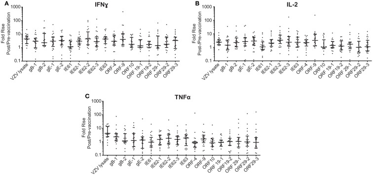
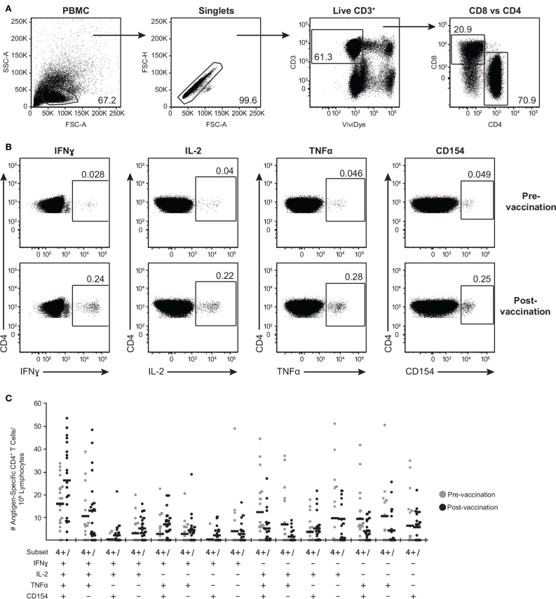
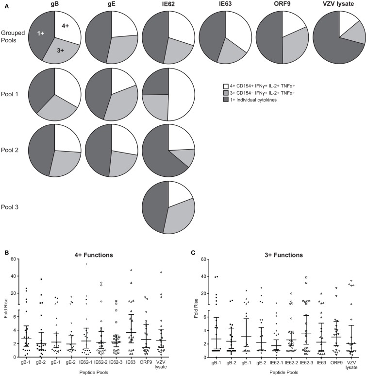
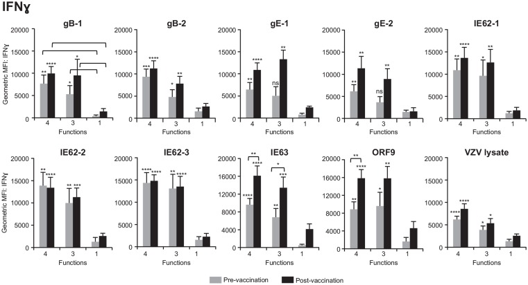
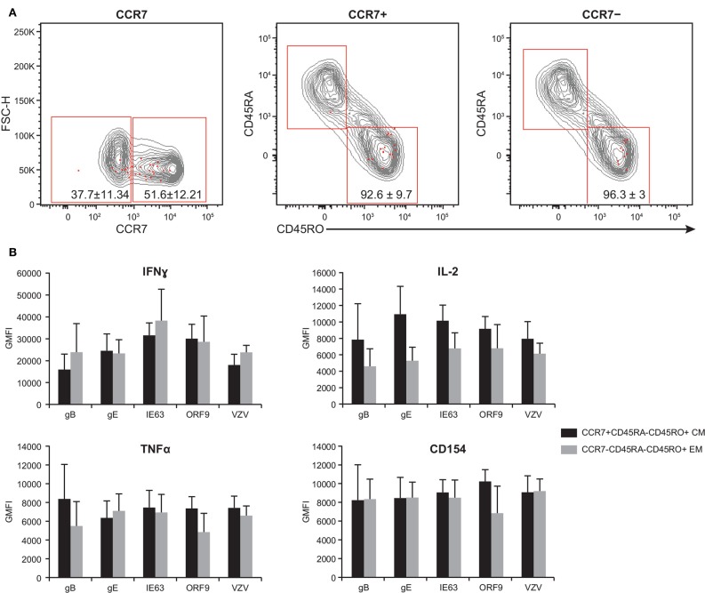
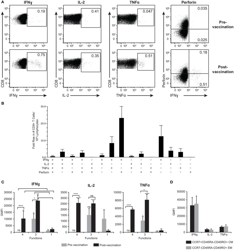
ZOSTAVAX(®) is a live attenuated varicella-zoster virus (VZV) vaccine that is licensed for the protection of individuals ≥50 years against shingles and its most common complication, postherpetic neuralgia. While IFNγ responses increase upon vaccination, the quality of the T cell response has not been elucidated. By using polychromatic flow cytometry, we characterized the breadth, magnitude, and quality of ex vivo CD4(+) and CD8(+) T cell responses induced 3-4 weeks after ZOSTAVAX vaccination of healthy adults. We show, for the first time that the highest frequencies of VZV-specific CD4(+) T cells were poly-functional CD154(+)IFNγ(+)IL-2(+)TNFα(+) cells, which were boosted upon vaccination. The CD4(+) T cells were broadly reactive to several VZV proteins, with immediate early (IE) 63 ranking the highest among them in the fold rise of poly-functional cells, followed by IE62, gB, open reading frame (ORF) 9, and gE. We identified a novel poly-functional ORF9-specific CD8(+) T cell population in 62% of the subjects, and these were boosted upon vaccination. Poly-functional CD4(+) and CD8(+) T cells produced significantly higher levels of IFNγ, IL-2, and TNFα compared to mono-functional cells. After vaccination, a boost in the expression of IFNγ by poly-functional IE63- and ORF9-specific CD4(+) T cells and IFNγ, IL-2, and TNFα by ORF9-specific poly-functional CD8(+) T cells was observed. Responding poly-functional T cells exhibited both effector (CCR7(-)CD45RA(-)CD45RO(+)), and central (CCR7(+)CD45RA(-)CD45RO(+)) memory phenotypes, which expressed comparable levels of cytokines. Altogether, our studies demonstrate that a boost in memory poly-functional CD4(+) T cells and ORF9-specific CD8(+) T cells may contribute toward ZOSTAVAX efficacy.

Keywords: VZV antigens; ZOSTAVAX; flow cytometry; memory response; poly-functional T cells.


Figures

Figure 1
Figure 1
Boost in IFNγ, IL-2, and TNFα following ZOSTAVAX vaccination. Whole blood was drawn from 21 subjects prior to and following ZOSTAVAX (3–4 weeks) vaccination. Within 2 h of blood draw, the specimen was stimulated with VZV peptide pools, VZV lysate (positive control), MRC5, and DMSO (negative controls) for 24 h. Plasma was then harvested and tested for the presence of (A) effector cytokines – IFNγ, IL-2, and TNFα; (B) pro-inflammatory cytokines – IL-1β and IL-10; and (C) anti-inflammatory cytokine IL-10, by using the Meso Scale Discovery kits. Each dot represents one subject. The geometric mean ± 95% confidence interval is shown for all data sets. Horizontal broken line denotes the lowest limit of detection for all cytokines.
Figure 2
Figure 2
Fold rise in (A) IFNγ, (B) IL-2, and (C) TNFα levels following ZOSTAVAX vaccination. The fold rise (post/pre) was calculated for each vaccinated subject. Graph shows the median ± interquartile range for all 21 subjects.
Figure 3
Figure 3
Poly-functional CD4+ T cells are boosted following ZOSTAVAX vaccination. PBMC were thawed and rested overnight before VZV antigen stimulation. One million PBMC/sample were stimulated with antigens for 5.5 h at 37°C/5% CO2, with brefeldin A and monensin added 30 min after the initial incubation. Cells were stained with Vividye, then fixed, permeabilized, and stained with antibodies to identify (A) CD3+ T cells, expressing either CD4 or CD8 that were producing (B) IFNγ, IL-2, TNFα, and CD154. Figure shows IE62-specific responses from one individual, and numbers on dot plots represent percentages of cells (DMSO subtracted) expressing each molecule. (C) Shows the heterogeneity of IE63-specific poly-functional CD4+ T cells for all 21 subjects as determined by Boolean-gating strategy. Each dot represents one subject, and the mean is shown for all phenotypes.
Figure 4
Figure 4
Hierarchy of VZV antigens stimulating poly-functional CD4+ T cells. (A) Grouped pie charts comparing the fold rise in poly-functional CD4+ T cells after stimulating PBMC with gB, gE, IE62, IE63, ORF9, and VZV lysate. Pie charts display 4+ functions (CD154+IFNγ+IL-2+TNFα+), 3+ functions (CD154IFNγ+IL-2+TNFα+), or mono-functional cells. gB, gE, and IE62 were fractioned into multiple pools and poly-functional T cells were also assessed. Fold rise in the number of (B) 4+ (CD154+IFNγ+IL-2+TNFα+) or (C) 3+ (CD154IFNγ+IL-2+TNFα+) CD4+ T cells for each VZV peptide pool. Each dot represents one subject, and the geometric mean ± 95% confidence interval is shown.
Figure 5
Figure 5
Poly-functional CD4+ T cells express higher levels of IFNγ than mono-functional cells. Geometric mean fluorescence intensity (GMFI) of IFNγ was determined in 4+ (CD154+IFNγ+IL-2+TNFα+), 3+ (CD154IFNγ+IL-2+TNFα+), and mono-functional antigen-specific CD4+ T cells prior to and following ZOSTAVAX vaccination. The mean ± SEM for the 21 subjects is shown for all data sets.
Figure 6
Figure 6
Poly-functional CD4+ T cells display both TEM and TCM phenotypes. PBMC isolated from five subjects following ZOSTAVAX vaccination were thawed and rested overnight before VZV antigen stimulation. PBMC were stimulated with various VZV antigens for 5.5 h at 37°C/5% CO2, with brefeldin A and monensin added 30 min after the initial incubation. Cells were stained with Vividye, surface stained with fluorescent primary antibodies against CCR7, CD45RA, and CD45RO. After washing off excess antibodies, PBMC were fixed, permeabilized, and stained with fluorescent primary antibodies to identify T cells producing IFNγ, IL-2, TNFα, and CD154 as described in Figure 3. Boolean analysis was performed to identify poly-functional CD4+ T cells with 4+ functions. (A) CCR7, CD45RA, and CD45RO expressions of 4+ (CD154+IFNγ+IL-2+TNFα+) antigen-specific CD4+ T cells (red dots) overlayed on CD3+ T cells (gray contours). Numbers in dot plots represent percentage mean ± SEM for the five subjects. (B) The GMFI of IFNγ, IL-2, TNFα, and CD154 was assessed in both TCM (CCR7+CD45RACD45RO+) and TEM (CCR7CD45RACD45RO+) 4+ antigen-specific CD4+ T cells.
Figure 7
Figure 7
ORF9-specific CD8+ T cells are poly-functional, produce more cytokines than mono-functional cells, and display both TEM and TCM phenotypes. (A) Representative gating strategy of PBMC prior to and following ZOSTAVAX vaccination that were stimulated with ORF9 peptide pool. Numbers in dot plots denotes percentages of CD8+ T cells producing IFNγ, IL-2, TNFα, and perforin. (B) Fold rise in the number of poly-functional CD8+ T cells demonstrating the heterogeneity of the T cell population. (C) The expression levels (GMFI) of IFNγ, IL-2, and TNFα by 4+ (IFNγ+IL-2+TNFα+perforin+), 3+ (IFNγ+IL-2+TNFα+), and mono-functional cells prior to and following ZOSTAVAX vaccination. (D) GMFI of IFNγ, IL-2, and TNFα was assessed in both ORF9-specific CD8+ TCM (CCR7+CD45RACD45RO+) and TEM (CCR7CD45RACD45RO+).

Similar articles

Cited by

References

    1. Mueller NH, Gilden DH, Cohrs RJ, Mahalingam R, Nagel MA. Varicella zoster virus infection: clinical features, molecular pathogenesis of disease, and latency. Neurol Clin (2008) 26:675–697,viii.10.1016/j.ncl.2008.03.011 - DOI - PMC - PubMed
    1. Hope-Simpson RE. The nature of herpes zoster: a long-term study and a new hypothesis. Proc R Soc Med (1965) 58:9–20. - PMC - PubMed
    1. Kumagai T, Chiba Y, Wataya Y, Hanazono H, Chiba S, Nakao T. Development and characteristics of the cellular immune response to infection with varicella-zoster virus. J Infect Dis (1980) 141:7–13.10.1093/infdis/141.1.7 - DOI - PubMed
    1. Arvin AM, Koropchak CM, Williams BR, Grumet FC, Foung SK. Early immune response in healthy and immunocompromised subjects with primary varicella-zoster virus infection. J Infect Dis (1986) 154:422–9.10.1093/infdis/154.3.422 - DOI - PubMed
    1. Hope-Simpson RE. Postherpetic neuralgia. J R Coll Gen Pract (1975) 25:571–5. - PMC - PubMed