

T Cell Motility as Modulator of Interactions with Dendritic Cells
- PMID: 26579132
- PMCID: PMC4629691
- DOI: 10.3389/fimmu.2015.00559
T Cell Motility as Modulator of Interactions with Dendritic Cells
Abstract
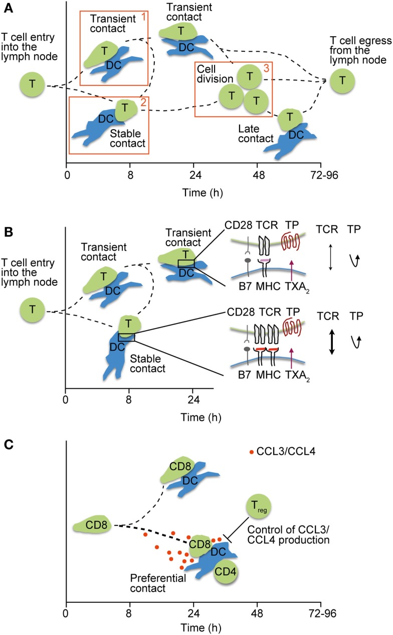
It is well established that the balance of costimulatory and inhibitory signals during interactions with dendritic cells (DCs) determines T cell transition from a naïve to an activated or tolerant/anergic status. Although many of these molecular interactions are well reproduced in reductionist in vitro assays, the highly dynamic motility of naïve T cells in lymphoid tissue acts as an additional lever to fine-tune their activation threshold. T cell detachment from DCs providing suboptimal stimulation allows them to search for DCs with higher levels of stimulatory signals, while storing a transient memory of short encounters. In turn, adhesion of weakly reactive T cells to DCs presenting peptides presented on major histocompatibility complex with low affinity is prevented by lipid mediators. Finally, controlled recruitment of CD8(+) T cells to cognate DC-CD4(+) T cell clusters shapes memory T cell formation and the quality of the immune response. Dynamic physiological lymphocyte motility therefore constitutes a mechanism to mitigate low avidity T cell activation and to improve the search for "optimal" DCs, while contributing to peripheral tolerance induction in the absence of inflammation.
Keywords: Myo1g; T cell motility; T cell–DC interactions; intravital imaging; thromboxane A2.
Figures
Similar articles
-
Thromboxane A2 acts as tonic immunoregulator by preferential disruption of low-avidity CD4+ T cell-dendritic cell interactions.J Exp Med. 2014 Dec 15;211(13):2507-17. doi: 10.1084/jem.20140137. Epub 2014 Dec 8. J Exp Med. 2014. PMID: 25488981 Free PMC article.
-
Dendritic type, accessory cells within the mammalian thymic microenvironment. Antigen presentation in the dendritic neuro-endocrine-immune cellular network.In Vivo. 1997 Jul-Aug;11(4):351-70. In Vivo. 1997. PMID: 9292303
-
Late dendritic cells are still able to evoke a potent alloreactive CTL response.Immunobiology. 2008;213(1):51-64. doi: 10.1016/j.imbio.2007.06.005. Epub 2007 Aug 6. Immunobiology. 2008. PMID: 18207027
-
Immunodeficiency virus exploitation of dendritic cells in the early steps of infection.J Leukoc Biol. 2003 Nov;74(5):683-90. doi: 10.1189/jlb.0403178. Epub 2003 Jul 22. J Leukoc Biol. 2003. PMID: 12960236 Review.
-
Involvement of dendritic cells in the pathogenesis of inflammatory bowel disease.Adv Exp Med Biol. 2006;579:117-32. doi: 10.1007/0-387-33778-4_8. Adv Exp Med Biol. 2006. PMID: 16620015 Review.
Cited by
-
Role of Mechanotransduction and Tension in T Cell Function.Front Immunol. 2018 Nov 15;9:2638. doi: 10.3389/fimmu.2018.02638. eCollection 2018. Front Immunol. 2018. PMID: 30519239 Free PMC article. Review.
-
CCR7 Is Recruited to the Immunological Synapse, Acts as Co-stimulatory Molecule and Drives LFA-1 Clustering for Efficient T Cell Adhesion Through ZAP70.Front Immunol. 2019 Jan 14;9:3115. doi: 10.3389/fimmu.2018.03115. eCollection 2018. Front Immunol. 2019. PMID: 30692994 Free PMC article.
-
In silico evaluation of the immunogenic profile of lung cancers with SMARCA4 genetic alterations.Sci Rep. 2025 May 22;15(1):17832. doi: 10.1038/s41598-025-02494-x. Sci Rep. 2025. PMID: 40404793 Free PMC article.
-
Taking up Cancer Immunotherapy Challenges: Bispecific Antibodies, the Path Forward?Antibodies (Basel). 2015 Dec 26;5(1):1. doi: 10.3390/antib5010001. Antibodies (Basel). 2015. PMID: 31557983 Free PMC article. Review.
References
Publication types
LinkOut - more resources
Full Text Sources
Other Literature Sources
Research Materials
Miscellaneous