Photoreceptor Inner and Outer Segment Junction Reflectivity after Vitrectomy for Macula-Off Rhegmatogenous Retinal Detachment
- PMID: 26579234
- PMCID: PMC4633586
- DOI: 10.1155/2015/451408
Photoreceptor Inner and Outer Segment Junction Reflectivity after Vitrectomy for Macula-Off Rhegmatogenous Retinal Detachment
Abstract
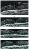
Purpose. To evaluate the spatial distribution of photoreceptor inner and outer segment junction (IS/OS) reflectivity changes after successful vitrectomy for macula-off retinal detachment (PPV-mOFF) using spectral domain optical coherence tomography (SdOCT). Methods. Twenty eyes after successful PPV-mOFF were included in the study. During a mean follow-up period of 15.3 months, SdOCT was performed four times. To evaluate the IS/OS reflectivity a four-grade scale was used. Results. At the first follow-up visit the IS/OS had very similar reflectivity in entire length of the central scan with total average value of 1,05. At the second visit the most significant increase of the reflectivity was observed in temporal and nasal parafovea with average values of 2,17 and 2,22, respectively. The third region of increased reflectivity of an average value of 2,33 appeared during the third follow-up visit and was located in the foveola. At the last follow-up visit in entire central cross section the IS/OS reflectivity exceeded grade 2 reaching the highest average values in nasal and temporal parafovea and foveola. Conclusions. A gradual increase of the IS/OS reflectivity was observed in eyes after PPV-mOFF. The process is not random and starts independently in the peripheral and central part of the macula which may be attributed to the variable regenerative potential of cones and rods.
Figures


Similar articles
-
Morphologic and Functional Assessment of Photoreceptors After Macula-Off Retinal Detachment With Adaptive-Optics OCT and Microperimetry.Am J Ophthalmol. 2020 Jun;214:72-85. doi: 10.1016/j.ajo.2019.12.015. Epub 2019 Dec 25. Am J Ophthalmol. 2020. PMID: 31883465
-
Foveal microstructure and visual acuity after retinal detachment repair: imaging analysis by Fourier-domain optical coherence tomography.Ophthalmology. 2009 Mar;116(3):519-28. doi: 10.1016/j.ophtha.2008.10.001. Epub 2009 Jan 14. Ophthalmology. 2009. PMID: 19147231
-
Spectral-domain optical coherence tomography imaging of the detached macula in rhegmatogenous retinal detachment.Retina. 2009 Feb;29(2):232-42. doi: 10.1097/IAE.0b013e31818bcd30. Retina. 2009. PMID: 18997641
-
Outer retinal thickness and retinal sensitivity in macula-off rhegmatogenous retinal detachment after successful reattachment.Eur J Ophthalmol. 2012 Nov-Dec;22(6):1032-8. doi: 10.5301/ejo.5000148. Epub 2012 Apr 11. Eur J Ophthalmol. 2012. PMID: 22505048
-
Anatomical and functional macular changes after rhegmatogenous retinal detachment with macula off.Am J Ophthalmol. 2012 Jan;153(1):128-36. doi: 10.1016/j.ajo.2011.06.010. Epub 2011 Sep 19. Am J Ophthalmol. 2012. PMID: 21937016
Cited by
-
Clinical and spectral-domain optical coherence tomography findings and changes in new-onset macular edema after silicone oil tamponade.BMC Ophthalmol. 2025 Mar 27;25(1):153. doi: 10.1186/s12886-025-03986-0. BMC Ophthalmol. 2025. PMID: 40148832 Free PMC article.
References
LinkOut - more resources
Full Text Sources
Other Literature Sources

