. 2015:2015:235720.
doi: 10.1155/2015/235720. Epub 2015 Oct 22.

A Simple and Safe Technique for CT Guided Lung Nodule Marking prior to Video Assisted Thoracoscopic Surgical Resection Revisited

Affiliations

A Simple and Safe Technique for CT Guided Lung Nodule Marking prior to Video Assisted Thoracoscopic Surgical Resection Revisited

James A Stephenson et al. Lung Cancer Int. 2015.

Abstract

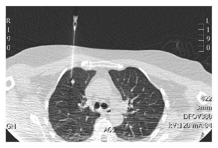
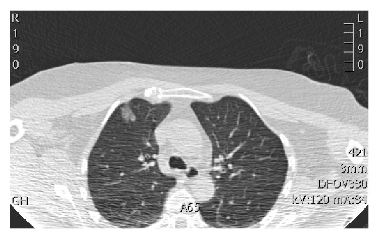
Aim. We describe our experience of a simple, safe, and reproducible technique for lung nodule marking prethoracoscopic metastasectomy. Thoracoscopic lung nodule resection reduces patient discomfort, complications, higher level of care, hospital stay, and cost; however, small deeply placed lung nodules are difficult to locate and resect thoracoscopically. Materials and Methods. We describe and review the success of our novel technique, where nodules are identified on a low dose CT and marked with methylene blue using CT fluoroscopy guidance immediately prior to surgery. Results. 30 nodules were marked with a mean size of 8 mm (4-18 mm) located at a mean depth of 17 mm, distributed through both lungs. Dye was detected at the pleural surface in 97% of the patients and at the nodule in 93%. There were no major complications. Thoracoscopic resection was possible in 90%. Conclusion. This is a simple and safe method of lung nodule marking to facilitate thoracoscopic resection in cases where this may not be technically possible due to nodule location.


Figures

Figure 1
Figure 1
Image capture from CT fluoroscopy: needle inserted under CT fluoroscopic guidance to a position adjacent to the target nodule.
Figure 2
Figure 2
Image capture from CT fluoroscopy: the needle has been retracted and removed. The high-density methylene blue/blood track to the pleural surface can be identified.

References

    1. Mack M. J., Aronoff R. J., Acuff T. E., Douthit M. B., Bowman R. T., Ryan W. H. Present role of thoracoscopy in the diagnosis and treatment of diseases of the chest. The Annals of Thoracic Surgery. 1992;54(3):403–409. doi: 10.1016/0003-4975(92)90428-7. - DOI - PubMed
    1. Miller D. L., Allen M. S., Trastek V. F., Deschamps C., Pairolero P. C. Videothoracoscopic wedge excision of the lung. The Annals of Thoracic Surgery. 1992;54(3):410–414. doi: 10.1016/0003-4975(92)90429-8. - DOI - PubMed
    1. Suzuki K., Nagai K., Yoshida J., et al. Video-assisted thoracoscopic surgery for small indeterminate pulmonary nodules: indications for preoperative marking. Chest. 1999;115(2):563–568. doi: 10.1378/chest.115.2.563. - DOI - PubMed
    1. Nakas A., Klimatsidas M. N., Entwisle J., Martin-Ucar A. E., Waller D. A. Video-assisted versus open pulmonary metastasectomy: the surgeon's finger or the radiologist's eye? European Journal of Cardio-Thoracic Surgery. 2009;36(3):469–474. doi: 10.1016/j.ejcts.2009.03.050. - DOI - PubMed
    1. Lenglinger F. X., Schwarz C. D., Artmann W. Localization of pulmonary nodules before thoracoscopic surgery: value of percutaneous staining with methylene blue. American Journal of Roentgenology. 1994;163(2):297–300. doi: 10.2214/ajr.163.2.7518642. - DOI - PubMed

LinkOut - more resources