. 2016 Jul;117(7):1556-67.
doi: 10.1002/jcb.25447. Epub 2016 Feb 19.

Calcium Sensing Receptor Function Supports Osteoblast Survival and Acts as a Co-Factor in PTH Anabolic Actions in Bone

Affiliations

Calcium Sensing Receptor Function Supports Osteoblast Survival and Acts as a Co-Factor in PTH Anabolic Actions in Bone

Saja A Al-Dujaili et al. J Cell Biochem. 2016 Jul.

Abstract

Anabolic actions of PTH in bone involve increased deposition of mineralizing matrix. Regulatory feedback of the process may be important to maintain calcium homeostasis and, in turn, calcium may inform the process. This investigation clarified the role of calcium availability and the calcium sensing receptor (CaSR) in the anabolic actions of PTH. CaSR function promoted osteoblastic cell numbers, with lower cell numbers in post-confluent cultures of primary calvarial cells from Col1-CaSR knock-out (KO) mice, and for calvarial cells from wild-type (WT) mice treated with a calcilytic. Increased apoptosis of calvarial cells with calcilytic treatment suggested CaSR is critical for protection against stage-dependent cell death. Whole and cortical, but not trabecular, bone parameters were significantly lower in Col1-CaSR KO mice versus WT littermates. Intact Col1-CaSR KO mice had lower serum P1NP levels relative to WT. PTH treatment displayed anabolic actions in WT and, to a lesser degree, KO mice, and rescued the lower P1NP levels in KO mice. Furthermore, PTH effects on whole tibiae were inhibited by osteoblast-specific CaSR ablation. Vertebral body implants (vossicles) from untreated Col1-CaSR KO and WT mice had similar bone volumes after 4 weeks of implantation in athymic mice. These findings suggest that trabecular bone formation can occur independently of the CaSR, and that the CaSR plays a collaborative role in the PTH anabolic effects on bone. J. Cell. Biochem. 117: 1556-1567, 2016. © 2015 Wiley Periodicals, Inc.

Keywords: CALCIUM; CALCIUM SENSING RECEPTOR; OSTEOBLASTS; PARATHYROID HORMONE.


Figures

Fig. 1
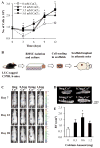
Fig. 1. Osteoblasts and bone volume are influenced by calcium availability
(A) Calvarial osteoblast numbers are dependent on exogenous calcium concentration in vitro. Cell number increased dose-dependently and reached a maximum with 3.5 mM CaCl2 treatment (* p<0.05 relative to no calcium, n=4/gp). (B) Schematic illustration of ossicle experiment. BMSCs were isolated from long bones of luciferase-tagged C57BL/6 mice, expanded in vitro and seeded (1×10⁶ cells/ossicle) in PLLA-based scaffolds. Scaffolds were implanted in athymic mice and left for 28 days, with regular in vivo monitoring using bioluminescence (BLI). (C) BLI imaging was used to monitor cell number post-implantation. Representative microCT images (D) and analysis (E) of scaffolds after harvest indicated a significant increase in bone volume in the 0.6 mg Ca2+ scaffold. (Data is mean ± SEM, n=8/gp; * p<0.05 relative to 0 mg Ca2+).
Fig. 2
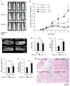
Fig. 2. Calcium enhanced PTH effects on osteoblast number in vivo
(A) Representative BLI images of mice implanted with scaffolds (0 mg vs. 0.6 mg Ca2+) seeded with luciferase-tagged BMSCs. (B) PTH administration significantly increased luminescence reflecting increased cell number relative to vehicle which was further enhanced by calcium (0.6 mg Ca2+) relative to calcium-free (0 mg Ca2+) scaffolds. Representative images (C) and analysis (D) of scaffolds on day 40 using microCT suggested a PTH effect in scaffolds with no calcium. (E) BLI/BV, reflecting the number of cells per bone volume, was significantly increased by the combination of calcium and PTH. (F) Histological analysis of osseous matrix area relative to total scaffold area using H&E staining revealed an increase in mineralized tissue in Ca-free scaffolds with PTH treatment but not in calcium scaffolds. (G) There was no difference in cell number (BLI) per bone area (histology) between calcium and calcium-free scaffolds within treatment groups, but a significant increase was found in calcium scaffolds with PTH. (H) Representative images of scaffolds. Unstained (grey/white) areas indicate remaining scaffold particles, whereas pink denotes eosin-positive bone matrix. (Data is mean ± SEM, n = 8/gp; * p<0.05 PTH + 0.6 mg Ca2+ vs. Veh + 0.6 mg Ca2+, ** p<0.05 PTH + 0.6 mg Ca2+ vs. PTH + 0 mg Ca2+, *** p<0.05 PTH + 0 mg Ca2+ vs. Veh + 0 mg Ca2+).
Fig. 3
Fig. 3. Presence of the CaSR supports osteoblast survival in vitro
Calvarial cell culture from osteoblast-specific CaSR knock-out mice (Col-BoneCaSRΔflox/Δflox; KO) and control littermates (WT). (A) There were no differences between the number of KO and WT calvarial osteoblasts during the initial days of culture. However, after cells reached confluence the number of WT cells continued to increase, whereas KO cells plateaued. (n=7–11/gp, * p<0.05 KO relative to WT cells). (B) Pharmacologic treatment of WT cells with a CaSR agonist (calcimimetic, R-568) or antagonist (calcilytic, NPS 2143) had no effect on cell number during the initial days of culture, but NPS 2143 caused cell number to decline after cells reached confluence. (C) Measurement of DNA fragmentation revealed an increase in apoptosis with NPS 2143 treatment on day 14 relative to the other treatment groups. (n=4/gp for B–C, * p<0.05 relative to both vehicle and calcimimetic treatment). All data is mean ± SEM.
Fig. 4
Fig. 4. PTH effects on body weight, serum P1NP and tibial radiography in WT vs. Col-BoneCaSRΔflox/Δflox mice
(A) Images of 21-day-old WT and KO littermates after 17 days of vehicle or PTH treatment. (B) Mouse total body weight was significantly lower in KO mice, with no PTH effect in either WT or KO mice. (C) Serum P1NP levels were significantly reduced in KO vs. WT vehicle-treated mice. PTH treatment increased P1NP levels in WT mice, and normalized the effect of the Col-BoneCaSRΔflox/Δflox KO to WT-vehicle levels. (n=8–9 for B–C; * p<0.05 relative to WT control in each treatment group.) (D) KO tibiae appeared smaller and more radiolucent than WT. PTH treatment increased radiopacity of both KO and WT bones. (E) Histologic analysis revealed that osseous bone matrix area was not affected by the Col1-CaSR KO, and was increased with PTH treatment in both WT and KO mice (n=8–9/gp; # p<0.01 relative to vehicle treatment). (F) Representative images of H&E staining of mouse tibiae. All data are mean ± SEM.
Fig. 5
Fig. 5. PTH effects on microCT parameters in WT vs. Col1-CaSR KO mice
MicroCT reconstruction (A) and analysis (B–J) of Col1-CaSR WT and KO tibiae revealed significant differences between WT and KO mice in both vehicle and PTH-treated mice. (B–C) The anabolic actions of PTH on whole tibiae (W.B. – whole bone) were diminished in Col1-CaSR KO mice. (D–H) No differences in trabecular bone (Tb.) parameters (BV/TV, BMD, Tb.N. – trabecular number, Tb.Th. trabecular thickness, Tb.Sp. – trabecular spacing) between vehicle-treated WT and KO mice. A consistent PTH effect was seen in trabecular bone parameters of WT mice, and to a lesser extent in the KO mice but not in trabecular BV/TV of KO mice. (I–J) Cortical bone (Ct.) parameters (BV/TV, BMD) were significantly compromised in Col1-CaSR KO mice, and PTH did not alter cortical parameters in either group. (Data are mean ± SEM, n = 8/gp; * p<0.05 relative to WT control in the same treatment group, unless otherwise indicated).

Similar articles

Cited by

References

    1. Bashutski JD, Eber RM, Kinney JS, Benavides E, Maitra S, Braun TM, Giannobile WV, McCauley LK. Teriparatide and osseous regeneration in the oral cavity. New England Journal of Medicine. 2010;363:2396–405. - PMC - PubMed
    1. Bennett JH, Joyner CJ, Triffitt JT, Owen ME. Adipocytic cells cultured from marrow have osteogenic potential. Journal of Cell Science. 1991;99(Pt 1):131–9. - PubMed
    1. Beresford JN, Bennett JH, Devlin C, Leboy PS, Owen ME. Evidence for an inverse relationship between the differentiation of adipocytic and osteogenic cells in rat marrow stromal cell cultures. Journal of Cell Science. 1992;102(Pt 2):341–51. - PubMed
    1. Boden SD, Kaplan FS. Calcium homeostasis. Orthopedic Clinics of North America. 1990;21:31–42. - PubMed
    1. Brown EM. The calcium-sensing receptor: physiology, pathophysiology and CaR-based therapeutics. Subcellular Biochemistry. 2007;45:139–67. - PubMed

LinkOut - more resources