. 2016 Jun;14(6):1381-93.
doi: 10.1111/pbi.12502. Epub 2015 Nov 18.

Eucalyptus hairy roots, a fast, efficient and versatile tool to explore function and expression of genes involved in wood formation

Affiliations

Eucalyptus hairy roots, a fast, efficient and versatile tool to explore function and expression of genes involved in wood formation

Anna Plasencia et al. Plant Biotechnol J. 2016 Jun.

Abstract

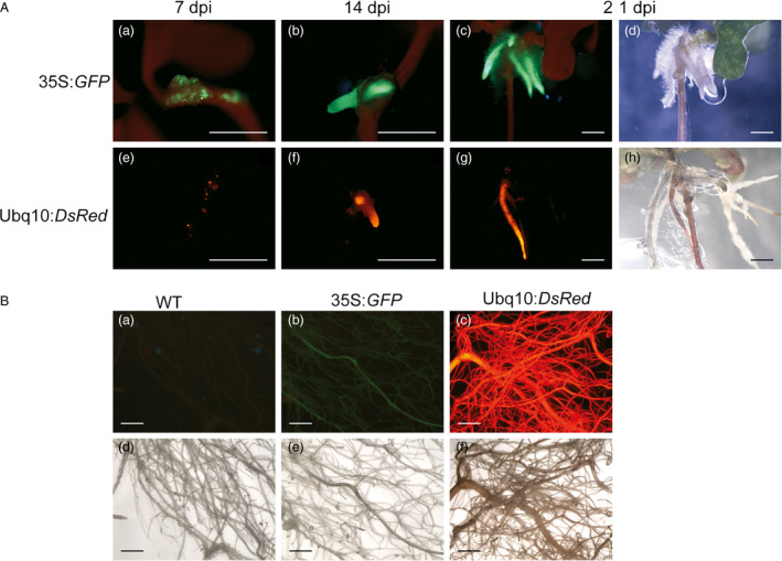
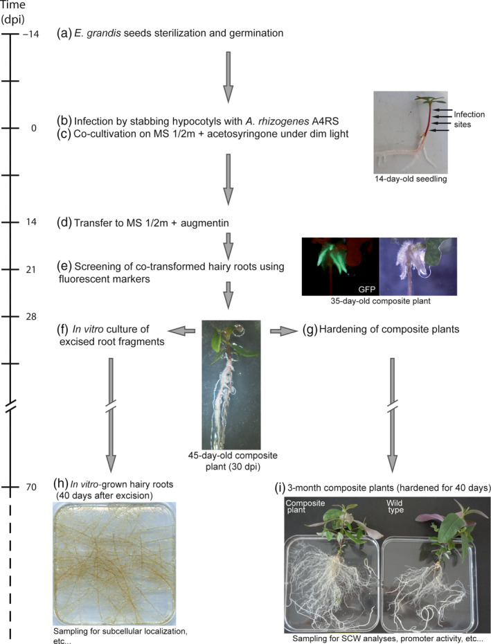
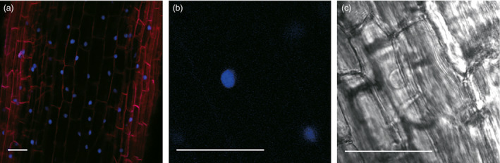
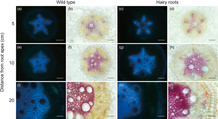
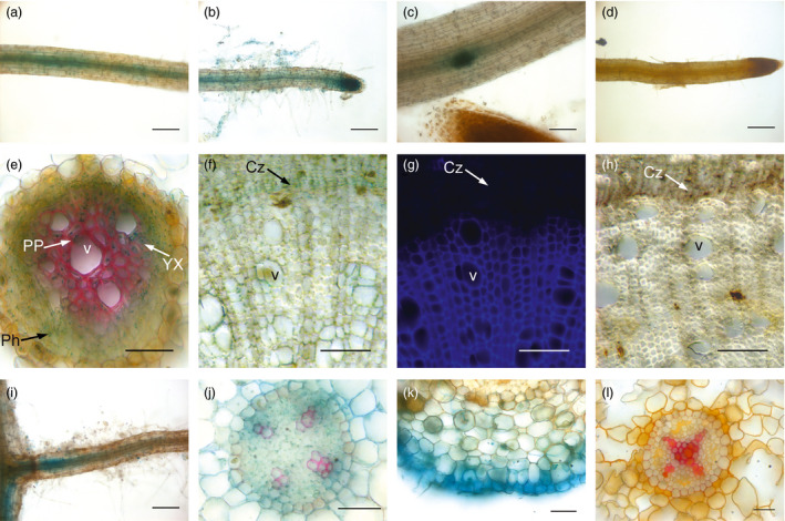
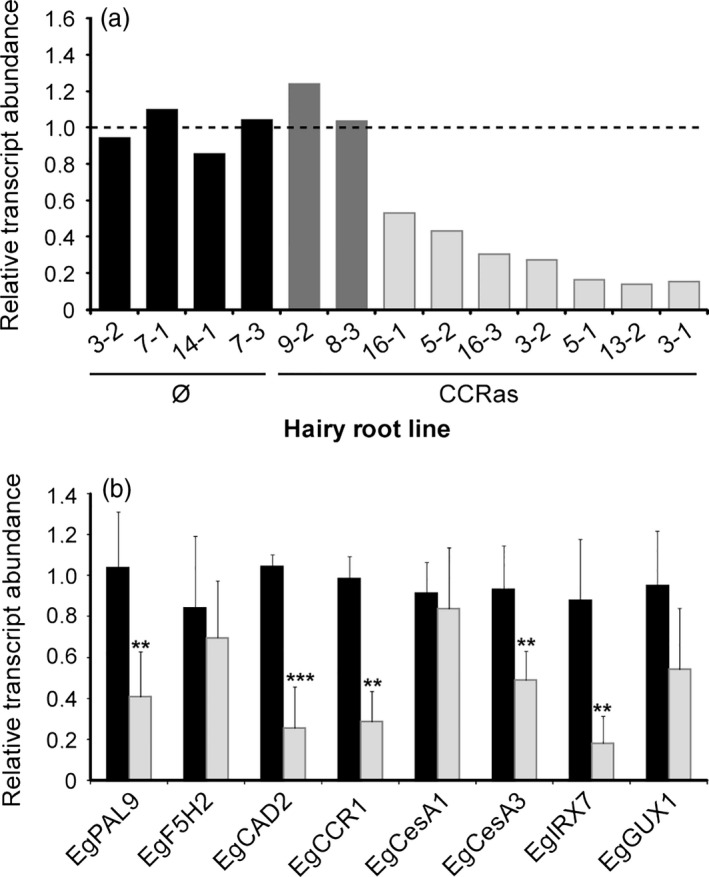
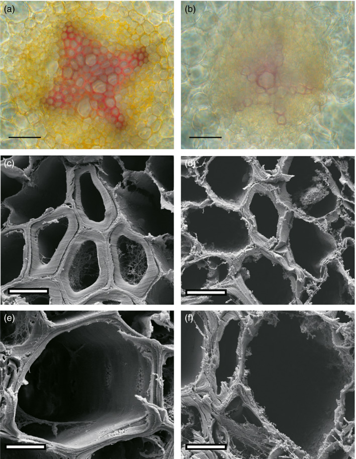
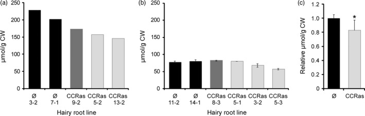
Eucalyptus are of tremendous economic importance being the most planted hardwoods worldwide for pulp and paper, timber and bioenergy. The recent release of the Eucalyptus grandis genome sequence pointed out many new candidate genes potentially involved in secondary growth, wood formation or lineage-specific biosynthetic pathways. Their functional characterization is, however, hindered by the tedious, time-consuming and inefficient transformation systems available hitherto for eucalypts. To overcome this limitation, we developed a fast, reliable and efficient protocol to obtain and easily detect co-transformed E. grandis hairy roots using fluorescent markers, with an average efficiency of 62%. We set up conditions both to cultivate excised roots in vitro and to harden composite plants and verified that hairy root morphology and vascular system anatomy were similar to wild-type ones. We further demonstrated that co-transformed hairy roots are suitable for medium-throughput functional studies enabling, for instance, protein subcellular localization, gene expression patterns through RT-qPCR and promoter expression, as well as the modulation of endogenous gene expression. Down-regulation of the Eucalyptus cinnamoyl-CoA reductase1 (EgCCR1) gene, encoding a key enzyme in lignin biosynthesis, led to transgenic roots with reduced lignin levels and thinner cell walls. This gene was used as a proof of concept to demonstrate that the function of genes involved in secondary cell wall biosynthesis and wood formation can be elucidated in transgenic hairy roots using histochemical, transcriptomic and biochemical approaches. The method described here is timely because it will accelerate gene mining of the genome for both basic research and industry purposes.

Keywords: Agrobacterium rhizogenes; Eucalyptus; hairy roots; lignin; secondary cell wall; xylem.


Figures

Figure 1
Figure 1
Developmental time course of Eucalyptus grandis hairy roots expressing either the GFP or the DsRed fluorescent markers. Panel A: stereomicrographs of roots emerging from infected sites on hypocotyls at 7 (a, e), 14 (b, f) or 21 (c, d, g, h) days after infection (dpi). GFP‐transformed calli or roots appeared in green, whereas autofluorescence appeared in red (a, b, c). DsRed‐transformed calli or roots appeared in red without noticeable autofluorescence (e, f, g). Panel B: stereomicrographs of hairy roots from wild‐type (a, d) and composite plants overexpressing GFP (b, e) or DsRED (c, f) hardened for 1.5 month (75 dpi). Bright field was used to show root morphology and to localize untransformed roots (Panel A: d, h; Panel B: d–f). Scale bars = 2 mm.
Figure 2
Figure 2
Sequential steps of the transformation of 14‐day‐old Eucalyptus grandis seedlings by A4RS harbouring either a GFP‐ or DsRed‐based binary vector. (a) E. grandis seeds were sterilized and germinated in 1/4 strength MS medium. (b) 14‐day‐old seedlings were infected by stabbing the hypocotyl with a needle swabbed with Agrobacterium rhizogenes. (c) Infected plants were co‐cultivated with agrobacteria for 14 days on MS medium with 1/2 strength macroelements (MS 1/2m) supplemented with acetosyringone under dim light. (d) Plants were transferred to MS 1/2m medium supplemented with augmentin. (e) The hairy roots generated were examined at 21 days after infection (dpi) under a stereo fluorescence microscope. (f) Co‐transformed roots were excised to be cultivated in vitro on MER media. (g) Before hardening, nontransformed roots were removed and the resulting composite plants were placed in pots and cultivated in a phytotron. (i) After 40 days of hardening, roots were sampled for secondary cell wall and promoter activity analyses, whereas in vitro‐grown excised roots (h) could be used for other purposes such as subcellular localization of proteins.
Figure 3
Figure 3
Nuclear localization of histone linker (H1) fused to CFP in Eucalyptus grandis root cells. Confocal images of E. grandis hairy root cells expressing H1‐CFP fusion protein. All nuclei fluoresced in blue and the cell walls in red (a). Detail of fluorescent nuclei under CFP emission (b) and bright field (c). Scale bars = 30 μm.
Figure 4
Figure 4
Comparison of xylem development and lignified secondary cell walls between transgenic and wild‐type roots. Transversal root sections made at 5 (a–d), 10 (e–h) and 20 cm (i–l) from the root apex for wild‐type (a, b, e, f, i, j) and hairy (c, d, g, h, k, l) roots. Lignified cell walls are visualized in blue by UV autofluorescence (a, c, e, g, i, k) and in red by using phloroglucinol–HCl (b, d, f, h, j, l). Scale bars = 30 μm.
Figure 5
Figure 5
Histochemical localization of GUS activity in Eucalyptus grandis hairy roots transformed with the EgCCR1 and EgCAD2 promoters. Roots from hardened composite plants (3 months old) showing GUS activity for EgCCR1 (a–c, e–g) and EgCAD2 (i–k) promoters. Control roots are also shown (d, h, l). GUS activity is observed in the vascular cylinder (a, b, c, i), the root tip (b) and at the emergence of a lateral root (c). Transversal cross‐sections were performed at 2 (j, l), 15 (e, k) and 30 cm (f–h) from the root apex. Lignified cell walls are visualized in red using phloroglucinol–HCl performed after GUS staining (e, j, l) or, in the case of roots at 30 cm (f), are alternatively visualized under UV light (g). GUS activity is observed in secondary xylem cells (e, f), cells from the cambial zone (f) and cells from epidermis (k). No GUS activity is observed in control roots (d, h, l). YX: young xylem cells; PP: paratracheal parenchyma cell; Ph: phloem; Cz: cambial zone; v: vessel. Scale bars = 100 μm (a–d, f–i) and 30 μm (e, j–l).
Figure 6
Figure 6
Relative transcript levels of EgCCR1 and secondary cell wall (SCW)‐related genes in antisense EgCCR1 ( CCR as) hairy root lines. (a) Mean relative transcript levels of EgCCR1 analysed by RTqPCR. Black bars: control roots transformed with an empty pGWAY‐1 vector (Ø); dark grey bars: nonsilenced CCR as lines (9‐2; 8‐3), light grey bars: CCR as lines exhibiting different degrees of silencing (16‐1; 5‐2; 16‐3; 3‐2; 5‐1; 13‐2; 3‐1). (b) Transcript levels of several SCW‐related biosynthetic genes were assessed in control (black bars) and silenced CCR as roots (light grey bars). For each gene, mean relative transcript levels (±SD) for the four controls and for the seven silenced CCR as lines are shown. **P < 0.01, ***P < 0.001. EgPAL9: phenylalanine ammonia lyase 9, EgF5H2: ferulate 5‐hydroxylase 2, EgCCR1: cinnamoyl‐CoA reductase 1, EgCAD2: cinnamyl alcohol dehydrogenase 2, EgCesA: cellulose synthase, EgIRX7: glucuronoxylan glucuronosyltransferase, EgGUX1: glucuronyltransferase.
Figure 7
Figure 7
Transversal sections of EgCCR1‐down‐regulated hairy roots. Sections were made at 5 cm from the root apex both for control roots transformed with pGWAY‐0 empty vector (a, c, e) and for the EgCCR1‐down‐regulated hairy roots (b, d, f). (a, b) Phloroglucinol staining; scale bars = 30 μm. (c–f) Scanning electron microscopy images of xylem fibres (c, d) and vessels (e, f); scale bars = 5 μm.
Figure 8
Figure 8
Thioacidolysis lignin yields in CCR as roots. Thioacidolysis lignin yields, expressed in μmoles per gram of extract‐free sample, for each individual line using older roots fragments (a) and young roots fragments (b). Normalized thioacidolysis lignin yields (mean values ± SD) are also shown for control (4 lines) and down‐regulated CCR as (5 lines) roots (c). Black bars: control roots transformed with pGWAY‐0 empty vector (Ø); dark grey bars, nonsilenced CCR as roots; light grey bars, silenced CCR as roots. *P < 0.05.

Similar articles

Cited by

References

    1. Alpizar, E. , Dechamp, E. , Espeout, S. , Royer, M. , Lecouls, A.C. , Nicole, M. , Bertrand, B. et al. (2006) Efficient production of Agrobacterium rhizogenes‐transformed roots and composite plants for studying gene expression in coffee roots. Plant Cell Rep. 25, 959–967. - PubMed
    1. Baghdady, A. , Blervacq, A.‐S. , Jouanin, L. , Grima‐Pettenati, J. , Sivadon, P. and Hawkins, S. (2006) Eucalyptus gunnii CCR and CAD2 promoters are active in lignifying cells during primary and secondary xylem formation in Arabidopsis thaliana . Plant Physiol. Biochem. 44, 674–683. - PubMed
    1. Balasubramanian, A. , Venkatachalam, R. , Selvakesavan, K.R. , Mary, A. , Gherbi, H. , Svistoonoff, S. , Franche, C. et al. (2011) Optimisation of methods for Agrobacterium rhizogenes mediated generation of composite plants in Eucalyptus camaldulensis . BMC Proc. 5, O45.
    1. Bécard, G. and Fortin, J.A. (1988) Early events of vesicular‐arbuscular mycorrhiza formation on Ri T‐DNA transformed roots. New Phytol. 108, 211–218. - PubMed
    1. Boisson‐Dernier, A. , Chabaud, M. , Garcia, F. , Bécard, G. , Rosenberg, C. and Barker, D.G. (2001) Agrobacterium rhizogenes‐transformed roots of Medicago truncatula for the study of nitrogen‐fixing and endomycorrhizal symbiotic associations. Mol. Plant Microbe Interact. 14, 695–700. - PubMed

Publication types

MeSH terms

Associated data

LinkOut - more resources