

Compromised blood-brain barrier permeability: novel mechanism by which circulating angiotensin II signals to sympathoexcitatory centres during hypertension
- PMID: 26580484
- PMCID: PMC4799983
- DOI: 10.1113/JP271584
Compromised blood-brain barrier permeability: novel mechanism by which circulating angiotensin II signals to sympathoexcitatory centres during hypertension
Abstract
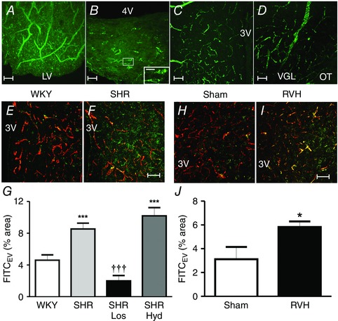
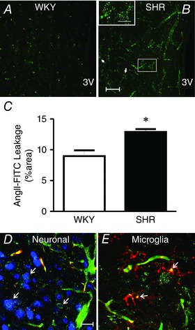
Angiotensin II (AngII) is a pivotal peptide implicated in the regulation of blood pressure. In addition to its systemic vascular and renal effects, AngII acts centrally to modulate the activities of neuroendocrine and sympathetic neuronal networks, influencing in turn sympatho-humoral outflows to the circulation. Moreover, a large body of evidence supports AngII signalling dysregulation as a key mechanism contributing to exacerbated sympathoexcitation during hypertension. Due to its hydrophilic actions, circulating AngII does not cross the blood-brain barrier (BBB), signalling to the brain via the circumventricular organs which lack a tight BBB. In this review, we present and discuss recent studies from our laboratory showing that elevated circulating levels of AngII during hypertension result in disruption of the BBB integrity, allowing access of circulating AngII to critical sympathoexcitatory brain centres such as the paraventricular nucleus of the hypothalamus and the rostral ventrolateral medulla. We propose the novel hypothesis that AngII-driven BBB breakdown constitutes a complementary mechanism by which circulating AngII, working in tandem with the central renin-angiotensin system, further exacerbates sympatho-humoral activation during hypertension. These results are discussed within the context of a growing body of evidence in the literature supporting AngII as a pro-inflammatory signal, and brain microglia as key cell targets mediating central AngII actions during hypertension.
© 2015 The Authors. The Journal of Physiology © 2015 The Physiological Society.
Figures
References
-
- Abbott NJ, Patabendige AA, Dolman DE, Yusof SR & Begley DJ (2010). Structure and function of the blood‐brain barrier. Neurobiol Dis 37, 13–25. - PubMed
-
- Abbott NJ, Ronnback L & Hansson E (2006). Astrocyte‐endothelial interactions at the blood‐brain barrier. Nat Rev Neurosci 7, 41–53. - PubMed
-
- Anderson JW, Smith PM & Ferguson AV (2001). Subfornical organ neurons projecting to paraventricular nucleus: whole‐cell properties. Brain Res 921, 78–85. - PubMed
-
- Bains JS & Ferguson AV (1995). Paraventricular nucleus neurons projecting to the spinal cord receive excitatory input from the subfornical organ. Am J Physiol 268, R625–R633. - PubMed
-
- Bains JS, Potyok A & Ferguson AV (1992). Angiotensin II actions in paraventricular nucleus: functional evidence for neurotransmitter role in efferents originating in subfornical organ. Brain Res 599, 223–229. - PubMed
Publication types
MeSH terms
Substances
Grants and funding
LinkOut - more resources
Full Text Sources
Medical