. 2016 Jan:487:273-84.
doi: 10.1016/j.virol.2015.10.024. Epub 2015 Nov 12.

Phosphorylation of mouse SAMHD1 regulates its restriction of human immunodeficiency virus type 1 infection, but not murine leukemia virus infection

Affiliations

Phosphorylation of mouse SAMHD1 regulates its restriction of human immunodeficiency virus type 1 infection, but not murine leukemia virus infection

Feifei Wang et al. Virology. 2016 Jan.

Abstract

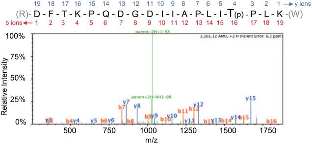
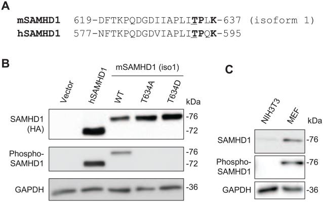
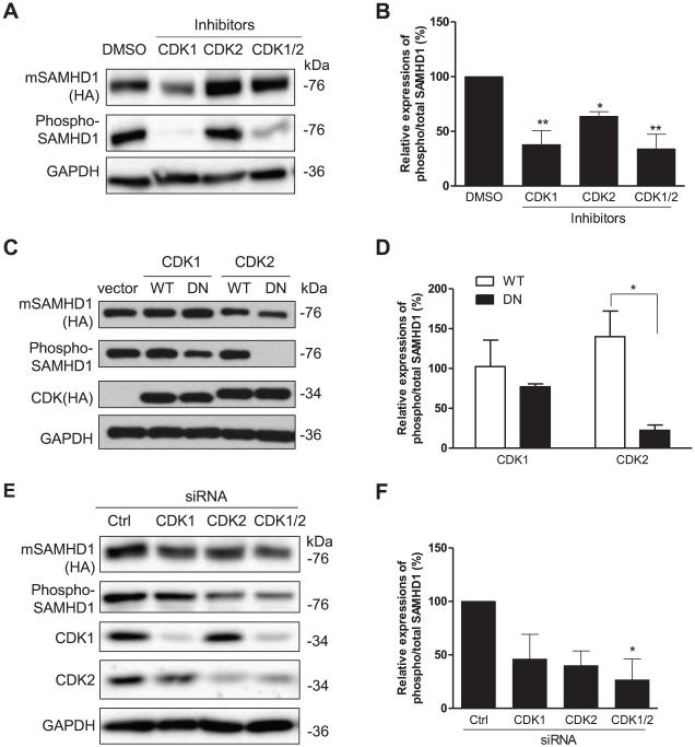
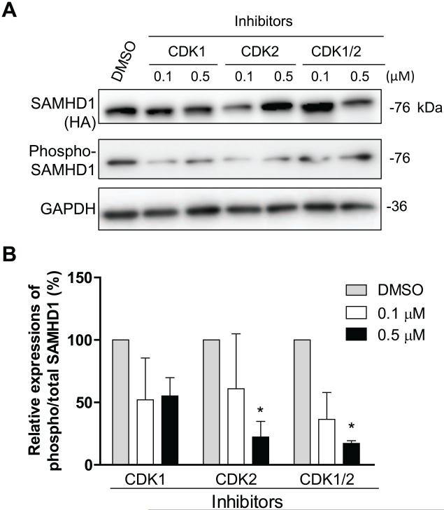
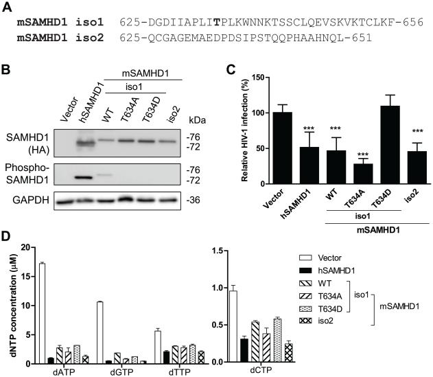
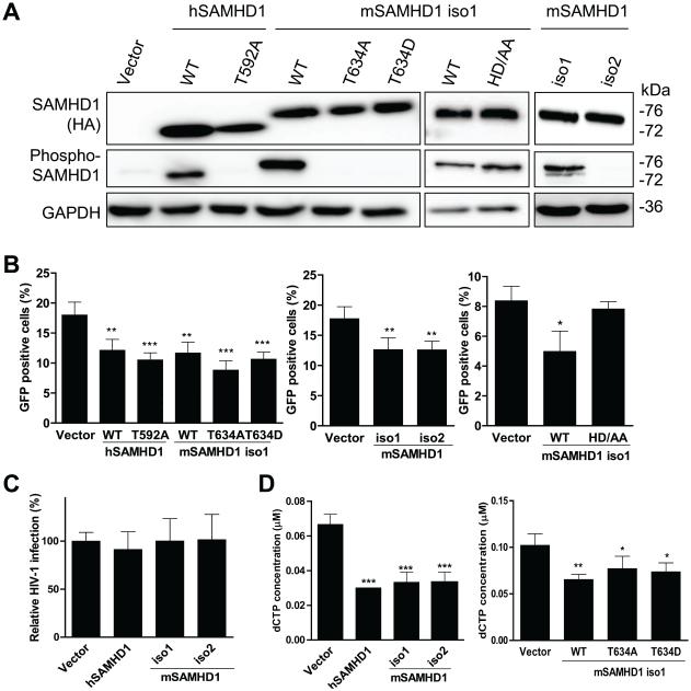
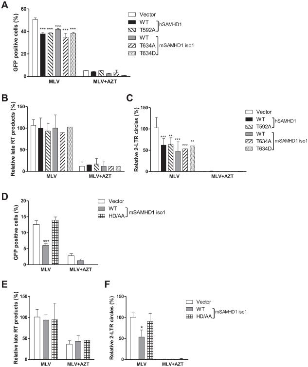
Human SAMHD1 (hSAMHD1) restricts HIV-1 infection in non-dividing cells by depleting intracellular dNTPs to limit viral reverse transcription. Phosphorylation of hSAMHD1 at threonine (T) 592 by cyclin-dependent kinase (CDK) 1 and CDK2 negatively regulates HIV-1 restriction. Mouse SAMHD1 (mSAMHD1) restricts HIV-1 infection in non-dividing cells, but whether its phosphorylation regulates retroviral restriction is unknown. Here we identified six phospho-sites of mSAMHD1, including T634 that is homologous to T592 of hSAMHD1 and phosphorylated by CDK1 and CDK2. We found that wild-type (WT) mSAMHD1 and a phospho-ablative mutant, but not a phospho-mimetic mutant, restricted HIV-1 infection in differentiated U937 cells. Murine leukemia virus (MLV) infection of dividing NIH3T3 cells was modestly restricted by mSAMHD1 WT and phospho-mutants, but not by a dNTPase-defective mutant. Our results suggest that phosphorylation of mSAMHD1 at T634 by CDK1/2 negatively regulates its HIV-1 restriction in differentiated cells, but does not affect its MLV restriction in dividing cells.

Keywords: HIV-1; Infection; MLV; Mouse and human SAMHD1; Phosphorylation; Restriction.


Figures

Fig. 1
Fig. 1
Residue T634 in the mSAMHD1 protein sequence is a phosphosite as identified by tandem mass spectrometry. Position T634 was identified as a phosphosite by MS/MS analysis. An example MS/MS spectrum is shown for the indicated tryptic peptide of mSAMHD1 (residues 619 to 637) that identified the T634 phosphorylation site, which is highlighted in the sequence and denoted by a “(p)”. Assigned b ions are shown in red, and y ions are shown in blue. Green peaks represent loss of a 98 Da fragment that corresponds to loss of a phosphate group as indicated.
Fig. 2
Fig. 2
Confirmation of the phosphorylation of mSAMHD1 at position T634 using a phospho-specific antibody. (A) Partial protein sequence alignment of mSAMHD1 isoform 1 and human (hSAMHD1) depicting conserved phosphosites at residue positions T634 and T592 in mSAMHD1 and hSAMHD1 sequences, respectively. The phosphorylated residue threonine (T) is underlined and the residues encompassing a consensus sequence (TPXK) for CDK interaction are highlighted in bold. (B) HEK293T cells were transiently transfected to overexpress hSAMHD1, mSAMHD1 wild-type (WT), phospho-ablative mutant (T634A) or phospho-mimetic mutant (T634D). Immunoblotting was performed on cell lysates to confirm position T634 of mSAMHD1 is a phosphosite. Total SAMHD1 protein levels were probed using an anti-HA antibody (top panel). (C) Endogenous mSAMHD1 in mouse embryonic fibroblasts (MEFs) is phosphorylated at T634. The total SAMHD1 was probed with a specific SAMHD1 antibody. (B) and (C) The phospho-SAMHD1 levels were detected using a phospho-specific SAMHD1 antibody (middle panel). GAPDH was used as a loading control. A representative immunoblot result from three independent experiments is shown.
Fig. 3
Fig. 3
CDK1 and CDK2 contribute to T634 phosphorylation of mSAMHD1 in HEK293T cells. (A) Cells were pre-treated with DMSO or inhibitors specific to CDK1 or CDK2 at 1 μM for 6 h and transiently transfected to overexpress mSAMHD1. The effect of each inhibitor on the phosphorylation of mSAMHD1 at T634 was assessed at 24 h post-transfection by immunoblotting. (B) An average of three independent experiments described in (A). (C) mSAMHD1 was overexpressed together with wild-type (WT) or dominant negative (DN) mutants of CDK1 or CDK2. Empty vector was used as a negative control. At 24 h post-transfection, cells were harvested for immunoblotting to determine total SAMHD1, phosphorylated SAMHD1 and CDK expression levels. Total mSAMHD1 and CDK1/2 protein levels were detected using an HA-specific antibody and phosphorylation of T634 was detected using a phospho-specific SAMHD1 antibody. (D) An average of three independent experiments described in (C). (E) Individual and combined siRNA-mediated knockdown of endogenous CDK1 and CDK2 were performed in the presence of ectopically expressed mSAMHD1 using a two-round transfection protocol. Cells were harvested 24 h following the second round of siRNA transfection and cell lysates were subjected to immunoblotting to determine the effect of CDK1/2 knockdown on the phosphorylation of mSAMHD1 at T634. The efficiency of siRNA-mediated knockdown of CDK1 and CDK2 was determined by probing with their respective antibodies. (F) An average of three independent experiments described in (E). In (A, C and E), total SAMHD1 levels were detected using an HA-specific antibody. The levels of phospho-SAMHD1 were detected using a phospho-SAMHD1 specific antibody. GAPDH was used as a loading control. In (B, D and F), the protein levels were quantified based on the band densities. The levels of total mSAMHD1 and phospho-SAMHD1 were normalized to GAPDH respectively, and then the ratio of phospho/total mSAMHD1 was calculated and the ratio of the vector group was set as 1. Results are shown as mean ± SD (n=3). The data was analyzed by T-test or one-way ANOVA with Dunnett’s test (*, P < 0.05, **, P < 0.01).
Fig. 4
Fig. 4
Endogenous CDK1 and CDK2 phosphorylate mSAMHD1 at T634 in mouse fibroblast cells. (A) NIH3T3 cells stably expressing mSAMHD1 were treated with inhibitors specific to CDK1 or CDK2 at the indicated concentrations. The effects of each inhibitor on the phosphorylation of mSAMHD1 at T634 were assessed at 24 h post-transfection by immunoblotting with a phospho-specific antibody. (B) An average of three independent experiments. The protein levels were quantified based on the band densities. The levels of total mSAMHD1 and phospho-SAMHD1 were normalized to GAPDH, and then the ratio of phospho/total mSAMHD1 was calculated and the ratio of the vector group was set as 100%. Results are shown as mean ± SD (n=3). The data was analyzed by one-way ANOVA with Dunnett’s test (*, P < 0.05).
Fig. 5
Fig. 5
The effect of T634 phosphorylation of mSAMHD1 on its restriction of HIV-1 infection in PMA-differentiated U937 cells. (A) The C-terminus alignment of mSAMHD1 isoform 1 (iso1) and isoform 2 (iso2) proteins illustrating the T634 region is lacking in the iso2 protein. (B) The expression levels of total and phosphorylated SAMHD1 (WT and mutants) in U937 stable cell lines were determined by immunoblotting with an anti-HA antibody and specific phospho-SAMHD1 antibody, respectively. GAPDH was used as a loading control. (C) U937 cells expressing hSAMHD1 WT, mSAMHD1 WT, phospho-ablative mutant (T634A), phospho-mimetic mutant (T634D) or an empty vector were infected with a single-cycle HIV-1 luc/VSV-G at a multiplicity of infection (MOI=1). At 24 h post-infection, cells were harvested for luciferase assay to measure HIV-1 infection. An average of three independent infection experiments is presented (n=12). The infection level in the vector control group was set as 100% for normalization. (D) The levels of intracellular dNTPs were significantly decreased by hSAMHD1, mSAMHD1 WT or mutants compared to the vector control (P < 0.0001). The results are shown as mean ± SD (n =2). The dNTP concentrations were calculated based on the cell volume of human primary macrophages (2,660 μm3). For (C-D), statistical analysis was performed by one-way ANOVA and Dunnett’s test (***, P < 0.0001).
Fig. 6
Fig. 6
MLV infection in dividing NIH3T3 cells is reduced by mSAMHD1 independently of its T634 phosphorylation. (A) Overexpression of HA-tagged mSAMHD1 isoform 1 (iso1) and isoform 2 (iso2) proteins and iso1 mutants in NIH3T3 stable cells lines was confirmed by immunoblotting using anti-HA antibody. Vector-transduced cells were used as a negative control. Phosphorylation status of hSAMHD1 (T592) and mSAMHD1 (T634) was assessed using a phospho-specific antibody. GAPDH was used as a loading control. (B) Overexpression of SAMHD1 WT or iso1 phospho-mutants, but not the dNTPase-defective mSAMHD1 iso1 mutant H238A/D239A (HD/AA), in NIH3T3 cells modestly reduces MLV infection. (C) HIV-1 infection is not restricted by WT hSAMHD1, WT mSAMHD1 iso1, or WT mSAMHD1 iso2 in NIH3T3 cells. NIH3T3 cells were infected with a single-cycle MLV-GFP/VSV-G reporter virus (B) or an HIV-1-GFP/VSV-G reporter virus (C) (MOI=1). At 48 h post-infection, cells were harvested to determine the percentage of GFP-positive cells by flow cytometry. (D) Intracellular dCTP levels were reduced in NIH3T3 cells expressing WT hSAMHD1, WT mSAMHD1 or phospho-mutants of mSAMHD1. The concentrations calculated based on the average cell size of NIH3T3 (diameter is 18 μm) and the spherical cell assumption. The data were analyzed by one-way ANOVA and Dunnett’s test (*, P < 0.05, **, P < 0.01, ***, P < 0.0001).
Fig. 7
Fig. 7
MLV infection in dividing NIH3T3 cells is inhibited by mSAMHD1 at the viral cDNA nuclear import stage but not the late RT step. (A and D) MLV infection in cells expressing WT or mutant SAMHD1 was reduced by AZT treatment (5 μM). (B and E) The levels of late reverse transcription (RT) products in MLV infected cells were not affected by overexpression of SAMHD1 WT or mutants in NIH3T3 cells. (C and F) The levels of 2-LTR circles were reduced by overexpression of SAMHD1 WT or phospho-mutants in NIH3T3 cells, the AZT treatment, but not overexpression of the HD/AA mutant of mSAMHD1 isoform 1 (iso1). (A-F) Cells were infected with MLV-GFP/VSV-G (MOI=1) in the presence of control media or AZT (5 μM). Cells were harvested at 24 h post-infection and used for genomic DNA extraction. The levels of MLV late RT products (B and E) and of 2-LTR circles (C and F) were measured using real-time PCR. An average of three independent experiments was presented. After normalization to GAPDH, the levels of late reverse transcription products or 2-LTR circles in MLV-infected vector control cells were set as 100%. The data were analyzed by one-way ANOVA and Dunnett’s test (*, P < 0.05, **, P < 0.01, ***, P < 0.0001).

References

    1. Baldauf HM, Pan X, Erikson E, Schmidt S, Daddacha W, Burggraf M, Schenkova K, Ambiel I, Wabnitz G, Gramberg T, Panitz S, Flory E, Landau NR, Sertel S, Rutsch F, Lasitschka F, Kim B, Konig R, Fackler OT, Keppler OT. SAMHD1 restricts HIV-1 infection in resting CD4(+) T cells. Nat Med. 2012;18:1682–1687. - PMC - PubMed
    1. Behrendt R, Schumann T, Gerbaulet A, Nguyen LA, Schubert N, Alexopoulou D, Berka U, Lienenklaus S, Peschke K, Gibbert K, Wittmann S, Lindemann D, Weiss S, Dahl A, Naumann R, Dittmer U, Kim B, Mueller W, Gramberg T, Roers A. Mouse SAMHD1 has antiretroviral activity and suppresses a spontaneous cell-intrinsic antiviral response. Cell Rep. 2013;4:689–696. - PMC - PubMed
    1. Bramson HN, Corona J, Davis ST, Dickerson SH, Edelstein M, Frye SV, Gampe RT, Harris PA, Hassell A, Holmes WD, Hunter RN, Lackey KE, Lovejoy B, Luzzio MJ, Montana V, Rocque WJ, Rusnak D, Shewchuk L, Veal JM, Walker DH, Kuyper LF. Oxindole-based inhibitors of cyclin-dependent kinase 2 (CDK2): Design, synthesis, enzymatic activities, and X-ray crystallographic analysis. Journal of Medicinal Chemistry. 2001;44:4339–4358. - PubMed
    1. Connor RI, Chen BK, Choe S, Landau NR. Vpr is required for efficient replication of human immunodeficiency virus type-1 in mononuclear phagocytes. Virology. 1995;206:935–944. - PubMed
    1. Cribier A, Descours B, Valadao AL, Laguette N, Benkirane M. Phosphorylation of SAMHD1 by Cyclin A2/CDK1 Regulates Its Restriction Activity toward HIV-1. Cell Rep. 2013;3:1036–1043. - PubMed

Publication types

MeSH terms

LinkOut - more resources