Review
. 2015 Nov 16;8(4):711-57.
doi: 10.3390/ph8040711.

Antimicrobial Peptides from Plants

Affiliations
Review

Antimicrobial Peptides from Plants

James P Tam et al. Pharmaceuticals (Basel). .

Abstract

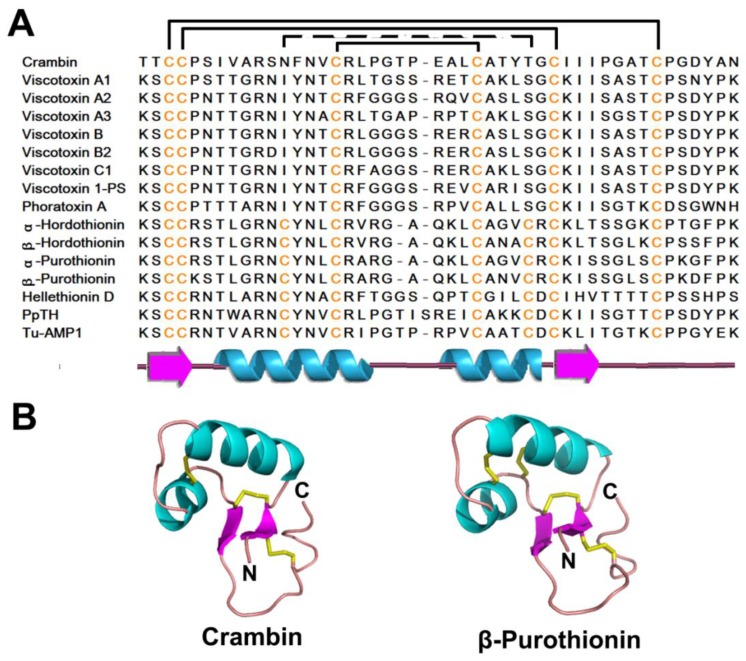
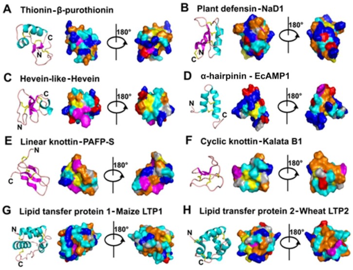
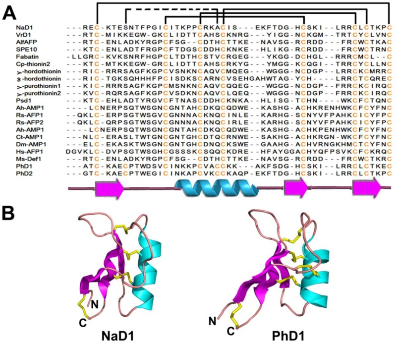
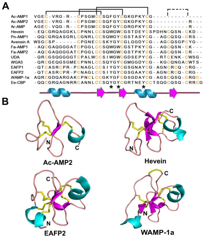
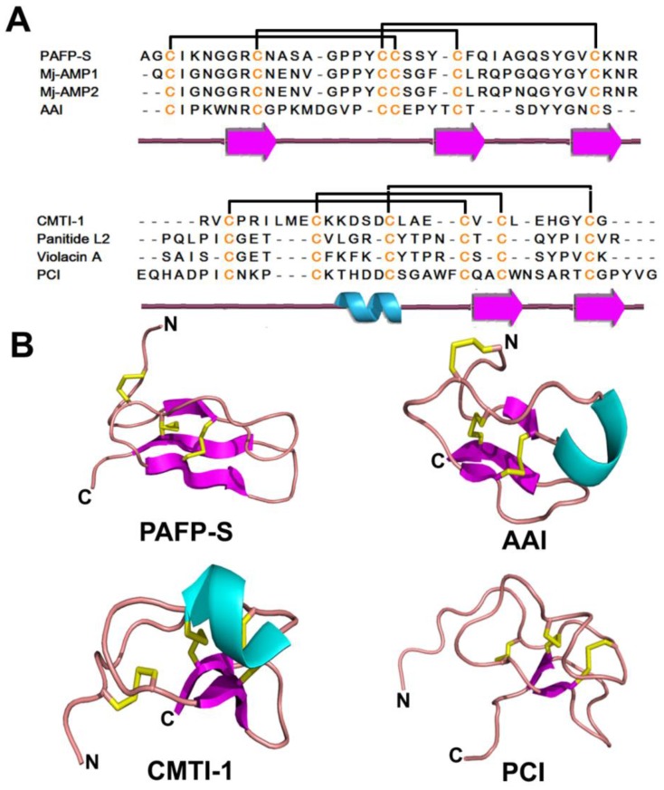
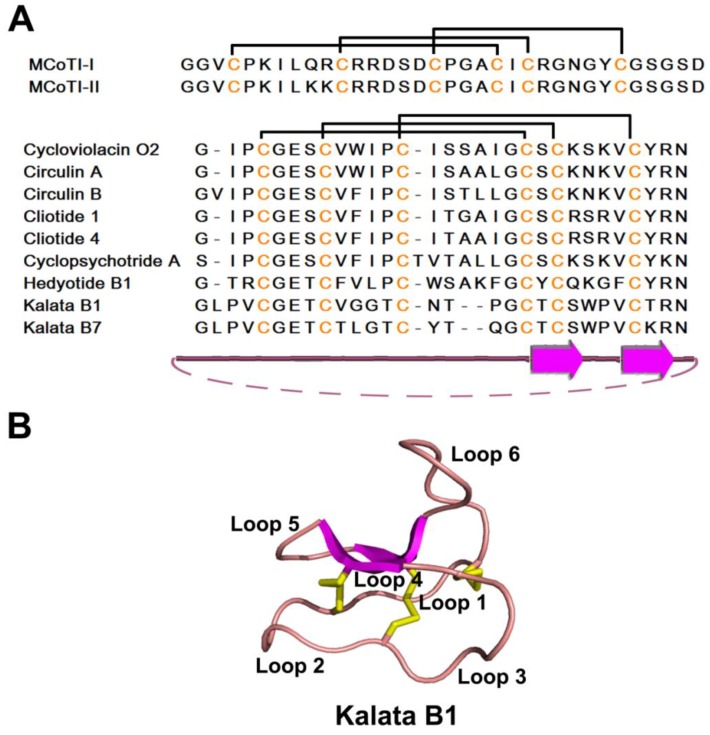
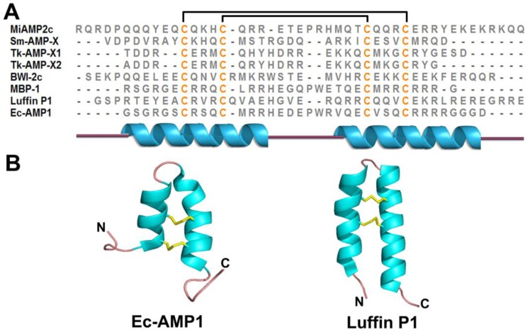
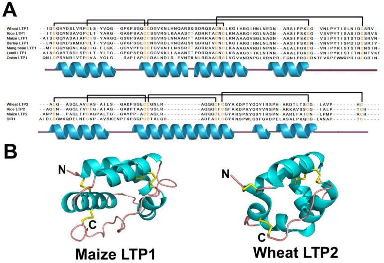
Plant antimicrobial peptides (AMPs) have evolved differently from AMPs from other life forms. They are generally rich in cysteine residues which form multiple disulfides. In turn, the disulfides cross-braced plant AMPs as cystine-rich peptides to confer them with extraordinary high chemical, thermal and proteolytic stability. The cystine-rich or commonly known as cysteine-rich peptides (CRPs) of plant AMPs are classified into families based on their sequence similarity, cysteine motifs that determine their distinctive disulfide bond patterns and tertiary structure fold. Cystine-rich plant AMP families include thionins, defensins, hevein-like peptides, knottin-type peptides (linear and cyclic), lipid transfer proteins, α-hairpinin and snakins family. In addition, there are AMPs which are rich in other amino acids. The ability of plant AMPs to organize into specific families with conserved structural folds that enable sequence variation of non-Cys residues encased in the same scaffold within a particular family to play multiple functions. Furthermore, the ability of plant AMPs to tolerate hypervariable sequences using a conserved scaffold provides diversity to recognize different targets by varying the sequence of the non-cysteine residues. These properties bode well for developing plant AMPs as potential therapeutics and for protection of crops through transgenic methods. This review provides an overview of the major families of plant AMPs, including their structures, functions, and putative mechanisms.

Keywords: cysteine-rich peptides; cystine knot; defensin; hevein; knottin; plant antimicrobial peptides; thionin.


Figures

Figure 1
Figure 1
Sequences (A) and structures (B) of representative thionins. The secondary structure is represented by different colors: cyan-α helix; magenta-β strand; pink-random coil and yellow-disulfide bonds.
Figure 2
Figure 2
Cartoon illustration and surface plot of representative members of different antimicrobial peptides. The orientation of surface plot (left) of each representative member is the same as the cartoon illustration, while that of the right one is evolved from the left one by 180° turn around the y axis. Color representation rules for different amino acids are as follows: blue-positively charged (R, K & H), red-negatively charged (D & E), orange-hydrophobic (I, L, V, A, P & M), cyan-hydrophilic (S, T, E & Q), magenta-aromatic (F, Y & W), yellow-cysteine (C) and gray-glycine (G).
Figure 3
Figure 3
Sequences (A) and structures (B) of representative plant defensins. The secondary structure is represented by different colors: cyan-α helix; magenta-β strand; pink-random coil and yellow-disulfide bonds.
Figure 4
Figure 4
Sequences (A) and structures (B) of representative hevein-like peptides. The secondary structure is represented by different colors: cyan-α helix; magenta-β strand; pink-random coil and yellow-disulfide bonds.
Figure 5
Figure 5
Sequences (A) and structures (B) of representative linear knottin-type peptides. The secondary structure is represented by different colors: cyan-α helix; magenta-β strand; pink-random coil and yellow-disulfide bonds.
Figure 6
Figure 6
Sequences (A) and structures (B) of representative cyclic knottin-type peptides. The secondary structure is represented by different colors: magenta-β strand; pink-random coil and yellow-disulfide bonds.
Figure 7
Figure 7
Sequences (A) and structures (B) of representative α-hairpinins. The secondary structure is represented by different colors: cyan-α helix; pink-random coil and yellow-disulfide bonds.
Figure 8
Figure 8
Sequences (A) and structures (B) of representative lipid transfer proteins. The secondary structure is represented by different colors: cyan-α helix; pink-random coil and yellow-disulfide bonds.

References

    1. Stintzi A., Heitz T., Prasad V., Wiedemann-Merdinoglu S., Kauffmann S., Geoffroy P., Legrand M., Fritig B. Plant “pathogenesis-related” proteins and their role in defense against pathogens. Biochimie. 1993;75:687–706. doi: 10.1016/0300-9084(93)90100-7. - DOI - PubMed
    1. Sinha M., Singh R.P., Kushwaha G.S., Iqbal N., Singh A., Kaushik S., Kaur P., Sharma S., Singh T.P. Current overview of allergens of plant pathogenesis related protein families. Sci. World J. 2014;2014:543195. doi: 10.1155/2014/543195. - DOI - PMC - PubMed
    1. Ebrahim S., Usha K., Singh B. Pathogenesis related (pr) proteins in plant defense mechanism. Sci. Against Microb. Pathog. 2011;2:1043–1054.
    1. Sels J., Mathys J., De Coninck B.M., Cammue B.P., De Bolle M.F. Plant pathogenesis-related (pr) proteins: A focus on pr peptides. Plant Physiol. Biochem. 2008;46:941–950. doi: 10.1016/j.plaphy.2008.06.011. - DOI - PubMed
    1. Egorov T.A., Odintsova T.I., Pukhalsky V.A., Grishin E.V. Diversity of wheat anti-microbial peptides. Peptides. 2005;26:2064–2073. doi: 10.1016/j.peptides.2005.03.007. - DOI - PubMed