Review
. 2015 Nov 13;7(11):9453-74.
doi: 10.3390/nu7115475.

The Subtle Balance between Lipolysis and Lipogenesis: A Critical Point in Metabolic Homeostasis

Affiliations
Review

The Subtle Balance between Lipolysis and Lipogenesis: A Critical Point in Metabolic Homeostasis

Chiara Saponaro et al. Nutrients. .

Abstract

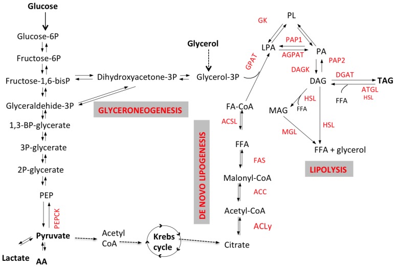
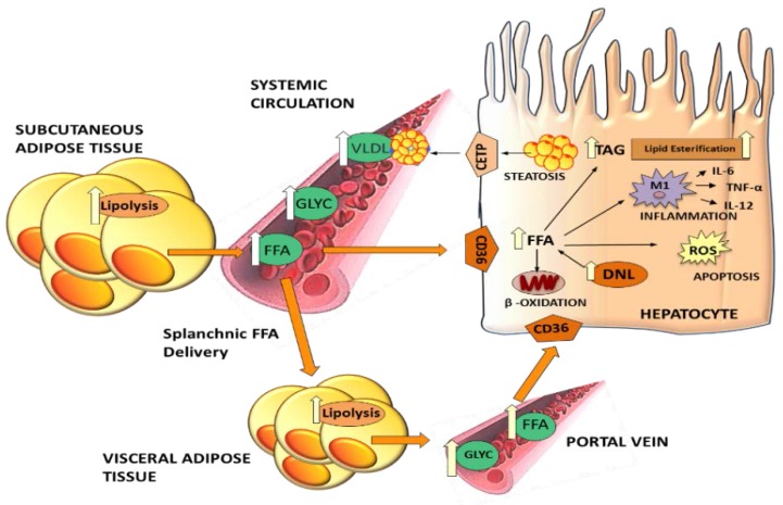
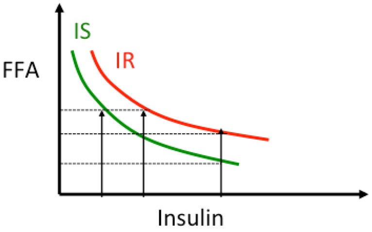
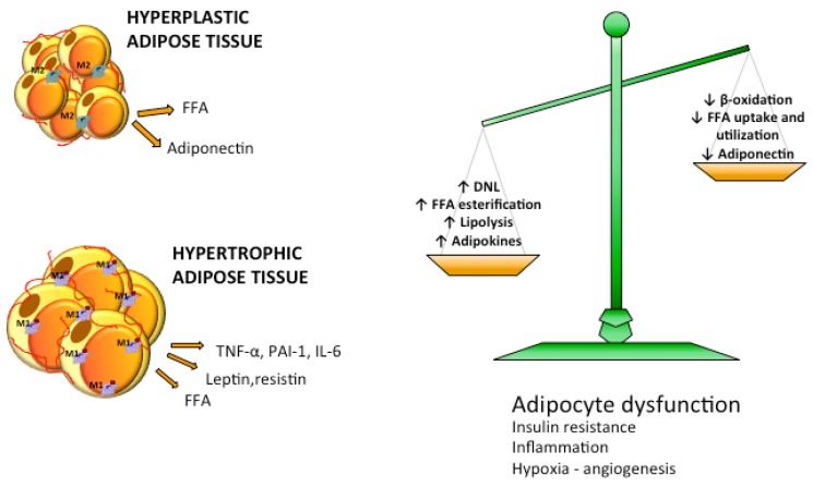
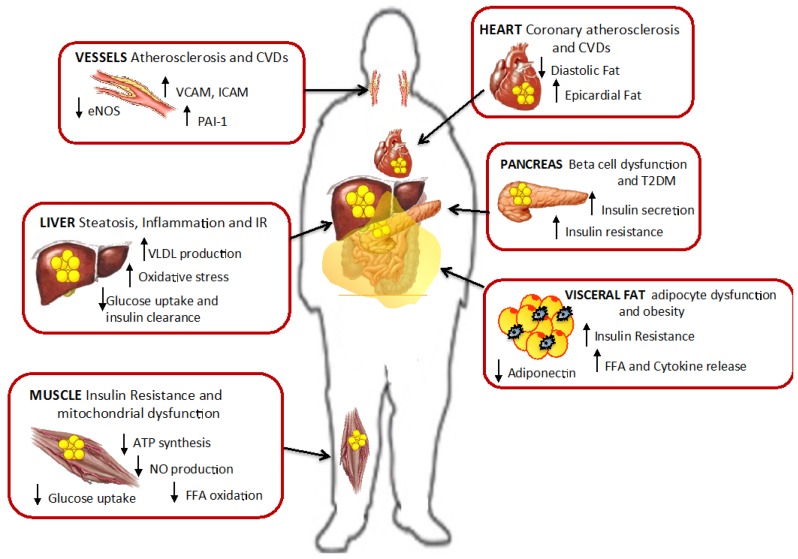
Excessive accumulation of lipids can lead to lipotoxicity, cell dysfunction and alteration in metabolic pathways, both in adipose tissue and peripheral organs, like liver, heart, pancreas and muscle. This is now a recognized risk factor for the development of metabolic disorders, such as obesity, diabetes, fatty liver disease (NAFLD), cardiovascular diseases (CVD) and hepatocellular carcinoma (HCC). The causes for lipotoxicity are not only a high fat diet but also excessive lipolysis, adipogenesis and adipose tissue insulin resistance. The aims of this review are to investigate the subtle balances that underlie lipolytic, lipogenic and oxidative pathways, to evaluate critical points and the complexities of these processes and to better understand which are the metabolic derangements resulting from their imbalance, such as type 2 diabetes and non alcoholic fatty liver disease.

Keywords: HCC; NAFLD; SCD-1; de novo lipogenesis; ectopic fat; fatty liver; glyceroneogenesis; lipolysis; lipotoxicity; saturated fat.


Figures

Figure 1
Figure 1
Schematic representation of lipolytic and lipogenic pathways. Triacyglycerol (TAG) synthesis requires the activation of free fatty acids (FFA) into Acyl-CoA by enzyme acyl-CoA synthetase. FFA-CoA and G3P are transformed via acylation, by glycerol-3-phosphate acyltransferase (GPAT) and acylCoA acylglycerol-3-phosphate acyltransferases (AGPAT), to phosphatidic acid (PA); then, after a dephosphorylation by phosphohydrolase (PAP2), diacylglycerols (DAG) are formed. Diacylglycerol acyltransferase (DGAT) catalyzes the conversion of DAG into TAG. In the adipocyte, G3P might come either from glycolysis or from non-carbohydrate substrates via the enzyme phosphoenolpyruvate carboxykinase (PEPCK), through a process named glyceroneogenesis. In the liver G3P can also be synthesized from plasma glycerol. De novo fatty acids synthesis (also referred to as de novo lipogenesis or DNL) occurs in the cytoplasm of various cells (e.g., adipocytes and hepatocytes) where citric acid is converted to acetyl-CoA by ATP-citrate lyase (ACL) and subsequently to malonyl-CoA by acetyl-CoA carboxylase (ACC). DNL occurs mainly in the liver, but it might occur in adipose tissue as well, although with low rates. This process requires the two enzymes ATP-citrate lyase (ACL), acetyl-CoA carboxylase (ACC) and the multi-enzymatic complex fatty acid synthase (FAS). G3P can be synthesized directly from non-carbohydrate substrates such as pyruvate, lactate or amino acids in oxaloacetate, that is converted to G3P either directly from phoenolpyruvate (PEP), via the key enzyme phosphoenolpyruvate carboxykinase (PEPCK), or through synthesis of dihydroxyacetone (DHA). TAG catabolism (i.e., lipolysis) involves several lipases, adipose triglyceride lipase (ATGL), hormone-sensitive lipase (HSL) and monoacylglycerol lipase (MGL) and produces the release of three free fatty acids (FFA) and one glycerol molecule.
Figure 2
Figure 2
Effects of increased lipolysis on liver dysfunction. Excess lipolysis results in high free fatty acid (FFA) flux into the liver, where FFAs cause steatosis and exert lipotoxic effects. Triglycerides (TAG) synthetized in the liver are secreted into the plasma circulation as very low density lipoproteins (VLDL) causing dyslipidemia. Visceral fat has a preferential role in hepatic fat accumulation since released FFA reach the liver via the portal vein. Also increased hepatic de novo lipogenesis (DNL), inflammation and oxidative stress contribute to liver damage and hepatocyte dysfunction.
Figure 3
Figure 3
Relationship insulin-lipolysis. As insulin concentration increases, lipolysis, and thus plasma free fatty acids (FFA) concentration, is suppressed following a non-linear curve [65,69,70]. In presence of insulin resistance the curve is shifted to the right indicating that for the same insulin levels lipolysis is less suppressed and circulating FFA levels are higher. The product FFA × Insulin is used as an index of adipose tissue-insulin resistance.
Figure 4
Figure 4
Imbalance in lipid metabolism causes increased efflux of FFA to adipose tissue. Reduced free fatty acids (FFA) utilization and β-oxidation and increased lipogenic and lipolytic pathways lead to overflow of FFA in the circulation. Adipose tissue activates adipogenesis and increases the number of adipocytes becoming hyperplastic or enlarges adipocyte size becoming hypertrophic. Hyperplastic adipose tissue is normally metabolically healthy while hypertrophic adipose tissue is characterized by dysfunctional adipocytes, insulin resistance, hypoxia and inflammation.
Figure 5
Figure 5
Pathways of β-oxidation. β-oxidation is the catabolic pathway that occurs in mitochondria and produces energy from TG hydrolysis. (1) FFA are transformed to Acyl-CoA in cytosol; (2) protein Carnitine Palmitoyl Transferase-1 (CPT1) catalizes the transfer of the acyl group of a long-chain fatty acyl-CoA to carnitine to form acylcarnitines (mainly Palmitoylcarnitine); (3) Carnitine Acyltranferase (CACT) transfers acylcarnitine across outer mitochondrial membrane; (4) Carnitine Palmitoyl Transferase-2 (CPT2) reconverts acylcarnitine in acylCoA and carnitine; (5) Acyl-CoA enters in β-oxidation cycle and is degraded in several Acetyl-CoA molecules; (6) Acetyl-CoA enters in Krebs cycle to produce energy as Adenosine Triphosphate (ATP).
Figure 6
Figure 6
Ectopic fat accumulation and effect of lipotoxicity. Fat accumulation in non-adipose tissues promotes cell dysfunction, insulin resistance and inflammation in liver, muscle, pancreas and visceral fat. Also in vessels and heart lipotoxicity leads to increased risk for cardiovascular diseases and atherosclerosis. Modified from Gaggini M. et al. [2].

References

    1. Morelli M., Gaggini M., Daniele G., Marraccini P., Sicari R., Gastaldelli A. Ectopic fat: The true culprit linking obesity and cardiovascular disease? Thromb. Haemost. 2013;110:651–660. doi: 10.1160/TH13-04-0285. - DOI - PubMed
    1. Gaggini M., Saponaro C., Gastaldelli A. Not all fats are created equal: Adipose vs. Ectopic fat, implication in cardiometabolic diseases. Horm. Mol. Biol. Clin. Investig. 2015;22:7–18. doi: 10.1515/hmbci-2015-0006. - DOI - PubMed
    1. Gaggini M., Morelli M., Buzzigoli E., DeFronzo R.A., Bugianesi E., Gastaldelli A. Non-alcoholic fatty liver disease (NAFLD) and its connection with insulin resistance, dyslipidemia, atherosclerosis and coronary heart disease. Nutrients. 2013;5:1544–1560. doi: 10.3390/nu5051544. - DOI - PMC - PubMed
    1. Yki-Jarvinen H. Non-alcoholic fatty liver disease as a cause and a consequence of metabolic syndrome. Lancet Diabetes Endocrinol. 2014;2:901–910. doi: 10.1016/S2213-8587(14)70032-4. - DOI - PubMed
    1. Gastaldelli A. Visceral adipose tissue and ectopic fat deposition. In: Bray G.A., Bouchard C., editors. Handbook of Obesity. Volume 1. CRC Press; Boca Raton, FL, USA: 2014. pp. 237–248.

Publication types

MeSH terms