Review
. 2016 Jan;17(1):55-64.
doi: 10.1038/nrm.2015.3. Epub 2015 Nov 18.

Beyond β-catenin: prospects for a larger catenin network in the nucleus

Affiliations
Review

Beyond β-catenin: prospects for a larger catenin network in the nucleus

Pierre D McCrea et al. Nat Rev Mol Cell Biol. 2016 Jan.

Abstract

β-catenin is widely regarded as the primary transducer of canonical WNT signals to the nucleus. In most vertebrates, there are eight additional catenins that are structurally related to β-catenin, and three α-catenin genes encoding actin-binding proteins that are structurally related to vinculin. Although these catenins were initially identified in association with cadherins at cell-cell junctions, more recent evidence suggests that the majority of catenins also localize to the nucleus and regulate gene expression. Moreover, the number of catenins reported to be responsive to canonical WNT signals is increasing. Here, we posit that multiple catenins form a functional network in the nucleus, possibly engaging in conserved protein-protein interactions that are currently better characterized in the context of actin-based cell junctions.


Figures

Figure 1
Figure 1. An overview of vertebrate catenins
Most vertebrates have twelve genes that encode different types of catenins, with the genomes of some teleost fishes having certain catenins duplicated,,. a | Vertebrate catenins containing Armadillo domains were first identified at cell–cell contacts in association with classic cadherins (β-catenin and p120 catenin subfamily members), or with desmosomal cadherins (plakophilin catenin subfamily members and γ-catenin (plakoglobin)). Subsequently, catenins were found to have key roles in the cytoplasm and the nucleus. The large Armadillo domain of each catenin is shown in grossly simplified form (different shades of blue). It varies from 9–12 Armadillo repeats, depending on the catenin (each repeat is ~42 amino acids in length), with limited unique sequences interspersed. As the different repeat units within the same larger Armadillo domain are only weakly homologous to one another, different protein-protein interactions are supported, depending on the domain region in question. Additional features on certain catenins include phosphorylation domains, alternative translation start sites, alternative splicing events at the RNA level, nuclear localization signals (NLS), nuclear export signals (NES) and PDZ motifs, and amino-terminal ‘destruction boxes’ that are responsive to canonical WNT signals. Shading reflects the relatedness of protein families; for example, β-catenin is more closely related to plakoglobin than to the p120 or plakophilin catenin families. An Armadillo catenin (p120) depicted with additional features is presented in FIG. 3; the reader is also referred to more comprehensive reviews noted in the text. b | α-catenins are filamentous actin (F-actin)-binding proteins that are structurally related to vinculin. The N-terminal dimerization domain engages in mutually exclusive β-catenin (heteromeric) or α-catenin (homomeric) binding, whereas the central mechanosensitive and carboxy-terminal F-actin-binding domains are together composed of four to five α-helical bundles.
Figure 2
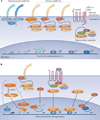
Figure 2. Conditions that promote catenin nuclear signalling
a | When WNT signalling through the Frizzled–low-density lipoprotein receptor-related protein 5 (LRP5) or Frizzlled–LRP6 receptor is low or absent and/or cadherin expression levels are high, catenins typically do not enter the nucleus owing to their inhibition via the Axin-adenomatous polyposis coli (APC)–casein kinase 1α (CK1α)–glycogen synthase kinase 3β (GSK3β) destruction complex and their associations with cadherins, respectively. Plakophilin 1 (PKP1) 1,PKP2 or PKP3 interact with desmosomal cadherins indirectly through γ-catenin (plakoglobin) and desmoplakin (grey circles), whereas β-catenin and p120 catenins, as well as (indirectly) α-catenin, interact with classic cadherins. In the absence of nuclear catenins, the DNA-binding factors shown repress gene targets in a number of cases, although some transcription factors are activators even in the absence of catenins (for example, ETS translocation variant 1 (ETV1)). Likewise, a recent genome-wide study suggests the association of Kaiso with gene activation (not shown). b | WNT signalling or cadherin loss promotes nuclear accumulation of β-catenin, p120 catenin (isoform 1) and possibly other catenin isoforms. p120 catenin derepresses Kaiso to activate genes. p120 catenin also dissociates the RE1-silencing transcription factor (REST)–CoREST complex from genes, resulting in target derepression (activation). Also, other p120 catenin family members (for example, δ-catenin or PKP3) have been shown to enter the nucleus and regulate transcription, but which pathways impact their nuclear residence has been less explored. In the case of δ-catenin, caspase 3 (CASP3)-mediated cleavage seems to promote the nuclear accumulation of its carboxyl fragment, followed by zinc-finger transcriptional repressor ZIFCAT association and unknown gene regulatory effects. δ-catenin can also enter the nucleus in the uncleaved form and has been demonstrated to bind to and modulate the Kaiso repressor (not shown) . Nuclear PKP3 has been shown to bind to ETV1 and enhance its activity. Additionally, α-catenin accumulates in the nucleus and negatively modulates β-catenin-driven transcription (see FIG. 5). X, Y and Z indicate additional nuclear binding partners of β-catenin, such as SOX proteins, nuclear hormone receptors and FOXO. X,Y,Z also reflects the concept that each catenin probably binds to multiple gene regulatory proteins. TCF, T cell factor.
Figure 3
Figure 3. p120 catenin isoform 1 is responsive to canonical WNT signals
p120 catenin isoform 1, but not the shorter p120 catenin isoforms 2–4, contains an amino-terminal destruction box (shown in black within the coiled-coil (CC) domain); this is subject to phosphorylation by casein kinase 1α (CK1α) and glycogen synthase kinase 3β (GSK3β), leading to the ubiquitylation and proteasome-mediated destruction of p120 catenin isoform 1 when canonical WNT ligands are not bound to their Frizzled–low-density lipoprotein receptor-related protein 5 (LRP5) or Frizzled-LRP6 receptor pairs. The WNT receptors and destruction complex are shown in simplified form. As with β-catenin (not shown), WNT activity results in the stabilization of p120 catenin isoform 1 through inhibition of the Axin–adenomatous polyposis coli (APC)–CK1α –GSK3β destruction complex, leading to increased p120 catenin isoform 1 function in the nucleus through association with regulators of gene activity. p120 catenin is involved in additional cellular processes (not depicted) such as the regulation of cadherins and small GTPases; although conjectural, this may enable the regulation by WNT of p120 catenin isoform 1 to affect a number of cellular compartments coordinately. Arrows with digits 1–4 indicate alternative protein translation start sites; digits 1–9 indicate Armadillo repeats; green underlining indicates areas in the Armadillo repeat domain containing primary sequence stretches distinct from the typical Armadillo repeat; capital letters A to D indicate regions where optional splicing events occur at the RNA level. PD, phosphorylation domain.
Figure 4
Figure 4. p120 catenin modulates gene transcription via various zinc-finger domain transcriptional repressors
a | Analogous to the case for β-catenin, p120 catenin isoform 1 is stabilized in response to WNT signalling. This enables the signalling pool of p120 catenin isoform 1 to increase and relieve Kaiso-mediated repression, thereby activating transcription. Alternative, shorter p120 catenin isoforms (that is, isoforms 2–4; see FIG. 3) may also relieve Kaiso-mediated repression, but they are not as sensitive to canonical WNT regulation. A number of genes seem to be co-regulated by β-catenin–T cell factor (TCF) and p120 catenin–Kaiso. An alternative model of p120 catenin function in regulating Kaiso and TCF-mediated gene expression is referenced in the text. b | p120 catenin can be released into the cytoplasm by E-cadherin loss or E-cadherin downregulation, or via changes in cadherin or p120 catenin phosphorylation states. Nuclear p120 displaces the RE1-silencing transcription factor (REST)–CoREST repressive complex from REST target genes to activate transcription (pathway labelled 1 in the scheme). Additionally, GLIS2 enhances nuclear entry of p120 catenin, which in turn enhances the cleavage of GLIS2 within its DNA-binding zinc-finger domain by an unknown mechanism. This may reduce GLIS2 repressor function, resulting in gene activation mediated by GLIS2 (pathway labelled 2 in the scheme). APC, adenomatous polyposis coli; CK1α, casein kinase 1α; GSK3β, glycogen synthase kinase 3β; LEF, lymphoid enhancer-binding factor; LRP, low-density lipoprotein receptor-related protein.
Figure 5
Figure 5. Models for transcription inhibition by αE-catenin
αE-catenin might inhibit WNT target gene expression by directly binding to adenomatous polyposis coli (APC) to promote the turnover of β-catenin via the destruction complex, which enables its ubiquitylation and proteasome-mediated degradation (a) or the recruitment of transcriptional co-repressors such as C-terminal binding protein (CtBP), CoREST and Lys-specific demethylase 1 (LSD1), thereby inhibiting β-catenin-mediated transcription (b). Alternatively, αE-catenin might inhibit β-catenin–T cell factor (TCF)-mediated transcription by promoting the polymerization of globular actin (G-actin) in the nucleus, leading to the formation of filamentous actin (F-actin) and local depletion of nuclear actin monomers (or short polymers) that are needed for the full functionality of RNA polymerase II (Pol II) and chromatin remodelling complexes involved in transcriptional activity (c). LEF, lymphoid enhancer-binding factor.

References

    1. Carnahan RH, Rokas A, Gaucher EA, Reynolds AB. The molecular evolution of the p120-catenin subfamily and its functional associations. PLoS ONE. 2010;5:e15747. - PMC - PubMed
    1. McCrea PD, Maher MT, Gottardi CJ. Nuclear signaling from cadherin adhesion complexes. Curr. Top. Dev. Biol. 2015;112:129–196. - PMC - PubMed
    1. Hatzfeld M, Wolf A, Keil R. Plakophilins in desmosomal adhesion and signaling. Cell Commun. Adhes. 2014;21:25–42. - PubMed
    1. Johnson JL, Najor NA, Green KJ. Desmosomes: regulators of cellular signaling and adhesion in epidermal health and disease. Cold Spring Harb. Perspect. Med. 2014;4:a015297. - PMC - PubMed
    1. Hulpiau P, Gul IS, van Roy F. New insights into the evolution of metazoan cadherins and catenins. Prog. Mol. Biol. Transl. Sci. 2013;116:71–94. - PubMed

Publication types

LinkOut - more resources