. 2015 Dec 3;97(6):855-61.
doi: 10.1016/j.ajhg.2015.10.011. Epub 2015 Nov 12.

Disruptive SCYL1 Mutations Underlie a Syndrome Characterized by Recurrent Episodes of Liver Failure, Peripheral Neuropathy, Cerebellar Atrophy, and Ataxia

Affiliations

Disruptive SCYL1 Mutations Underlie a Syndrome Characterized by Recurrent Episodes of Liver Failure, Peripheral Neuropathy, Cerebellar Atrophy, and Ataxia

Wolfgang M Schmidt et al. Am J Hum Genet. .

Abstract

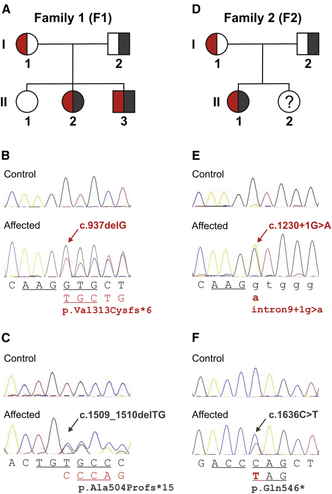
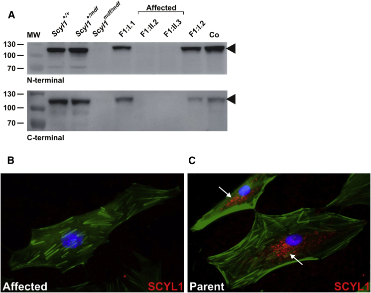
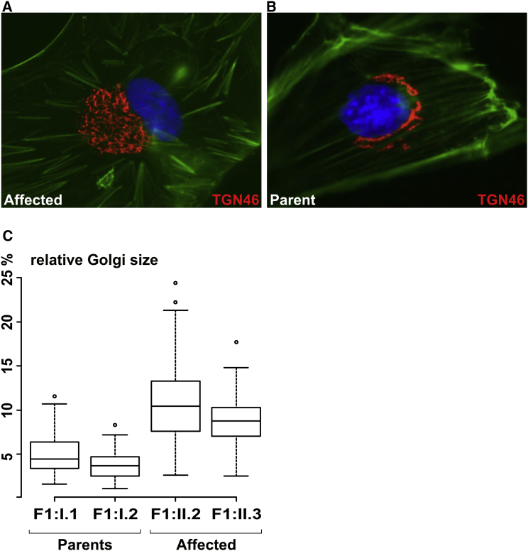
Hereditary ataxias comprise a group of genetically heterogeneous disorders characterized by clinically variable cerebellar dysfunction and accompanied by involvement of other organ systems. The molecular underpinnings for many of these diseases are widely unknown. Previously, we discovered the disruption of Scyl1 as the molecular basis of the mouse mutant mdf, which is affected by neurogenic muscular atrophy, progressive gait ataxia with tremor, cerebellar vermis atrophy, and optic-nerve thinning. Here, we report on three human individuals, from two unrelated families, who presented with recurrent episodes of acute liver failure in early infancy and are affected by cerebellar vermis atrophy, ataxia, and peripheral neuropathy. By whole-exome sequencing, compound-heterozygous mutations within SCYL1 were identified in all affected individuals. We further show that in SCYL1-deficient human fibroblasts, the Golgi apparatus is massively enlarged, which is in line with the concept that SCYL1 regulates Golgi integrity. Thus, our findings define SCYL1 mutations as the genetic cause of a human hepatocerebellar neuropathy syndrome.


Figures

Figure 1
Figure 1
Hepato- and Splenomegaly Represent Early-Onset Clinical Findings Abdominal computed tomography image (coronal reconstruction) showing significant hepato- and splenomegaly in the male affected individual (family 1) at age 8 years. The liver (L) was palpable 9 cm below the right costal margin and the spleen (S) was palpable 6 cm below the left costal margin.
Figure 2
Figure 2
Selective Atrophy of the Cerebellar Vermis and Thinning of the Optic Nerve (A–C) T2 weighted MRI scans from the male affected individual (F1:II.3) at age 16, revealing selective cerebellar vermis atrophy with spared hemispheres (and no brain stem or cerebral abnormalities). The signal of the cerebellar white and gray matter appears normal. Mid-sagittal cut showing upper vermis atrophy with markedly dilated interfolial fissures (black arrows) and normal pons (A), axial image (B), and coronal image (C) (note the normal hemispheres at this level). White arrows in (B) and (C) point to atrophic vermis. (D) Retro-bulbar coronal cut from T2 weighted MRI scan from the same affected individual, showing optic nerve thinning (optic nerves are indicated by white arrows, the inset shows a close-up of transverse-cut optic nerve with a horizontal diameter of 1.8 mm). Significant optic nerve thinning as revealed by retro-bulbar diameter measurements was present in both affected siblings (1.7–1.9 mm, compared to ∼3 mm in healthy age-matched probands12).
Figure 3
Figure 3
Compound Heterozygous SCYL1 Mutations in Three Individuals from Two Unrelated Families Panels (A)–(C) show family 1 and panels (D)–(F) show family 2. Pedigrees in (A) and (D) depict segregation of SCYL1 lesions detected by whole-exome sequencing and confirmed by conventional capillary DNA sequencing (B and C and E and F, respectively). DNA from the unaffected sister in family 2 (F2:II.2) was not available for analysis. Biallelic mutations are represented in red and dark gray. Reference sequences are shown below the traces from affected individuals, where consequences caused by the respective mutations (indicated by arrows) are shown in the second lines. Codon triplets are represented by underlined letters, exonic residues are shown in capitals, and intronic sequence is shown in lower case. Mutation positions refer to GenBank transcript NM_020680.3.
Figure 4
Figure 4
SCYL1-Null Mutations Lead to Complete Lack of SCYL1 in Cultured Skin Fibroblasts (A) Complete loss of SCYL1 in protein extracts from fibroblasts from both affected siblings (F1:II.2 and F1:II.3), in contrast to fibroblasts from their parents (F1:I.1 and F1:I.2), with both N-terminal (upper panel) and C-terminal antibodies (lower panel). Extracts prepared from human WI-38 fetal lung fibroblasts (Co) and murine fibroblasts from mdf (Scyl1mdf/mdf) and wild-type (Scyl1+/+) as well as heterozygous (Scyl1+/mdf) littermates were used as controls. MW; molecular weight marker (kDa). Arrowheads indicate full-length SCYL1 (∼105 kDa band). (B and C) Absent SCYL1 immunoreactivity (N-terminal antibody, goat anti-rabbit secondary antibody conjugated with Alexa Fluor 594, red fluorescence) in skin fibroblasts from female affected individual F1:II.2 (B) in comparison to a predominantly perinuclear vesicular SCYL1-staining pattern (white arrows) obtained in fibroblasts from her mother, F1:I.1 (C). The F-actin cytoskeleton is visualized by Phalloidin Atto 488 (green fluorescence) and nuclei are depicted with DAPI (blue).
Figure 5
Figure 5
Absence of SCYL1 Leads to Enlargement of the Golgi Apparatus in Human Fibroblasts By immunofluorescence, the Golgi network appeared enlarged in the skin fibroblasts from the male affected individual, F1:II.3, (A) when compared to the Golgi staining in his father’s, F1:I.2, fibroblasts (B). Anti-TGN46 (also known as TGOLN2, a trans-Golgi-network protein) antibody (Sigma-Aldrich; T7576) is shown as red fluorescence. The F-actin cytoskeleton is visualized by Phalloidin Atto 488 (green fluorescence) and nuclei are depicted with DAPI (blue). Morphometric analysis revealed enlargement of Golgi apparatuses in SCYL1-negative fibroblasts (C). Shown as a percentage of total cell size; p < 5e-14, Mann-Whitney-Wilcoxon test.

References

    1. Storey E. Genetic cerebellar ataxias. Semin. Neurol. 2014;34:280–292. - PubMed
    1. Mancuso M., Orsucci D., Siciliano G., Bonuccelli U. The genetics of ataxia: through the labyrinth of the Minotaur, looking for Ariadne’s thread. J. Neurol. 2014;261:S528–S541. - PMC - PubMed
    1. Jayadev S., Bird T.D. Hereditary ataxias: overview. Genet. Med. 2013;15:673–683. - PubMed
    1. Anheim M., Tranchant C., Koenig M. The autosomal recessive cerebellar ataxias. N. Engl. J. Med. 2012;366:636–646. - PubMed
    1. Schmidt W.M., Kraus C., Höger H., Hochmeister S., Oberndorfer F., Branka M., Bingemann S., Lassmann H., Müller M., Macedo-Souza L.I. Mutation in the Scyl1 gene encoding amino-terminal kinase-like protein causes a recessive form of spinocerebellar neurodegeneration. EMBO Rep. 2007;8:691–697. - PMC - PubMed

Publication types

MeSH terms

Substances