. 2015 Nov 19:5:16765.
doi: 10.1038/srep16765.

Influence of age on rat bone-marrow mesenchymal stem cells potential

Affiliations

Influence of age on rat bone-marrow mesenchymal stem cells potential

J Fafián-Labora et al. Sci Rep. .

Abstract

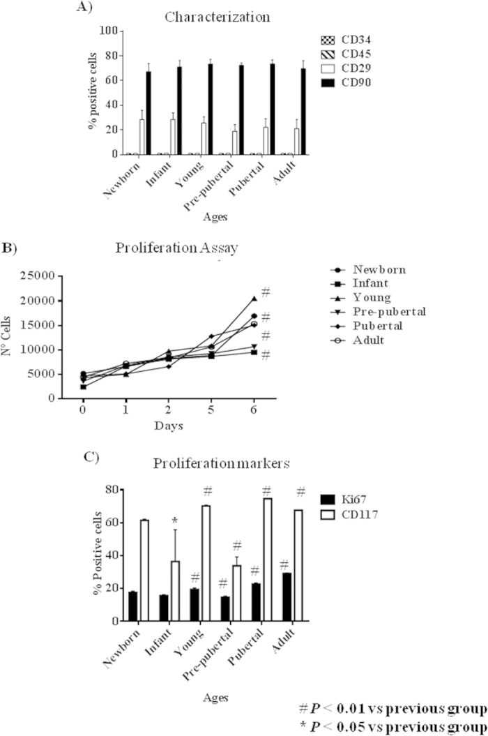
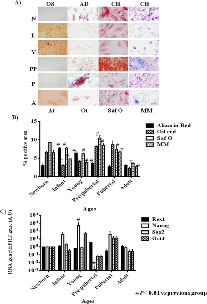
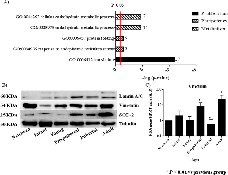
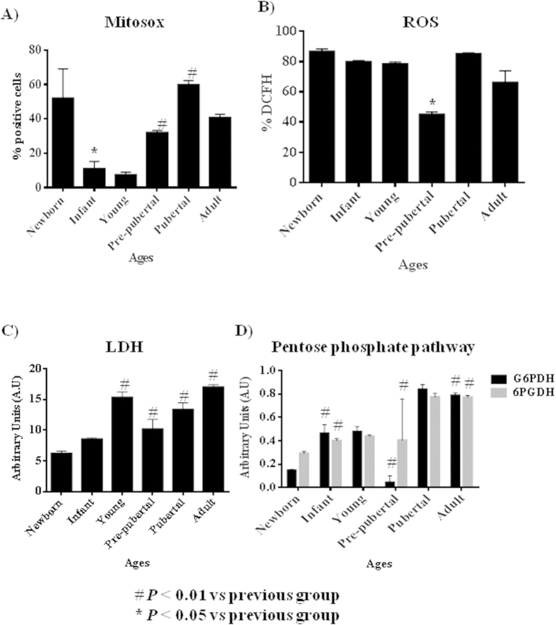
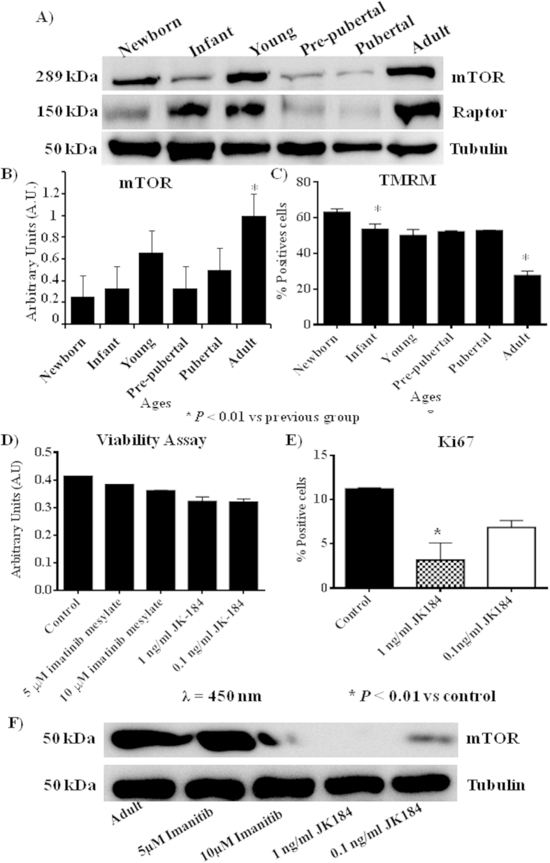
Mesenchymal stem cells promising role in cell-based therapies and tissue engineering appears to be limited due to a decline of their regenerative potential with increasing donor age. Six age groups from bone marrow mesenchymal stem cells of Wistar rats were studied (newborn, infant, young, pre-pubertal, pubertal and adult). Quantitative proteomic assay was performance by iTRAQ using an 8-plex iTRAQ labeling and the proteins differentially expressed were grouped in pluripotency, proliferative and metabolism processes. Proliferation makers, CD117 and Ki67 were measure by flow cytometry assay. Real time polymerase chain reaction analysis of pluripotency markers Rex1, Oct4, Sox2 and Nanog were done. Biological differentiation was realized using specific mediums for 14 days to induce osteogenesis, adipogenesis or chondrogenesis and immunostain analysis of differentiated cell resulting were done. Enzimoimmunoassay analysis of several enzymes as L-lactate dehydrogenase and glucose-6-phosphate isomerase were also done to validate iTRAQ data. Taking together these results indicate for the first time that mesenchymal stem cells have significant differences in their proliferative, pluripotency and metabolism profiles and those differences are age depending.


Figures

Figure 1
Figure 1. Proliferation profile from rat mesenchymal stem cells at different age.
(A) Characterization by flow cytometry assay of percentage of positives mesenchymal stem cells markers (CD29 and CD73) and negative hematopoietic markers (CD34 and CD45). (B) Proliferation assay of studied aging groups for 6 days. (C) Percentage of proliferation markers, DC117 and Ki67, from studied aging groups by flow cytometry assay. One representative experiment is shown. #p value less than 0.05 compared with previous group and *p value less than 0.01 compared with previous group, were considered statistically significant using Mann-Whitney-U tests.
Figure 2
Figure 2. Pluripotency profile from rats mesenchymal stem cells at different age.
(A) Representative pictures of mesenchymal stem cell from bone marrow of studied age group after 14 days with specific differentiation medium. On the top specific medium is indicated; OS=osteogenic medium; AD = adipogenic medium; CH = chondrogenic medium. Differentiation medium are indicated in Material and methods. On the bottom stain is indicated; Ar = alizarin red; Or = oil red; Saf O = safranine O and MM = Modified Masson’s stain. Straight size is 200 μM. (B) Densitometry study of mesenchymal stem cell from bone marrow of studied aging group after 14 days with specific differentiation medium after immunostaining assay. AnalySIS Image Processing computer was used to quantify the signal of different stain obtained. #p value less than 0.01 was considered statistically significant using Mann-Whitney-U tests. (C) Histogram represents gene expression of pluripotency markers, Rex1, Nanog, Sox2 and Oct4. Real-time reverse transcriptase PCR (qRT-PCR) analysis normalized by expression of HPRT gene used as housekeeping. #p value less than 0.01 compared with previous group was considered statistically significant using Mann-Whitney-U tests. Three replicates were made.
Figure 3
Figure 3. Validation of iTRAQ analysis.
(A) Significant activates pathways obtained by comparing modulated proteins obtained by iTRAQ analysis employing functional annotations according to the String 9.0 software. Small numbers on the right of each bar are the modulated protein involved in each process. (B) Western blot analysis of Lamin A/C, Vimentin and Superoxide dismutase 2 (SOD-2). Tubulin was used as housekeeping. On the left molecular weight of each protein is shown. On the bottom the group’s source of mesenchymal stem cells used. The gels have been run under the same experimental conditions. (C) Vinculin gene expression using real-time reverse transcriptase PCR (qRT-PCR) analysis normalized by expression of HPRT gene used as housekeeping.
Figure 4
Figure 4. Metabolism profile from rats mesenchymal stem cells at different age.
(A) 2′,7′-dichlorofluorescein signal measured by flow cytometry to check ROS intracellular (B) Mitosox signal measured by flow cytometry to check ROS mitochondrial. (C) Lactate-dehydrogenase (LDH) activity measured by spectrophotometer analysis. (D) Pentose phosphate pathway activity measured by spectrometer analysis. G6PDH = Glucose-6-phosphate 1-dehydrogenase; 6GPDH = 6-phosphogluconate-dehydrogenase. *p value less than 0.01 compared with previous group and #p value less than 0.05 compared with previous group, were considered statistically significant using Mann-Whitney-U tests.
Figure 5
Figure 5. mTOR pathway profile from rat mesenchymal stem cells at different age (A) Western blot of mTOR pathway, mTOR and raptor, Tubulin was used as housekeeping.
(B) Densitometry analysis of westerns of mTOR normalized with respect to Tubulin. *p value less than 0.01 compared with previous group. (C) Tetramethylrhodamine, methyl ester (TMRM) dye accumulated in active mitochondria with intact potentials, was used to detect functional mitochondria in the MSCs at different ages following functional mitochondrial staining protocol from commercial. (D) Viability assay of mesenchymal stem cells from adult group incubated with 10 μM or 5 μM of imatinib mesylate or 1 ng/ml or 0.1 ng/ml of JK184. (E) Flow cytometry of Ki67 from mesenchymal stem cells of adult group incubated with 1 ng/ml or 0.1 ng/ml of JK184 in the medium. F) Western blot of mTOR and Tubulin of mesenchymal stem cells from adult group after incubating with 10 μM or 5 μM of imatinib mesylate or 1 ng/ml or 0.1 ng/ml of JK184 in the medium. Control = mesenchymal stem cells incubated with growth medium alone. The gels have been run under the same experimental conditions. *p value less than 0.01 compared with control group was considered statistically significant using Mann-Whitney-U tests.

References

    1. Murphy M. B., Moncivais K. & Caplan A. I. Mesenchymal stem cells: environmentally responsive therapeutics for regenerative medicine. Experimental & Molecular Medicine. 45, e54 (2013). - PMC - PubMed
    1. Bach M., Schimmelpfennig C. & Stolzing A. Influence of murine mesenchymal stem cells on proliferation, phenotype, vitality, and cytotoxicity of murine cytokine-induced killer cells in coculture. PLoS One. 9, e88115 (2014). - PMC - PubMed
    1. Zhang L. et al. The Effects of Mesenchymal Stem Cells in Craniofacial Tissue Engineering. Current Stem Cell Research & Therapy. 9, 280–289 (2014). - PubMed
    1. Yeh H. Y. et al. Neocartilage formation from mesenchymal stem cells grown in type II collagen-hyaluronan composite scaffolds. Differentiation. 86, 171–183 (2014). - PubMed
    1. Song J. Q. et al. Effect of treadmill exercise timing on repair of full-thickness defects of articular cartilage by bone-derived mesenchymal stem cells: an experimental investigation in rats. PLoS One. 9, e90858 (2014). - PMC - PubMed

Publication types

Substances