. 2016 Jan 8;291(2):508-20.
doi: 10.1074/jbc.M115.701102. Epub 2015 Nov 18.

The Activation Mechanism of Glycoprotein Hormone Receptors with Implications in the Cause and Therapy of Endocrine Diseases

Affiliations

The Activation Mechanism of Glycoprotein Hormone Receptors with Implications in the Cause and Therapy of Endocrine Diseases

Antje Brüser et al. J Biol Chem. .

Abstract

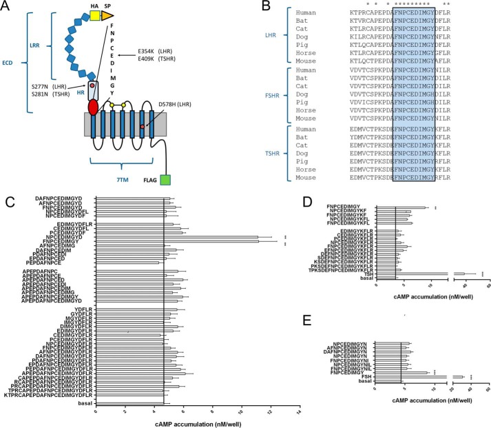
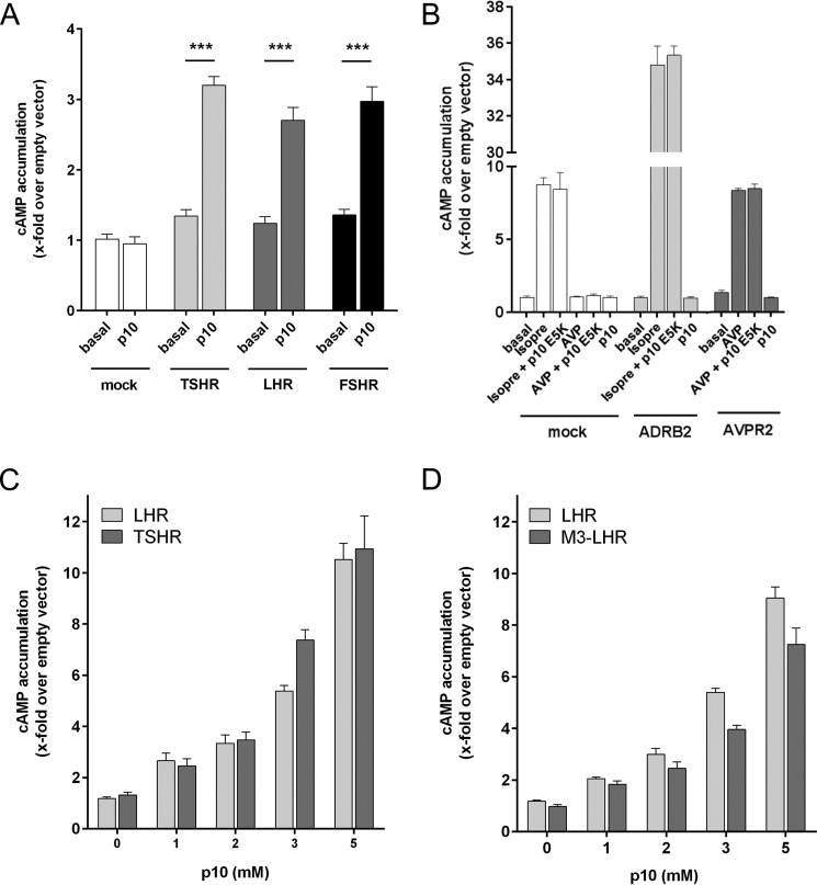
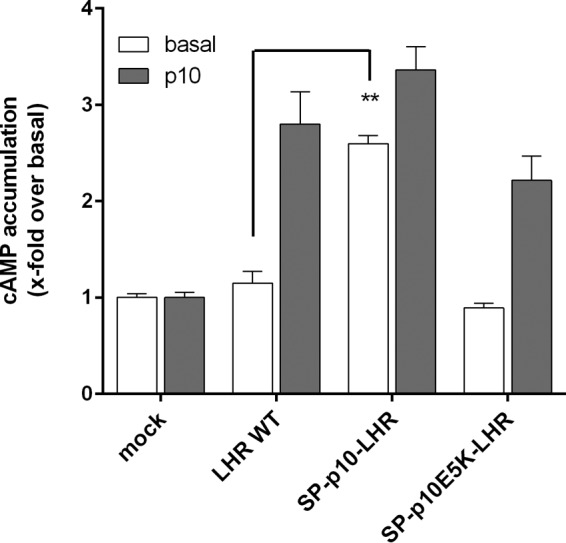
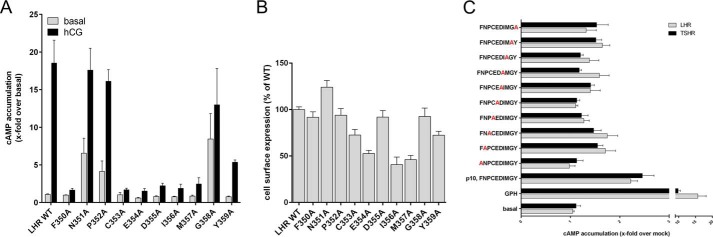
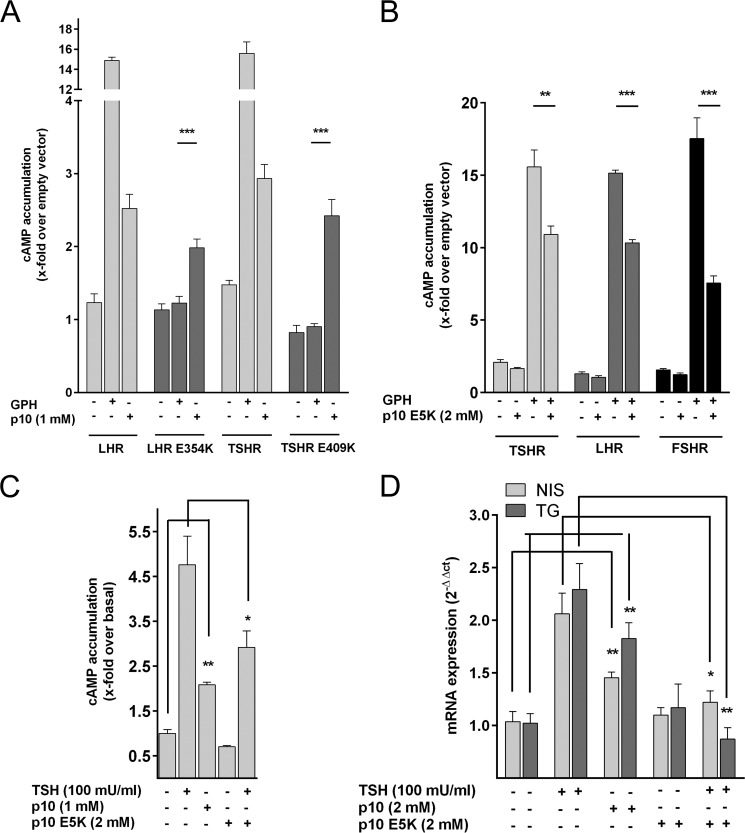
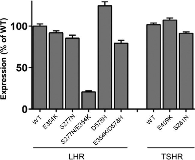
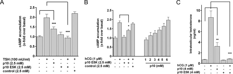
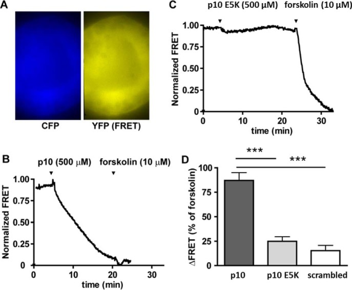
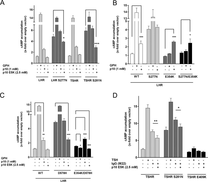
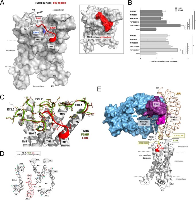
Glycoprotein hormones (GPHs) are the main regulators of the pituitary-thyroid and pituitary-gonadal axes. Selective interaction between GPHs and their cognate G protein-coupled receptors ensure specificity in GPH signaling. The mechanisms of how these hormones activate glycoprotein hormone receptors (GPHRs) or how mutations and autoantibodies can alter receptor function were unclear. Based on the hypothesis that GPHRs contain an internal agonist, we systematically screened peptide libraries derived from the ectodomain for agonistic activity on the receptors. We show that a peptide (p10) derived from a conserved sequence in the C-terminal part of the extracellular N terminus can activate all GPHRs in vitro and in GPHR-expressing tissues. Inactivating mutations in this conserved region or in p10 can inhibit activation of the thyroid-stimulating hormone receptor by autoantibodies. Our data suggest an activation mechanism where, upon extracellular ligand binding, this intramolecular agonist isomerizes and induces structural changes in the 7-transmembrane helix domain, triggering G protein activation. This mechanism can explain the pathophysiology of activating autoantibodies and several mutations causing endocrine dysfunctions such as Graves disease and hypo- and hyperthyroidism. Our findings highlight an evolutionarily conserved activation mechanism of GPHRs and will further promote the development of specific ligands useful to treat Graves disease and other dysfunctions of GPHRs.

Keywords: G protein-coupled receptor (GPCR); follicle-stimulating hormone (FSH); hormone receptor; luteinizing hormone; signal transduction; signaling; thyroid; thyroid-stimulating hormone.


Figures

FIGURE 1.
FIGURE 1.
Peptides derived from the ECD C terminus are agonists at GPHRs. A, the structural architecture of a prototypical GPHR is shown. An ECD consisting of a signal peptide (SP), an LRR domain, the HR, and the 7TM domain can be distinguished. All constructs were epitope-tagged with an N-terminal HA epitope (yellow) and a C-terminal FLAG epitope (green). Positions of mutations studied are given for LHR and TSHR. B, the ECD C terminus contains a block of 9 amino acids that is conserved among GPHRs. C–E, peptides (1 mm) of different lengths derived from the C-terminal part of the LHR (C), TSHR (D), and FSHR (E) hinge region were tested on LHR-, TSHR- and FSHR-transfected COS-7 cells, respectively. cAMP levels of transfected COS-7 cells were determined as described under “Experimental Procedures.” All peptides were controlled on empty vector-transfected cells and showed no cAMP signal above basal (cAMP level pcDps: 3.7 ± 0.3 nm/well; cAMP level LHR with 1 μm hCG: 56.8 ± 8.5 nm/well). **, p < 0.01, ***, p < 0.001, as compared with basal (without peptide), paired Student's t test. All data are means ± S.E. of three independent experiments performed in triplicate.
FIGURE 2.
FIGURE 2.
p10 specifically activates GPHRs. A, p10 (1 mm) was tested on COS-7 cells transfected with the LHR, FSHR, and TSHR. B, COS-7 cells were transfected with the human ADRB2 and the human V2 vasopressin receptor (AVPR2) and incubated with isoprenaline (isopre: 1 μm), arginine-vasopressin (AVP: 1 μm) and p10 (2 mm). Empty pcDps vector (mock) served as negative control (pcDps; cAMP level was 2.9 ± 0.4 nm/well). C and D, concentration-response curves of p10 on LHR, M3-LHR, and TSHR revealed an EC50 value >2 mm. Basal pcDps levels were 3.6 ± 0.3 nm/well. Data are means ± S.E. of three independent experiments performed in triplicate.
FIGURE 3.
FIGURE 3.
N-terminal truncation activates the LHR. COS-7 cells were transfected with the indicated LHR constructs, and cAMP levels were determined with p10 (2 mm) and without. Empty pcDps vector (mock) served as negative control (pcDps; cAMP level was 1.2 ± 0.2). Data are means ± S.E. of three independent experiments performed in triplicate.
FIGURE 4.
FIGURE 4.
Alanine-scanning mutagenesis of the p10 region in GPHRs and the p10 peptide. A, COS-7 cells were transfected with the indicated LHR constructs, and cAMP levels were determined with hCG (500 nm) and without. Empty pcDps vector (mock) served as negative control (pcDps; cAMP level was 2.6 ± 0.8). Data are means ± S.E. of three independent experiments performed in triplicate. B, cell surface expression of HA epitope-tagged receptors transiently expressed in COS-7 cells was determined by cell surface ELISA (see “Experimental Procedures”). Optical density (OD) is given as the percentage of LHR WT minus the OD of mock-transfected cells. The OD values for mock-transfected cells and LHR WT were 0.02 ± 0.007 and 0.642 ± 0.092, respectively. Data are given as means ± S.E. of three independent experiments performed in triplicate. C, alanine-scanning mutagenesis was performed on p10 and tested on COS-7 cells transfected with WT LHR and TSHR. GPH served as positive control (GPH; 100 milliunits/ml bTSH for TSHR and 500 nm hCG for LHR). Peptides (2 mm) were also tested on cells transfected with the empty vector (mock), and data are given as -fold over mock treated with the indicated peptide. Basal cAMP level of mock-transfected cells was 1.7 ± 0.3 nm/well. Means ± S.E. of three independent experiments performed in triplicate are presented.
FIGURE 5.
FIGURE 5.
Specificity of p10-mediated activation of GPHRs. A, COS-7 cells transfected with LHR and TSHR and their inactive mutants E354K and E409K, respectively, were incubated with their respective glycoprotein hormone (LHR: 500 nm hCG; TSHR: 100 milliunits/ml bTSH) and p10 and cAMP levels were determined. Basal cAMP level for pcDps was 1.8 ± 0.8 nm/well. B, COS-7 cells transfected with TSHR, LHR, and FSHR were incubated with their GPH (100 milliunits/ml bTSH, 500 nm hCG, 5 milliunits/ml FSH) and p10 E5K as indicated. Basal cAMP level for pcDps (empty vector) was 1.7 ± 0.5 nm/well. cAMP levels were determined (see “Experimental Procedures”). C and D, Nthy-ori 3-1 cells (40) were incubated with TSH, p10, and p10 E5K. In C and D, cAMP levels (basal cAMP levels: 1.1 ± 0.3 nm/well) (C) and mRNA expression levels (D) of the NIS and TG prior to and after stimulation were determined (see “Experimental Procedures”). Expression of NIS and TG was normalized to β2-microglobulin (Ct 15.7 ± 0.2). Gene regulation values are given as means of 2−ΔΔCt ± S.E. Data are means ± S.E. of three independent experiments performed in triplicate. *, p < 0.05, **, p < 0.01, ***, p < 0.001 (paired Student's t test).
FIGURE 6.
FIGURE 6.
Expression of WT and mutant GPHRs. COS-7-cells were transfected with either WT or mutant GPHRs, and expression of receptors was measured by cell surface ELISA (see “Experimental Procedures”). OD is given as the percentage of WT GPHR minus OD of mock-transfected cells. For surface ELISA, the OD value for mock-transfected cells, LHR WT, and TSHR WT was 0.025 ± 0.006, 0.895 ± 0.069, and 0.834 ± 0.072, respectively. Data are given as means ± S.E. of three independent experiments each performed in triplicate.
FIGURE 7.
FIGURE 7.
p10 activates GPHRs in tissues. A and B, cell suspensions from mouse thyroid (A) and mouse testis (B) were incubated with TSH and hCG, respectively, and with p10, p10 E5K, and a control peptide (scrambled amino acids of p10). Basal cAMP levels for thyroid and testis were 5.8 ± 2.3 and 30.1 ± 10.5 nm/well, respectively. C, cell suspension from testis was incubated with the indicated compounds, and testosterone levels were determined with an ELISA (see “Experimental Procedures”). Data are means ± S.E. of three (A and C) and five (B) independent experiments performed in triplicate. *, p < 0.05, **, p < 0.01, ***, p < 0.001 (paired Student's t test).
FIGURE 8.
FIGURE 8.
Real-time monitoring of cAMP levels in thyroid follicles isolated from the cAMP reporter mice. Thyroid follicles isolated from Epac1-camps mice were visualized by time-lapse fluorescence microscopy. A, CFP and corrected YFP images were recorded to determine YFP/CFP ratio images. B and C, the graphs show normalized YFP/CFP ratio values calculated from CFP and YFP images. D, FRET values were normalized to the basal level (set to 1) and the response to stimulation with 10 μm forskolin (set to 0). Data are means ± S.E. of three (scrambled peptide) and five (p10, p10 E5K) experiments. ***, p < 0.001 versus p10 (one-way analysis of variance followed by Bonferroni's post hoc test).
FIGURE 9.
FIGURE 9.
The mutant p10 E5K blocks mutation and antibody-induced GPHR activation. A–D, COS-7 cells were transfected with WT and the indicated mutant LHR and TSHR and incubated with 500 nm hCG or 100 milliunits/ml bTSH and p10 and inactivating p10 E5K (A–C) or 500 ng/ml of the monoclonal autoantibody M22 (D). cAMP levels were determined as described (see “Experimental Procedures”). cAMP level of mock-transfected cells was 2.3 ± 0.9 nm/well. Data are means ± S.E. of three experiments performed in triplicate. *, p < 0.05, **, p < 0.01, ***, p < 0.001 (paired Student's t test).
FIGURE 10.
FIGURE 10.
Structural homology model of the TSHR serpentine domain including the p10 region. A, the 7TM structure of the TSHR with the p10 region (red, backbone ribbon tube, side chains from Phe405–Tyr414), which equates in the TSHR exactly the transition between the extracellular HR and the TM1, is represented as a model based on crystal structures of homologous GPCRs (see “Experimental Procedures”). Ntt, N-terminal tail; Ctt, C-terminal tail. Inset: the surfaces of the 7TM and the p10 region are highlighted, showing that the flexibility of this fragment between the ECLs is spatially restricted by steric constraints. B, COS-7 cells transfected with TSHR and LHR were incubated with shortened p10 (1 mm) and p10 as positive control. cAMP levels of mock-transfected cells were 2.1 ± 1.1 nm/well. Data are means ± S.E. of three experiments performed in triplicate. *, p < 0.05, **, p < 0.01, ***, p < 0.001 (paired Student's t test). C, the positioning of the p10 region is shown in the overlay of the TSHR, LHR, and FSHR. Based on this model, the amino acid interface of p10 and its binding pocket is shown in D. Several positions of this interface were mutated and functionally tested. Positions marked with + lead to receptor activation, and positions marked with − lead to receptor inactivation. For references, see the TSHR database Sequence-Structure-Function-Analysis of Glycoprotein Hormone Receptors (46). E, the complex model of TSHR (white, backbone ribbon) with bound TSH and the activating antibody M22 visualizes a potential arrangement and the principle mechanism of GPHR activation. For comparison of binding sites, the LRR domain of TSHR complexed with the activating antibody M22 (9) was superimposed with the TSHR LRR domain/TSH model. The entire complex is derived by the arrangement of available structural information on GPHRs and GPCRs (see “Experimental Procedures”). ICL, intracellular loop.

References

    1. Parmentier M., Libert F., Maenhaut C., Lefort A., Gérard C., Perret J., Van Sande J., Dumont J. E., and Vassart G. (1989) Molecular cloning of the thyrotropin receptor. Science 246, 1620–1622 - PubMed
    1. McFarland K. C., Sprengel R., Phillips H. S., Köhler M., Rosemblit N., Nikolics K., Segaloff D. L., and Seeburg P. H. (1989) Lutropin-choriogonadotropin receptor: an unusual member of the G protein-coupled receptor family. Science 245, 494–499 - PubMed
    1. Sprengel R., Braun T., Nikolics K., Segaloff D. L., and Seeburg P. H. (1990) The testicular receptor for follicle stimulating hormone: structure and functional expression of cloned cDNA. Mol. Endocrinol. 4, 525–530 - PubMed
    1. Fan Q. R., and Hendrickson W. A. (2005) Structure of human follicle-stimulating hormone in complex with its receptor. Nature 433, 269–277 - PMC - PubMed
    1. Sangkuhl K., Schulz A., Schultz G., and Schöneberg T. (2002) Structural requirements for mutational lutropin/choriogonadotropin receptor activation. J. Biol. Chem. 277, 47748–47755 - PubMed

Publication types

MeSH terms