. 2016 Jan 8;291(2):593-604.
doi: 10.1074/jbc.M115.691097. Epub 2015 Nov 18.

A Single Glycan at the 99-Loop of Human Kallikrein-related Peptidase 2 Regulates Activation and Enzymatic Activity

Affiliations

A Single Glycan at the 99-Loop of Human Kallikrein-related Peptidase 2 Regulates Activation and Enzymatic Activity

Shihui Guo et al. J Biol Chem. .

Abstract

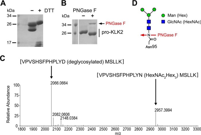
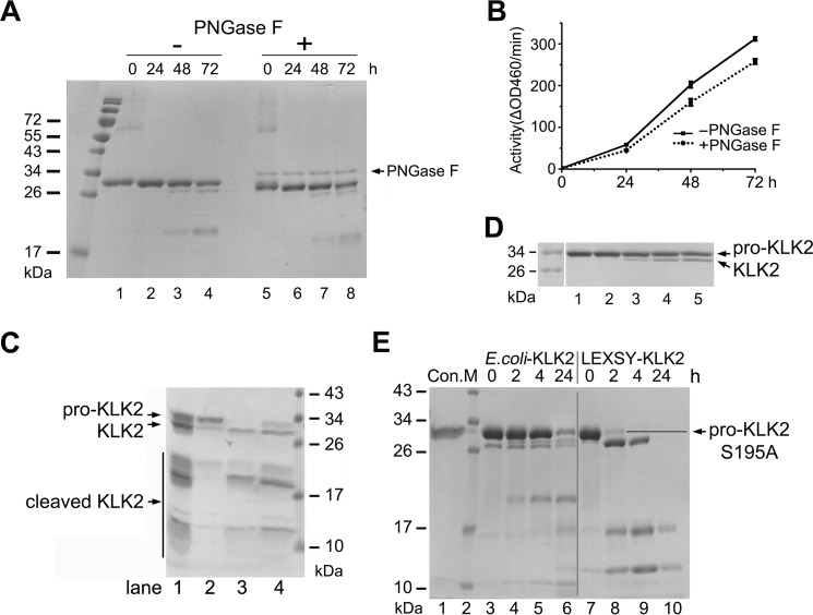
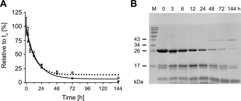
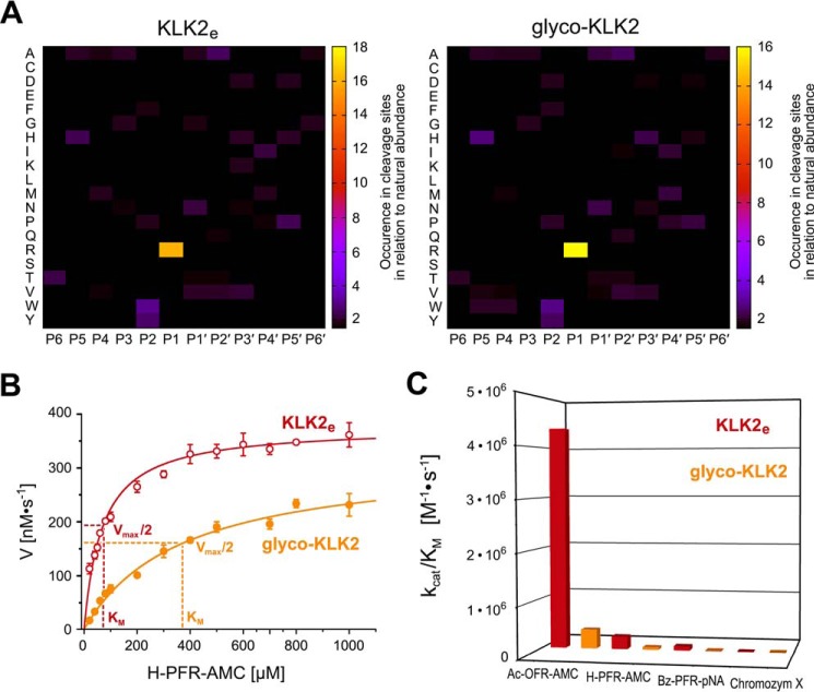
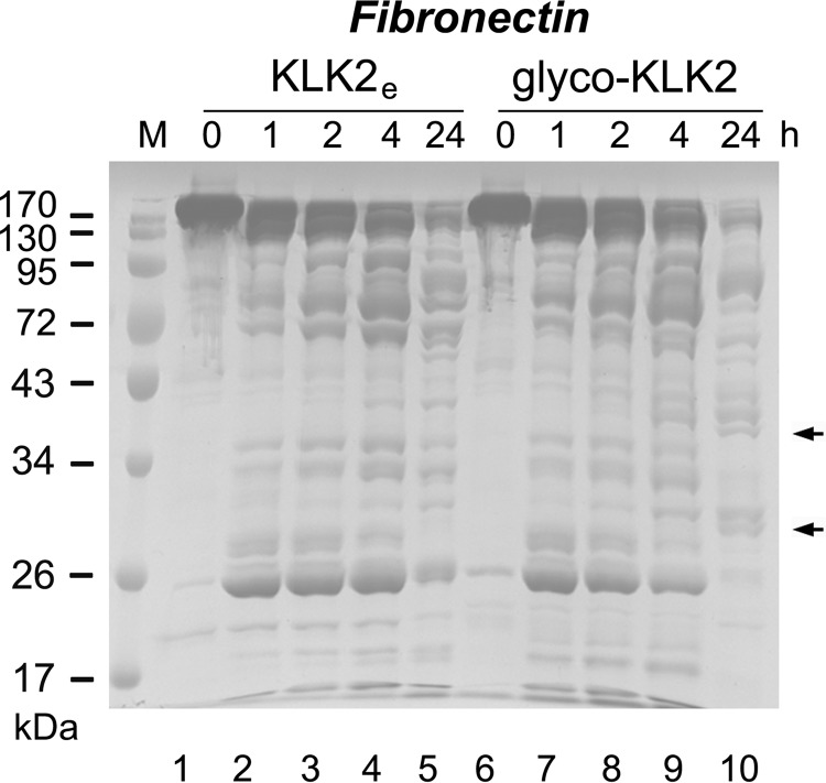
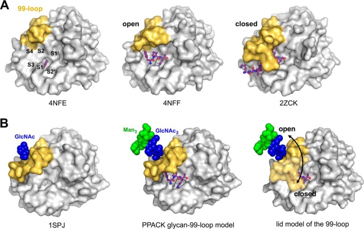
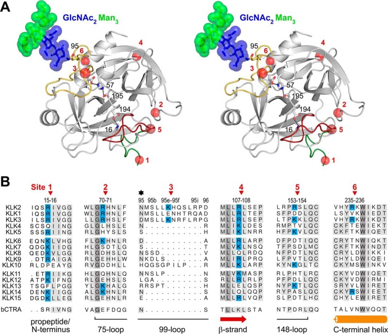
Human kallikrein-related peptidase 2 (KLK2) is a key serine protease in semen liquefaction and prostate cancer together with KLK3/prostate-specific antigen. In order to decipher the function of its potential N-glycosylation site, we produced pro-KLK2 in Leishmania tarentolae cells and compared it with its non-glycosylated counterpart from Escherichia coli expression. Mass spectrometry revealed that Asn-95 carries a core glycan, consisting of two GlcNAc and three hexoses. Autocatalytic activation was retarded in glyco-pro-KLK2, whereas the activated glyco-form exhibited an increased proteolytic resistance. The specificity patterns obtained by the PICS (proteomic identification of protease cleavage sites) method are similar for both KLK2 variants, with a major preference for P1-Arg. However, glycosylation changes the enzymatic activity of KLK2 in a drastically substrate-dependent manner. Although glyco-KLK2 has a considerably lower catalytic efficiency than glycan-free KLK2 toward peptidic substrates with P2-Phe, the situation was reverted toward protein substrates, such as glyco-pro-KLK2 itself. These findings can be rationalized by the glycan-carrying 99-loop that prefers to cover the active site like a lid. By contrast, the non-glycosylated 99-loop seems to favor a wide open conformation, which mostly increases the apparent affinity for the substrates (i.e. by a reduction of Km). Also, the cleavage pattern and kinetics in autolytic inactivation of both KLK2 variants can be explained by a shift of the target sites due to the presence of the glycan. These striking effects of glycosylation pave the way to a deeper understanding of kallikrein-related peptidase biology and pathology.

Keywords: N-linked glycosylation; autolytic inactivation; enzyme kinetics; kallikrein; prostate cancer; serine protease; substrate specificity; zymogen activation.


Figures

FIGURE 1.
FIGURE 1.
Analysis of KLK2 glycosylation. A, enterokinase-activated KLK2 from Leishmania expression (LEXSY) was analyzed under non-reducing and reducing conditions. Clipped products migrated as a single band under non-reducing conditions. Intact and clipped forms, which form fragments smaller than 17 kDa under reducing conditions (+DTT), were not separable and appeared as a single peak in gel filtration. B, removal of N-glycans from LEXSY-KLK2 with PNGase F (36 kDa) resulted in a shift of the apparent molecular mass by about 1 kDa. C, trypsin-digest mass spectrometry confirmed partially deglycosylated LEXSY-KLK2 by PNGase F under non-denaturing conditions. The deconvoluted mass spectrum is shown, and the indicated masses refer to uncharged molecules. The peak at 2957 was identified by sequencing and subsequent modification search as VPVSHSFPHPLYN-(Hex3HexNAc2)-MSLLK. The signal at 2066 represents the corresponding deglycosylated peptide (VPVSHSFPHPLYD-(deglycosylated)-MSLLK), with the expected core glycan mass of 891 Da. D, N-linked core glycans at Asn-95 of LEXSY KLK2, consisting most likely of two N-acetylglucosamine and three mannose units. PNGase F cleaves the N-glycan and generates an Asp-95.
FIGURE 2.
FIGURE 2.
Autoactivation and activation of pro-KLK2. A, time-dependent autoactivation of both glycosylated (lanes 1–4) and enzymatically deglycosylated pro-KLK2 (lanes 5–8) is monitored on an SDS-PAGE. Note the same shift of about 1 kDa as in Fig. 1B. B, activity assay of autoactivated KLK2 with the small peptide substrate H-PFR-AMC by glycosylated (black line) and enzymatically deglycosylated KLK2 (dotted line) at varying times (0, 24, 48, and 72 h), matching those in A. C, concentration-dependent autoactivation of pro-KLK2-S195A. The initial pro-KLK2 samples at concentrations of 250 (lane 1), 600 (lane 2), 800 (lane 3), and 900 (lane 4) μg/ml were kept at room temperature for 1 day and subsequently at 4 °C for 4 days, respectively. D, activation of pro-KLK2. Shown is incubation of 1 μg of pro-KLK2-S195A from LEXSY cells (lane 1) with 1% KLK2e from E. coli (lane 2), 1% glyco-KLK2 from LEXSY cells (lane 3), or 0.05% (lane 4) or 0.1% enterokinase (lane 5) overnight (14 h) at room temperature. E, pro-KLK2-S195A (lane 1) was digested by 20% KLK2e (lanes 3–6) or 20% glyco-KLK2 (lanes 7–10) for a time-dependent course. Samples were taken at 0, 2, 4, and 24 h, as indicated above the lanes, and then checked by Coomassie Blue-stained SDS-PAGE. The vertical gray line was inserted to indicate samples with different treatment. KLK2e shows a different cleavage pattern (e.g. by generating a 20-kDa fragment, which originates from the clipping at Arg-70↓His-71).
FIGURE 3.
FIGURE 3.
Time-dependent autoinactivation of KLK2. A, the decrease of intact glyco-KLK2 (black squares and line) correlates well with the loss of protease activity (open circles with dotted line). The loss of mature protein and activity follows a second-order reaction mechanism. B, time-dependent KLK2 autolysis was monitored by reducing SDS-PAGE.
FIGURE 4.
FIGURE 4.
Effects of glycosylation on KLK2 activity. A, PICS of KLK2e (left) and glyco-KLK2 (right) covering peptide substrates from residue P6 to P6′. Increased occurrence of an amino acid residue in cleavage sites of a random peptide library indicates the corresponding preference of the peptidase specificity sites S6–S6′. Both patterns are similar; P1-Arg dominates the specificity, whereas aromatic P2 residues and hydrophobic P4 residues are slightly favored. In P1′ and P2′, small or polar residues and small to medium hydrophobic residues are preferred, respectively. B, Michaelis-Menten plot of H-PFR-AMC turnover by KLK2e (open red circles) and glyco-KLK2 (filled orange circles). The velocity was normalized for 150 nm enzyme concentration, according to active site titrations. It is noteworthy that the Km of glyco-KLK2 is 5 times higher than that of KLK2e, despite a comparable Vmax and kcat of 17.0 and 13.6 s−1, respectively. C, catalytic efficiency depicted as columns for KLK2e (red) and glyco-KLK2 (orange) for fluorogenic and chromogenic substrates (Table 1). Chromozym X is Mco-d-Nle-Gly-Arg-pNA. Whereas the kcat varies considerably, the Km for all substrates is consistently about 5 times higher with glyco-KLK2 than with KLK2e.
FIGURE 5.
FIGURE 5.
Comparative degradation rates and fragmentation patterns of human fibronectin in the presence of KLK2e (lanes 2–6) glyco-KLK2 (lanes 7–11) as determined by SDS-PAGE. Aliquots were taken at representative intervals and analyzed by SDS-PAGE followed by Coomassie Blue staining. Fibronectin alone is stable at 37 °C for at least 24 h. Overall, the breakdown by glyco-KLK2 appears more efficient and results in some unique bands after 24-h incubation, as indicated by arrows.
FIGURE 6.
FIGURE 6.
Model of the 99-loop lid mechanism and possible regulation of KLK2 activity by glycosylation. A, the left panel shows the crystal structure of KLK2e (Protein Data Bank code 4NFE) with the small inhibitor benzamidine as a ball-and-stick model located in the S1 pocket, which is labeled like the other pockets from S4 to S2′, whereas the molecular surface of the wide open 99-loop is depicted in yellow. In the related KLK2e PPACK complex (Protein Data Bank code 4NFF), P1-Arg, P2-Pro, and d-Phe occupy the non-prime region, from the S1 to the S4 subsite, respectively. It exhibits an open 99-loop, which can hardly interfere with substrate binding. In the right panel, the structure of a human KLK3 acyl intermediate complex (Protein Data Bank code 2ZCK) represents a fully closed, non-glycosylated 99-loop, which is homologous to the KLK2e counterpart. Parts of the 99-loop interact tightly with the P2–P4 residues, contributing considerably to substrate specificity. B, in the left panel, the apo-KLK1 structure (Protein Data Bank code 1SPJ) contains a 99-loop with a glycosylated Asn-95, displaying a half-open conformation, most likely due to the presence of the stabilizing glycan, of which only a GlcNAc was visible. This structure is the basis of the PPACK glycan-99-loop model (middle), extended by one GlcNAc and three mannose units of a core glycan. PPACK and pNA or AMC substrate binding would be hampered by the rather occluding 99-loop conformation. The combined model of glycosylated 99-loop with a bound substrate in the S2–S4 region (right) suggests that the active site is less accessible in the closed state but also more stable during the following catalytic steps. A favored closed state will result in a lower kon rate and higher Km, consistent with the observed enzyme kinetics of glyco-KLK2 and KLK2e.
FIGURE 7.
FIGURE 7.
Inactivating cleavage sites of KLK2. A, stereo representation of a glyco-KLK2 model. The active site residues Asp-102, His-57, and Ser-195, and the stabilizing salt bridge Ile-16–Asp-194 are represented as sticks. The N terminus with the beginning of the propeptide (green), the 99-loop (yellow), and the 148-loop (red) are highlighted as specified. The six cleavage sites at Arg-15, Arg-70, Lys-95e, Arg-107, Arg-153, and Arg-226 are shown as red spheres. The glycan at Asn-95 that shields the 99-loop from cleavage is depicted with sticks and spheres. B, sequence alignment the six KLK2 cleavage sites, as shown in A, with the other 14 human tissue kallikrein family members and bovine chymotrypsin A (bCTRA) as a numbering reference. The red arrows indicate the autolysis sites on KLK2, whereas its secondary structure is depicted at the bottom, and the glycosylation site at Asn-95 is marked with a star. In contrast to KLK1 and -2, KLK3 is not glycosylated in the 99-loop but at Asn-61. Identical and conserved residues are highlighted with a gray background, whereby a darker gray indicates a higher degree of conservation. The basic P1 residues Arg and Lys in KLK2 and in the potential cleavage sites of other KLKs are highlighted with a blue background.

References

    1. Raspi G. (1996) Kallikrein and kallikrein-like proteinases: purification and determination by chromatographic and electrophoretic methods. J. Chromatogr. B Biomed. Appl. 684, 265–287 - PubMed
    1. Avgeris M., Mavridis K., and Scorilas A. (2012) Kallikrein-related peptidases in prostate, breast, and ovarian cancers: from pathobiology to clinical relevance. Biol. Chem. 393, 301–317 - PubMed
    1. Fukushima D., Kitamura N., and Nakanishi S. (1985) Nucleotide sequence of cloned cDNA for human pancreatic kallikrein. Biochemistry 24, 8037–8043 - PubMed
    1. Baker A. R., and Shine J. (1985) Human-kidney kallikrein: cDNA cloning and sequence-analysis. DNA 4, 445–450 - PubMed
    1. Schedlich L. J., Bennetts B. H., and Morris B. J. (1987) Primary structure of a human glandular kallikrein gene. DNA 6, 429–437 - PubMed

Publication types

LinkOut - more resources