. 2016 Jan 15;27(2):308-20.
doi: 10.1091/mbc.E15-02-0061. Epub 2015 Nov 18.

Arl13b and the exocyst interact synergistically in ciliogenesis

Affiliations

Arl13b and the exocyst interact synergistically in ciliogenesis

Cecília Seixas et al. Mol Biol Cell. .

Abstract

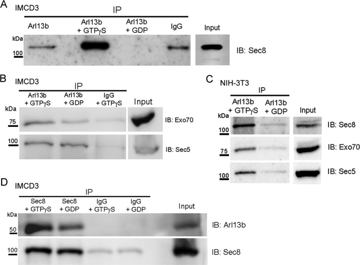
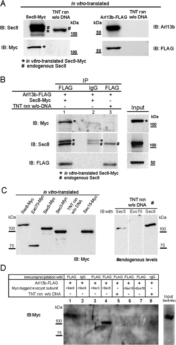
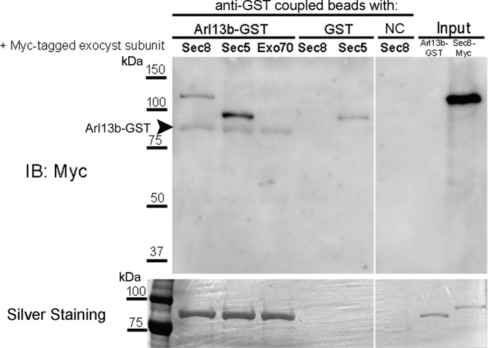
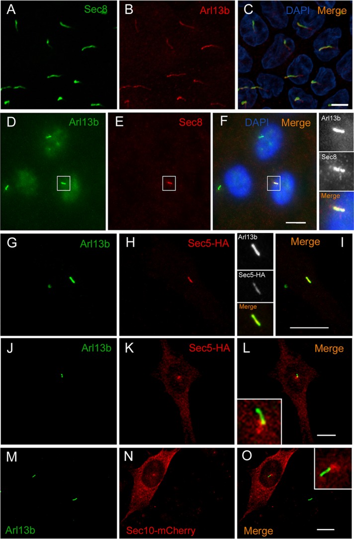
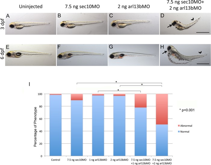
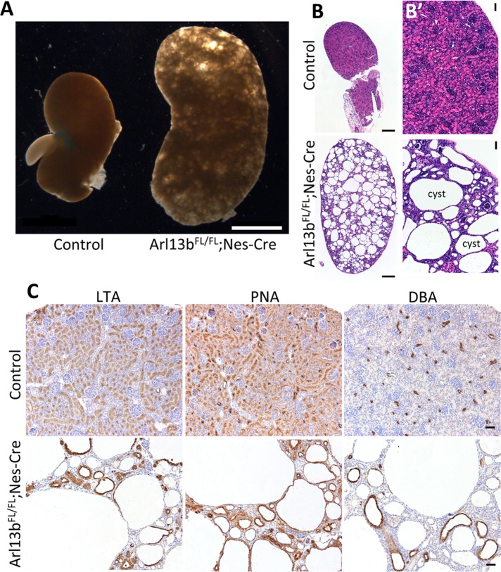
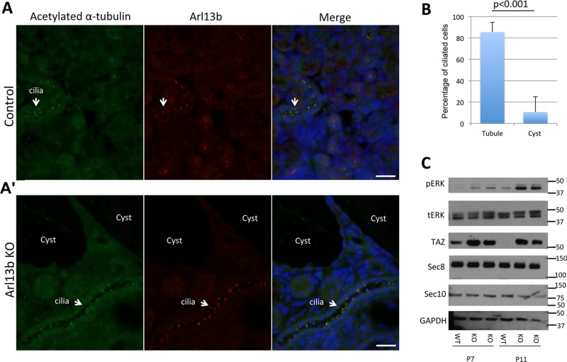
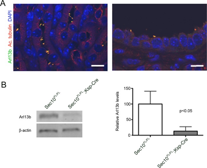
Arl13b belongs to the ADP-ribosylation factor family within the Ras superfamily of regulatory GTPases. Mutations in Arl13b cause Joubert syndrome, which is characterized by congenital cerebellar ataxia, hypotonia, oculomotor apraxia, and mental retardation. Arl13b is highly enriched in cilia and is required for ciliogenesis in multiple organs. Nevertheless, the precise role of Arl13b remains elusive. Here we report that the exocyst subunits Sec8, Exo70, and Sec5 bind preferentially to the GTP-bound form of Arl13b, consistent with the exocyst being an effector of Arl13b. Moreover, we show that Arl13b binds directly to Sec8 and Sec5. In zebrafish, depletion of arl13b or the exocyst subunit sec10 causes phenotypes characteristic of defective cilia, such as curly tail up, edema, and abnormal pronephric kidney development. We explored this further and found a synergistic genetic interaction between arl13b and sec10 morphants in cilia-dependent phenotypes. Through conditional deletion of Arl13b or Sec10 in mice, we found kidney cysts and decreased ciliogenesis in cells surrounding the cysts. Moreover, we observed a decrease in Arl13b expression in the kidneys from Sec10 conditional knockout mice. Taken together, our results indicate that Arl13b and the exocyst function together in the same pathway leading to functional cilia.


Figures

FIGURE 1:
FIGURE 1:
Arl13b interacts with the exocyst in ciliated cells. (A) Endogenous Arl13b was immunoprecipitated from cell lysates of ciliated IMCD3 cells after preincubation with either no nucleotide (left lane), GTPγS (second lane), or GDP (third lane). Nonspecific rabbit IgG was used as a negative control (fourth lane). Input is shown in the far right lane. Immunoprecipitates were resolved by SDS–PAGE and immunoblotted with Sec8 antibody. (B) Cell lysates of ciliated IMCD3 cells were immunoprecipitated with Arl13b after preincubation with GTPγS or GDP. Nonspecific rabbit IgG was used as a negative control. Immunoprecipitates were resolved by SDS–PAGE and immunoblotted with Exo70 or Sec5 antibodies. (C) Total cell lysates of ciliated NIH-3T3 cells were immunoprecipitated and analyzed as described in A. (D) Cell lysates of ciliated IMCD3 cells were immunoprecipitated with Sec8 antibody after incubation with GTPγS. An irrelevant mouse IgG1 (IgG) was used as a negative control. Immunoprecipitates were analyzed by SDS–PAGE and immunoblotted with Arl13b antibody. Results are representative of three independent experiments.
FIGURE 2:
FIGURE 2:
Arl13b interacts with the exocyst through the Sec5 and Sec8 subunits. (A) Sec8-Myc and Arl13b-FLAG were in vitro–translated using TNT T7 coupled reticulocyte lysate system. Five percent of the TNT reaction was resolved by SDS–PAGE and analyzed by immunoblotting with Sec8, Myc, Arl13b, or FLAG antibodies. As a control, a TNT reaction without DNA as template was performed and subsequently used in the immunoprecipitations to distinguish the in vitro–translated from the endogenous protein present in the reticulocyte lysate. (B) Immunoprecipitation with FLAG antibody was performed using the in vitro–translated proteins in the presence of GTPγS. Immunoprecipitates (lane 1) were analyzed by immunoblot with Myc, Sec8, or FLAG antibodies. As a negative control, immunoprecipitations were performed with an irrelevant mouse IgG1 (IgG; lane 2). In addition, as a control, immunoprecipitation with FLAG antibody was performed using as input a mixture of a TNT reaction made without DNA and the in vitro–translated Arl13b-FLAG in the presence of GTPγS (lane 3). Asterisks indicate in vitro–translated Sec8-Myc, and the pound signs indicate endogenous Sec8. (C) Left, Myc-tagged Sec3, Sec5, Sec8, Sec15, and Exo70 exocyst subunits were in vitro translated using the TNT system. Five percent of the TNT reaction was resolved by SDS–PAGE and analyzed by immunoblotting with Myc antibody. Right, endogenous levels of Sec5, Sec8, and Exo70 present on 5% of the TNT reaction were analyzed by immunoblotting with specific antibodies for each subunit. (D) Immunoprecipitation with FLAG antibody was performed using the in vitro–translated proteins described in C, in the presence of GTPγS. Immunoprecipitates were analyzed by immunoblot with Myc antibody or silver staining. Negative controls (lanes 2, 5. and 8) were performed as in B. Results are representative of three independent experiments.
FIGURE 3:
FIGURE 3:
Arl13b interacts directly with Sec5 and Sec8 exocyst subunits. Purified Myc-tagged, in vitro–translated Sec5, Sec8, or Exo70 was mixed with anti–GST-coupled agarose beads previously incubated with purified Arl13b-GST or GST. A negative control (NC) to detect nonspecific binding of exocyst subunits to the agarose beads was added, in which anti–GST-coupled beads were directly incubated with purified Sec8-Myc protein. Eluted products were analyzed by SDS–PAGE, followed by immunoblotting for Myc tag or silver staining. The latter shows that identical amounts of purified Arl13b-GST were incubated in the different conditions. The membrane was previously probed with GST antibody to detect Arl13b-GST (arrowhead). Five percent of the purified proteins used in the assay was run in the input. Results are representative of at least two independent experiments.
FIGURE 4:
FIGURE 4:
Arl13b colocalizes with the exocyst in cilia and the periciliary region. (A–C) Sec8 (green) colocalizes with Arl13b (red) along cilia of polarized MDCK cells. DAPI stains nuclei (blue). (D–F) In RPE1 cells, Sec8 (red) colocalizes with Arl13b (green) along cilia. Insets, higher magnification of the cilium staining. (G–I) In NIH-3T3 cells, Sec5-HA (red) colocalizes with Arl13b (green) along cilia. Insets, higher magnification of cilia showing colocalization of Sec5-HA and Arl13b. (J–L) Sec5-HA (red) is also found accumulated in the base of cilia marked by Arl13b (green) in NIH-3T3 cells. Inset, higher magnification of the periciliary region. (M–O) In NIH-3T3 cells, Sec10-mCherry accumulates in the base of cilia marked by Arl13b (green). Inset, higher magnification of the periciliary region. Scale bar, 5 μm (A–C), 10 μm (D–O).
FIGURE 5:
FIGURE 5:
arl13b and sec10 interact synergistically in cilia-related phenotypes. (A–H) Gross phenotypes of zebrafish embryo morphants at 3 d postfertilization (dpf) (A–D) and 6 dpf (E–H), on lateral view, after injection of MOs for arl13b and/or sec10. A synergistic interaction resulting in small eyes (arrow), pericardial edema (asterisk), and tail curvature (arrowhead) was observed upon coinjection of suboptimal doses of 2 ng of arl13b MO plus 7.5 ng of sec10 MO at both 3 and 6 dpf (D and H, respectively), which do not result in an abnormal phenotype when injected alone (B, C, F, and G); n = 535. (I) Histogram showing quantification of the effect of MOs at 3 dpf. Coinjection of suboptimal doses of 2 ng of arl13b MO plus 7.5 ng of sec10 MO results in a significant increase in the percentage of abnormal phenotypes (*p < 0.001) compared with injection of the MO alone (2 ng of arl13b MO or 7.5 ng of sec10 MO). Scale bars, 1 mm.
FIGURE 6:
FIGURE 6:
Conditional deletion of Arl13b in murine kidneys leads to the formation of cysts originating from different kidney tubule segments. (A) Images of kidneys from mice with a conditional deletion of Arl13b (Arl13bFL/FL;Nes-Cre), compared with a control littermate. (B, B′) Hematoxylin and eosin stainings of fixed sections from kidneys of control mice and mice with conditional deletion of Arl13b. (C) Kidney sections from control mice (top) or mice with conditional deletion of Arl13b (bottom) incubated with LTA lectin, which stains the apical membrane of proximal tubule epithelial cells; PNA lectin, which stains both distal and collecting tubules; and DBA lectin, which exclusively stains the collecting ducts. Scale bars, 2 mm (A), 200 μm (B), 20 μm (B′, C).
FIGURE 7:
FIGURE 7:
Arl13b conditional deletion inhibits ciliogenesis in the kidney and results in increased activation of the MAPK and Hippo pathways. (A, A′) Immunostaining of acetylated α-tubulin (red) and Arl13b (green) in kidneys from control mice (A) and mice with conditional deletion, Arl13bFL/FL;Nes-Cre (A′) at P7. The Arl13bFL/FL;Nes-Cre mutants lack cilia in cells surrounding the cysts, whereas cilia are present in normal-appearing tubules. (B) Quantification of ciliated cells from kidneys of Arl13bFL/FL;Nes-Cre mice shows a significant decrease in the number of cilia per cell in cysts compared with normal-appearing tubules (n = 325 cilia). Error bars represent SD. (C) Increased pERK and TAZ levels in Arl13bFL/FL;Nes-Cre mice. Immunoblot analysis of total cell extracts from whole kidneys of control mice and Arl13bFL/FL;Nes-Cre mice at P7 and P11 with phosphorylated ERK (pERK), total ERK (tERK), TAZ, Sec8, Sec10, or GAPDH antibodies. Note that distinct Arl13bFL/FL;Nes-Cre mice were analyzed at P7 and P11. Results are representative of four independent experiments. Scale bars, 10 μm.
FIGURE 8:
FIGURE 8:
Kidney-specific Sec10 conditional knockout results in ciliary defects and decreased Arl13b levels. (A) Immunofluorescence staining of adult kidney tubules of kidney-specific Sec10FL/FL;Ksp-Cre conditional knockout mouse for Arl13b (green) and the primary cilia marker acetylated α-tubulin (red). Cilia are fewer and shorter in Sec10-depleted tubules, although some Arl13b is still detected in the abnormal primary cilia. Scale bar, 10 μm. (B) Overall, Arl13b protein levels are dramatically decreased in Sec10FL/FL;Ksp-Cre kidneys compared with Sec10FL/FL controls, as measured by immunoblotting.

References

    1. Babbey CM, Bacallao RL, Dunn KW. Rab10 associates with primary cilia and the exocyst complex in renal epithelial cells. Am J Physiol Renal Physiol. 2010;299:F495–F506. - PMC - PubMed
    1. Badano JL, Mitsuma N, Beales PL, Katsanis N. The ciliopathies: an emerging class of human genetic disorders. Annu Rev Genomics Hum Genet. 2006;7:125–148. - PubMed
    1. Barral DC, Garg S, Casalou C, Watts GFM, Sandoval JL, Ramalho JS, Hsu VW, Brenner MB. Arl13b regulates endocytic recycling traffic. Proc Natl Acad Sci USA. 2012;109:21354–21359. - PMC - PubMed
    1. Blacque OE, Leroux MR. Bardet-Biedl syndrome: an emerging patho­mechanism of intracellular transport. Cell Mol Life Sci C. 2006;63:2145–2161. - PMC - PubMed
    1. Cantagrel V, Silhavy J, Bielas S. Mutations in the cilia gene ARL13B lead to the classical form of Joubert syndrome. Am J Hum Genet. 2008:170–179. - PMC - PubMed

Publication types

MeSH terms

Supplementary concepts