

Review
doi: 10.1155/2015/482582.
Epub 2015 Oct 25.
Mitochondrial Retrograde Signaling: Triggers, Pathways, and Outcomes
Affiliations
- PMID: 26583058
- PMCID: PMC4637108
- DOI: 10.1155/2015/482582
Item in Clipboard
Review
Mitochondrial Retrograde Signaling: Triggers, Pathways, and Outcomes
Oxid Med Cell Longev.
2015.
Abstract
Mitochondria are essential organelles for eukaryotic homeostasis. Although these organelles possess their own DNA, the vast majority (>99%) of mitochondrial proteins are encoded in the nucleus. This situation makes systems that allow the communication between mitochondria and the nucleus a requirement not only to coordinate mitochondrial protein synthesis during biogenesis but also to communicate eventual mitochondrial malfunctions, triggering compensatory responses in the nucleus. Mitochondria-to-nucleus retrograde signaling has been described in various organisms, albeit with differences in effector pathways, molecules, and outcomes, as discussed in this review.
Figures
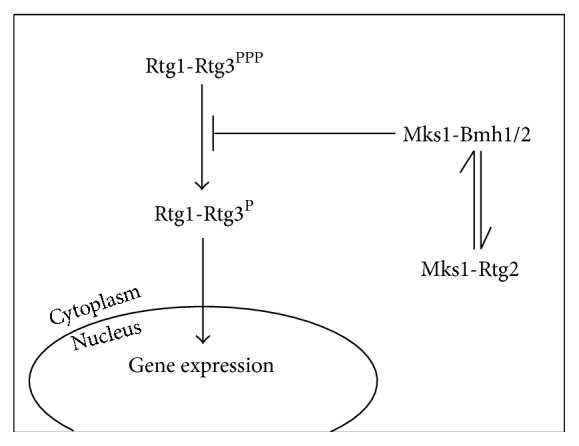
Simplified scheme of the RTG-dependent retrograde signaling pathway. In Saccharomyces cerevisiae this pathway depends on three proteins. Rtg1 and Rtg3 form a transcription factor that translocates to the nucleus when the pathway is activated. In the nucleus, Rtg1 and Rtg3 control the expression of a set of genes that code for mitochondrial proteins. Rtg2 is an activator of the pathway that allows the nuclear translocation of Rtg1 and Rtg3.
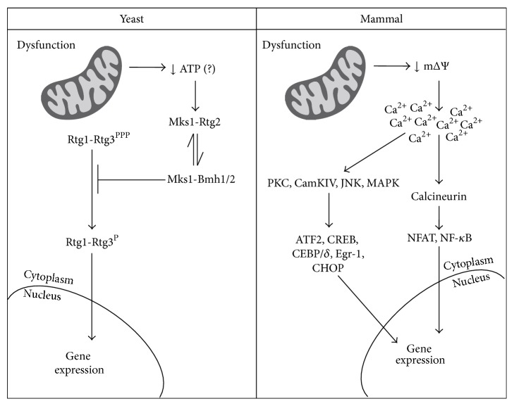
Scheme comparing the classical retrograde signaling pathways in yeast and mammals. In yeast, mitochondrial dysfunction leads to decreases in intracellular ATP concentration, which may favor Rtg2-Mks1 interaction [54] allowing Rtg1-Rtg3 activation. In mammals, mitochondrial dysfunction translates into drops in mitochondrial membrane potential, causing increments in intracellular calcium. Calcium-dependent kinases and phosphatases are then activated culminating with the activation of different transcription factors. Alternative retrograde signaling pathways in yeast, mammals, and other model organisms are discussed in the text.
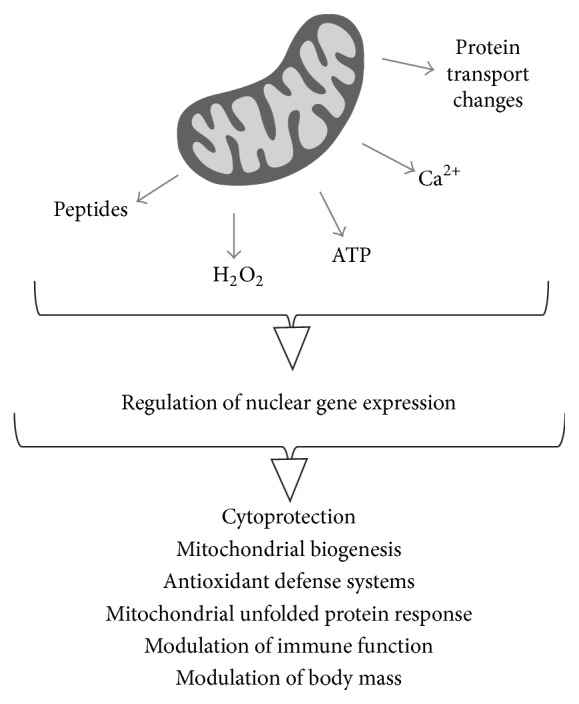
General view of mitochondrial signals and outcomes of retrograde communication. Diverse mitochondrial signals elicit varied responses, ranging from the increased synthesis of mitochondrial chaperones to improvement of immunity.
References
-
- Vasington F. D., Murphy J. V. Ca++ ion uptake by rat kidney mitochondria and its dependence on respiration and phosphorylation. The Journal of Biological Chemistry. 1962;237(8):2670–2677. - PubMed
Publication types
MeSH terms
Substances
LinkOut - more resources
Full Text Sources
Other Literature Sources
Molecular Biology Databases
