. 2016 Feb;79(2):272-87.
doi: 10.1002/ana.24559. Epub 2015 Dec 25.

Deep clinical and neuropathological phenotyping of Pick disease

Affiliations

Deep clinical and neuropathological phenotyping of Pick disease

David J Irwin et al. Ann Neurol. 2016 Feb.

Abstract

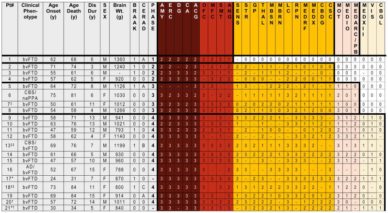
Objective: To characterize sequential patterns of regional neuropathology and clinical symptoms in a well-characterized cohort of 21 patients with autopsy-confirmed Pick disease.

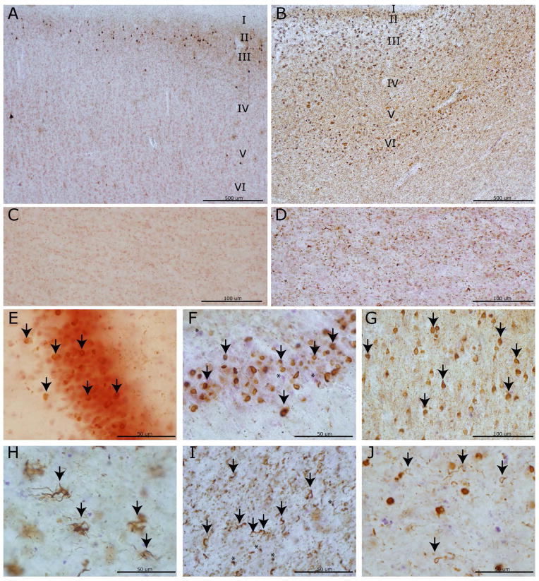
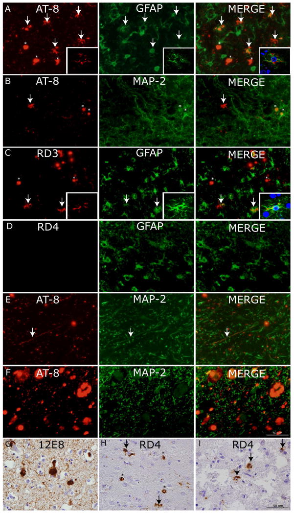
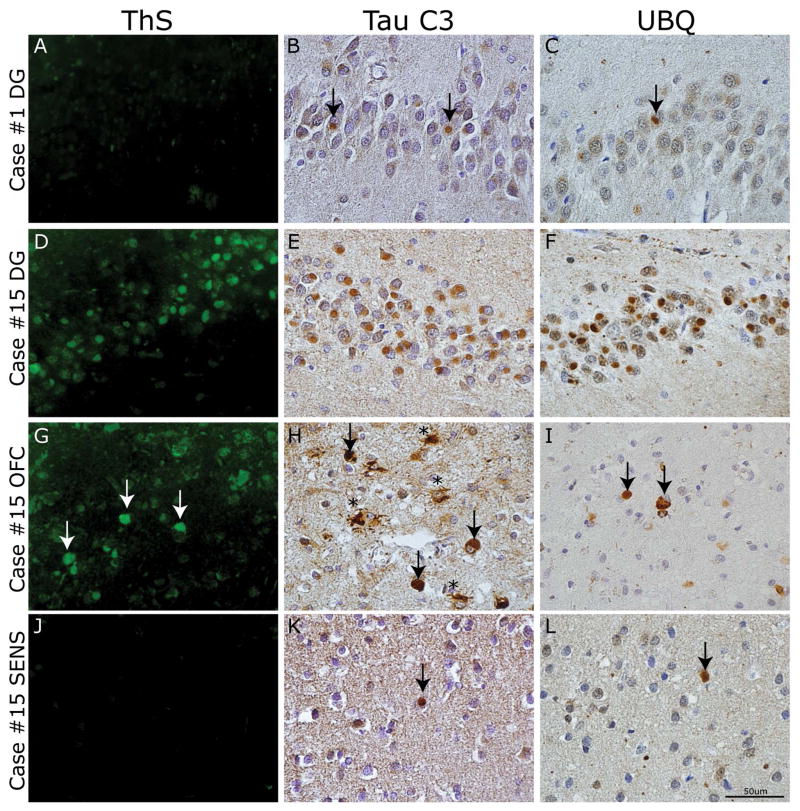
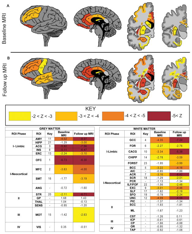
Methods: Detailed neuropathological examination using 70μm and traditional 6μm sections was performed using thioflavin-S staining and immunohistochemistry for phosphorylated tau, 3R and 4R tau isoforms, ubiquitin, and C-terminally truncated tau. Patterns of regional tau deposition were correlated with clinical data. In a subset of cases (n = 5), converging evidence was obtained using antemortem neuroimaging measures of gray and white matter integrity.

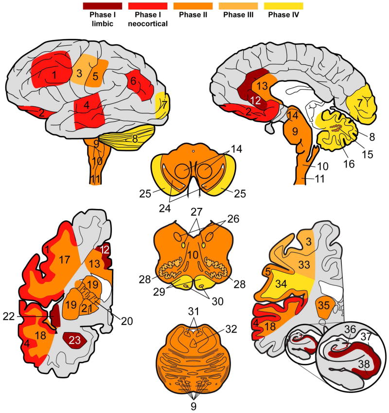
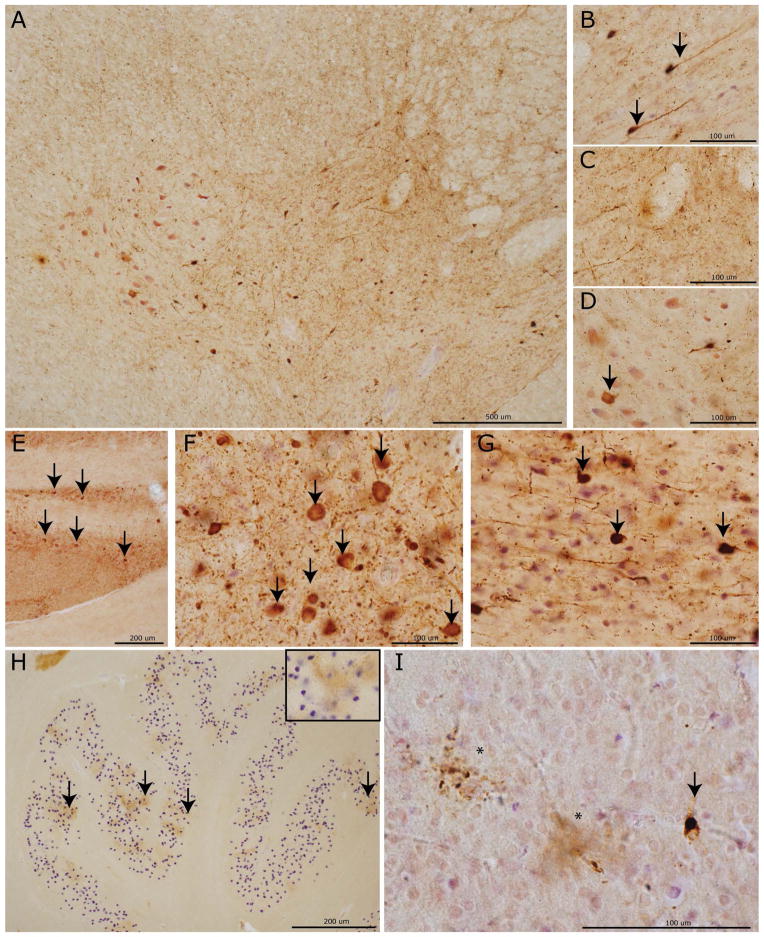
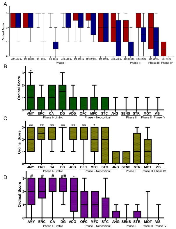
Results: Four sequential patterns of pathological tau deposition were identified starting in frontotemporal limbic/paralimbic and neocortical regions (phase I). Sequential involvement was seen in subcortical structures, including basal ganglia, locus coeruleus, and raphe nuclei (phase II), followed by primary motor cortex and precerebellar nuclei (phase III) and finally visual cortex in the most severe (phase IV) cases. Behavioral variant frontotemporal dementia was the predominant clinical phenotype (18 of 21), but all patients eventually developed a social comportment disorder. Pathological tau phases reflected the evolution of clinical symptoms and degeneration on serial antemortem neuroimaging, directly correlated with disease duration and inversely correlated with brain weight at autopsy. The majority of neuronal and glial tau inclusions were 3R tau-positive and 4R tau-negative in sporadic cases. There was a relative abundance of mature tau pathology markers in frontotemporal limbic/paralimbic regions compared to neocortical regions.

Interpretation: Pick disease tau neuropathology may originate in limbic/paralimbic cortices. The patterns of tau pathology observed here provide novel insights into the natural history and biology of tau-mediated neurodegeneration.


Conflict of interest statement

Potential Conflicts of Interest

The authors declared no potential conflicts of interest with respect to the research, authorship, and/or publication of this article.

Figures

Figure 1
Figure 1. Neuropathological Phases of Tau Deposition in PiD
Table demonstrates demographic and neuropathological data for each case. * Denotes pathogenic mutation in MAPT (p.L266V). ‡ secondary pathological diagnoses of Lewy body disease brainstem stage and anoxic encephalopathy, ‡‡ secondary pathological diagnosis of Alzheimer’s disease (low probability), † examined using 6 μm sections only. Ordinal scores: (0= none/rare, 1= mild, 2=moderate, 3=severe, -= not done). Braak 1= Braak tau stage I-II.
Figure 2
Figure 2. PiD tau pathology and neurodegeneration in cortical and limbic regions
Photomicrographs depict representative images of tau neuropathology (AT-8). A laminar distribution of tau pathology largely restricted to laminae II–III was seen in primary sensory cortex (A) with associated minimal white matter pathology (C) in a Phase II case compared to bi-laminar distribution in anterior cingulate gyrus (B) with layer V–VI involvement and severe white matter tau burden (D) in a Phase IV case. Note the collapse of the cortical layering from severe neuron loss in (B). PBs in dentate gyrus (arrows) in a Phase I case (E) with minimal neuron loss compared to a Phase IV case with mild neuron loss (F; asterisks denote extracellular purple Nissel substance from degenerated neurons). Severe PB burden (arrows) in the cornu ammonis of this hippocampus (G) of a Phase IV case. Glial tau inclusions in the orbitofrontal cortex are depicted (H-J). Ramified astrocytes (arrows) were often present in the orbitofrontal cortex (H). Oligodendrocytic tau pathology in coiled bodies (arrows) is present in both white matter near axonal projections (I) and near soma of affected neurons (J) (i.e. satellite oligodendrocytes) with less common “Pick-body like” oligodendroglial inclusions (I, asterisks).
Figure 3
Figure 3. Cellular localization of tau neuropathology
Photomicrographs depict double-label experiments in (A) using anti-phosphorylated tau (AT8) and glial-fibrillary associated protein (GFAP) find ramified astrocytes with tau pathology are largely co-localized with GFAP (arrows) while PBs are not (asterisks) (inset= confocal z-stack images, 63x). Conversely, in (B) AT-8 reactive PBs (asterisks) co-localize with dendritic marker (MAP-2) but ramified astrocytes with tau inclusions do not (arrow). To confirm the major tau isoform composition of tau pathology in ramified astrocytes we performed double label experiments in (C) using 3R tau specific MAb (RD3) and (D) 4R tau specific MAb (RD4) with GFAP which showed co-localization of GFAP (arrows) with tau inclusions in RD3 but not RD4 double label experiments (inset= confocal z-stack images, 63x). Cervical spinal cord upper Rexed layer tau pathology in (E) and locus coeruleus (F) are seen to co-localize to neuritic processes and cell bodies marked by MAP-2. (G) Robust 12E8 reactivity in PBs and threads in MFC of sporadic PiD. (H) RD4 reactivity was mild and largely found in PBs (asterisks) with occasional nearby ramified astrocyte (arrows). (I) The p.L266V cases showed consistent 4R tau reactive astrocytes (arrows).
Figure 4
Figure 4. Patterns of PiD tau neuropathology
Heat maps display neuroanatomic associations of sequential tau deposition in the central nervous system of PiD. Phases are defined by the extent of regional involvement of tau pathology across cases beginning with Phase I, where disease is restricted to limbic and neocortical frontotemporal regions and angular gyrus. Phase II has additional pathology in associated white matter tracts, subcortical structures and serotonergic/norandrenergic brainstem nuclei. Phase III tau neuropathology is characterized by additional pathology in primary motor cortex and pre-cerebellar nuclei in the medulla. Finally, Phase IV cases with the most severe tau pathology burden include additional tau deposits in the visual cortex and variably in the cerebellar granular layer and brainstem white matter. Key: 1=MFC, 2=OFC, 3= MOT, 4=SMT, 5=SENS, 6=ANG, 7=VIS, 8 CBGL, 9=Pons, 10, MEDRF, 11=CSC, 12=ACG, 13=CC, 14=MBRN, 15=CBDG, 16=CB WM, 17= MFC WM, 18= SMT WM, 19=STR, 20= Internal capsule, 21=GP, 22=INS, 23=AMY, 24= MBSN, 25=MB crus cerebri, 26=MEDX, 27=MEDXII, 28=MEDIO, 29=MED pyramids, 30=MED ARC/PB, 31=LC, 32=RPN, 33=MOT WM, 34=SENS WM, 35=THAL, 36= HIPP DG, 37= HIPP CA, 38= HIPP ERC.
Figure 5
Figure 5. Phase-specific regional tau neuropathology in PiD
Photomicrographs depict representative images from CNS areas sequentially involved in our hypothetical model of the progressive spread of tau pathology in PiD (AT-8). Cervical spinal cord (A) showing a moderate amount of tau neuropathology in the intermediolateral and upper Rexed layers of the spinal cord corresponding to Phase II–IV pathology. High power images reveal neuronal tau inclusions (arrows) in the upper Rexed layers (II–III) (B) and intermediolateral layer (VII) and (D) relative sparing on lower motor neurons (arrow) in layers VIII–IX. Tau inclusions in (E) pontine nuclei in the basis pontis with severe tau pathology (arrows) in the LC (F) and RPN (G) characteristic of Phases II–IV. The inferior olivary pre-cerebellar nucleus (H) with dot-like stippling pattern of tau reactivity (arrows; inset =40x magnification) seen in Phases III–V. Finally, a mild degree of tau positive PBs (arrows) and diffuse astrocytic tau inclusions (asterisks) in the primary visual cortex (I) representing end-stage Phase IV pathology.
Figure 6
Figure 6. Ordinal scores for severity of regional tau pathology and neurodegeneration in PiD Phases I–IV
Boxplots depict median, interquartile range and range of ordinal scores. (A) AT-8 reactive tau neuropathology (red) in 70 um sections was highly correlated with neuron loss (blue) for most regions. Overall there were higher levels of tau pathology in Phase I regions compared to additional regions affected in Phases II–IV. To further characterize Phase I regions we compared ordinal scores for markers of mature tau pathology. (B) ThS (green) reactivity was mild and most prominent in Phase I limbic regions. (C) The tau C3 C-terminal truncation specific epitope was more abundant in Phase I limbic and neocortical regions. (D) UBQ reactivity was more prominent in Phase I limbic regions. (B–D) K Kruskal-Wallis test finds a significant difference in ordinal scores between regions (p<0.0001). * denotes p<0.001 difference in post hoc region comparisons with VIS, ** with previous and ANG, SENS, # with previous and STR, MOT, ## with previous and MFC, SMT.
Figure 7
Figure 7. Spatial and temporal gradient of mature tau pathology in PiD
Photomicrographs depict panel of markers of mature tau pathology. Case #1 classified as Phase I had (A) an absence of ThS staining and minimal (B) Tau C3 and (C) UBQ reactivity in the dentate gyrus (arrows). In contrast, case #15 classified as Phase IV had (D) mild to moderate ThS stained PBs in the dentate gyrus of the hippocampus, (G) rare to mild reactivity in OFC (arrows) and (J) rare or no reactivity in SENS. The C-terminal truncation epitope labeled by Tau C3 MAb revealed most consistent tau reactivity in the dentate gyrus (E) and superficial neocortical layer (II–III) pick bodies (arrows) and ramified astrocytes (asterisks) in OFC (H) with mild amounts in SENS (arrow) (K). UBQ-reactive PBs showed consistent staining in DG (F), with less prominent reactivity in OFC (arrows) (I) and SENS (arrow) (L).
Figure 8
Figure 8. Longitudinal antemortem neuroimaging data in PiD
Heat maps depict neuroanatomical regions of interest (ROIs) tested with mean z-scores ≤ −2.0 from autopsied PiD patients (A) at baseline (n=5) and (B) follow up scan (n=4) compared with healthy controls for grey matter density and white matter mean diffusivity. There is time-dependent spread of neurodegeneration from frontal limbic and neocortical regions, largely reflecting proposed hypothetical phases of tau neuropathology based on histopathological analysis of regional tau burden. Key: 1=OFC, 2=MFC, 3=ACG, 4= genu of corpus callosum (GCC), 5= body corpus callosum (BCC), 6= body/column fornix (FOR), 7= anterior corona radiata (ACR), 8= anterior INS, 9= external capsule (EXC), 10= cingulum near ACG (CACG), 11= AMY, 12= uncinate fasciculus (UNC), 13=ERC, 14= cingulum near HIPP (CHIPP), 15= MOT, 16= SMT, 17= superior longitudinal fasciculus (SLF), 18= superior corona radiata (SCR), 19= superior fronto-occipial fasciculus (SFO),20= STR, 21= HIPP, 22= inferior longitudinal fasciculus/fronto-occipital fasciculus (ILF/FOF). Areas tested and not depicted include fornix crus/stria terminalis (FORST), primary sensory cortex (SENS), angular gyrus (ANG), splenium corpus callosum (SCC), primary visual cortex (VIS), corticospinal tract (CST), medial lemniscus (ML), anterior limb internal capsule (AIC), globus pallidus (GP), thalamus (THAL), cerebral peduncle (CP), posterior limb internal capsule (PIC). posterior corona radiata (PCR), inferior cerebellar peduncle (ICP), optic radiations (OR) and tapetum (TAP) (z scores all >−2.0).

References

    1. Pick A. Über die Beziehungen der senilen Hirnatrophie zur Aphasie. Prag Med Wochenschr. 1892;17:165–7.
    1. Kertesz A. Pick complex--historical introduction. Alzheimer disease and associated disorders. 2007 Oct-Dec;21(4):S5–7. - PubMed
    1. Mackenzie IR, Neumann M, Bigio EH, et al. Nomenclature and nosology for neuropathologic subtypes of frontotemporal lobar degeneration: an update. Acta neuropathologica. 2010 Jan;119(1):1–4. - PMC - PubMed
    1. Yokota O, Tsuchiya K, Arai T, et al. Clinicopathological characterization of Pick’s disease versus frontotemporal lobar degeneration with ubiquitin/TDP-43-positive inclusions. Acta neuropathologica. 2009 Apr;117(4):429–44. - PubMed
    1. Piguet O, Halliday GM, Reid WG, et al. Clinical phenotypes in autopsy-confirmed Pick disease. Neurology. 2011 Jan 18;76(3):253–9. - PubMed

Publication types