. 2015 Nov 19;6(11):e1984.
doi: 10.1038/cddis.2015.337.

Smad4 represses the generation of memory-precursor effector T cells but is required for the differentiation of central memory T cells

Affiliations

Smad4 represses the generation of memory-precursor effector T cells but is required for the differentiation of central memory T cells

J Cao et al. Cell Death Dis. .

Abstract

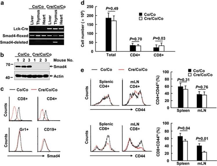
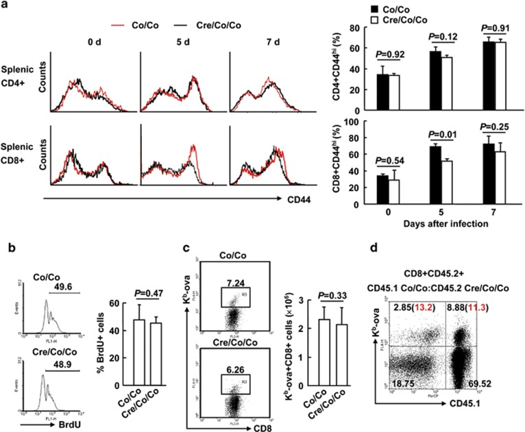
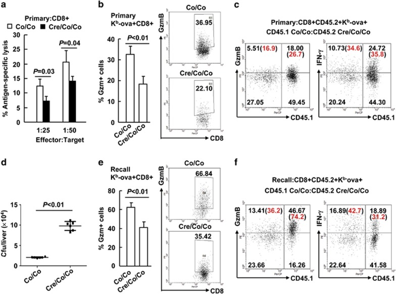
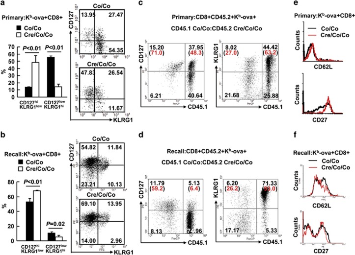
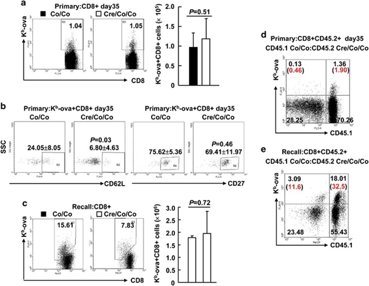
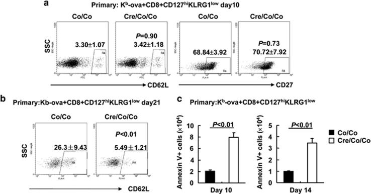
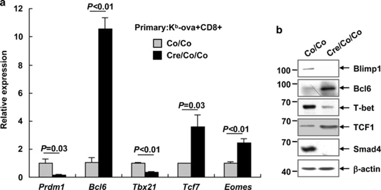
The transcriptional regulation underlying the differentiation of CD8(+) effector and memory T cells remains elusive. Here, we show that 18-month-old mice lacking the transcription factor Smad4 (homolog 4 of mothers against decapentaplegic, Drosophila), a key intracellular signaling effector for the TGF-β superfamily, in T cells exhibited lower percentages of CD44(hi)CD8(+) T cells. To explore the role of Smad4 in the activation/memory of CD8(+) T cells, 6- to 8-week-old mice with or without Smad4 in T cells were challenged with Listeria monocytogenes. Smad4 deficiency did not affect antigen-specific CD8(+) T-cell expansion but led to partially impaired cytotoxic function. Less short-lived effector T cells but more memory-precursor effector T cells were generated in the absence of Smad4. Despite that, Smad4 deficiency led to reduced memory CD8(+) T-cell responses. Further exploration revealed that the generation of central memory T cells was impaired in the absence of Smad4 and the cells showed survival issue. In mechanism, Smad4 deficiency led to aberrant transcriptional programs in antigen-specific CD8(+) T cells. These findings demonstrated an essential role of Smad4 in the control of effector and memory CD8(+) T-cell responses to infection.


Figures

Figure 1
Figure 1
Eighteen-month-old Smad4co/co;Lck-Cre mice exhibit impaired CD44 expression in CD8+ T cells. (a) Genotyping of Smad4co/co;Lck-Cre mice (Cre/Co/Co) and control littermates (Co/Co). (b) The expression of Smad4 and actin in the thymocytes of 6- to 8-week-old Smad4co/co;Lck-Cre mice and control littermates. IB, immunoblotting. (c) Flow cytometry analysis of Smad4 expression in splenic CD4+ T, CD8+ T, Gr1+, and CD19+ B cells of 6- to 8-week-old Smad4co/co;Lck-Cre mice and control littermates. (d) The absolute numbers of total white cells, CD4+ T, and CD8+ T cells in the spleen, as revealed by white cell count and flow cytometry analysis, in 18-month-old Smad4co/co;Lck-Cre mice and control littermates (n=6 per group). (e) Flow cytometry analysis of CD44 expression in CD4+ and CD8+ T cells in the spleen and mesenteric lymph node (mLN) of 18-month-old Smad4co/co;Lck-Cre mice and control littermates (n=6 per group)
Figure 2
Figure 2
Unchanged antigen-specific CD8+ T-cell expansion in the absence of Smad4. (ac) Six- to eight-week-old Smad4co/co;Lck-Cre mice and control littermates mice were infected with 5 × 10³ c.f.u. of LM-OVA (n=6 per group). (a) CD44 expression in CD4+ and CD8+ splenic T cells was analyzed by flow cytometry at days 0, 5, and 7 post infection. (b) Mice received 1 mg thymidine analog 5-bromo-2′-deoxyuridine (BrdU) in 0.1 ml PBS via i.p. injection at day 6 post infection; BrdU incorporation in CD8+ splenic T cells was analyzed by flow cytometry 24 h later. (c) The numbers of Kb-ova+CD8+ splenic T cells were analyzed by flow cytometry at day 7 post infection. (d) Bone marrow chimeric mice reconstituted with a mix of Smad4co/co;Lck-Cre (CD45.2CD45.2) and Smad4co/co (CD45.1CD45.2) cells were infected with 5 × 10³ c.f.u. of LM-OVA. Single-cell suspensions from the spleen were analyzed for the expression of CD45.1 and OVA specificity (Kb-ova) at day 7 post infection. Representative plot of gated CD8+CD45.2+ T cells from the spleen is shown (n=3). The number in the bracket indicates the percentage of the Kb-ova+ T cells in relation to CD8+ T cells of the same origin. Data shown in this figure are representative of at least three independent experiments
Figure 3
Figure 3
Smad4 is required for the cytotoxic function of CD8+ T cells. (a and b) Six- to eight-week-old Smad4co/co;Lck-Cre mice and their littermate controls were infected with 5 × 10³ c.f.u. of LM-OVA (n=6 per group). Single-cell suspensions from the spleen were prepared on day 7. (a) CD8+ cytotoxic activity was measured by specific killing of OVA peptide-loaded EL-4 cells. Peptide unloaded EL-4 cells were used as negative control. (b) GzmB expression in Kb-ova+CD8+ splenic T cells upon restimulation with the SIINFEKL peptide (10 nM, 6 h) was analyzed by intracellular staining and flow cytometry. (c) Bone marrow chimeric mice were prepared and treated as described in Figure 2d. The expression of CD45.1 and GzmB in Kb-ova+CD8+CD45.2+ splenic T cells at day 7 post infection upon OVA peptide restimulation was analyzed (n=3). (d and e) Smad4co/co;Lck-Cre mice and their littermates were rechallenged with 1 × 10⁵ c.f.u. of LM-OVA 35 days after primary infection (n=6 per group). (d) Bacterial burden in the liver was determined 2 days after the secondary infection. (e) GzmB expression in Kb-ova+CD8+ splenic T cells upon OVA peptide restimulation was analyzed 5 days after the secondary infection. (f) Bone marrow chimeric mice were rechallenged with 1 × 10⁵ c.f.u. of LM-OVA 35 days after primary infection. The expression of CD45.1 and GzmB in Kb-ova+CD8+CD45.2+ splenic T cells upon OVA peptide restimulation was analyzed 5 days after the secondary infection (n=3). Data shown in this figure are representative of at least three independent experiments
Figure 4
Figure 4
Smad4-deficient T cells show aberrant CD8+ T-cell differentiation. (a) The expression of CD127 and KLRG1 in Kb-ova+CD8+ splenic T cells of Smad4co/co;Lck-Cre mice and their littermate controls on day 7 of LM-OVA primary infection (n=6 per group). (b) The expression of CD127 and KLRG1 in Kb-ova+CD8+ splenic T cells of Smad4co/co;Lck-Cre mice and their littermate controls 5 days after the secondary infection (n=6 per group). (c) Bone marrow chimeric mice were prepared and treated as described in Figure 2d. The expression of CD45.1 and CD127 or KLRG1 in Kb-ova+CD8+CD45.2+ splenic T cells at day 7 post infection was analyzed by flow cytometry (n=3). (d) Bone marrow chimeric mice were rechallenged with 1 × 10⁵ c.f.u. of LM-OVA 35 days after primary infection. The expression of CD45.1 and CD127 or KLRG1 in Kb-ova+CD8+CD45.2+ splenic T cells 5 days after the secondary infection was analyzed by flow cytometry (n=3). (e) The expression of CD62L and CD27 in Kb-ova+CD8+ splenic T cells of Smad4co/co;Lck-Cre mice and their littermate controls on day 7 of LM-OVA primary infection (n=6 per group). (f) The expression of CD62L and CD27 in Kb-ova+CD8+ splenic T cells of Smad4co/co;Lck-Cre mice and their littermate controls 5 days after the secondary infection. Data shown in this figure are representative of at least three independent experiments
Figure 5
Figure 5
Smad4 deficiency leads to defective memory. (a) The percentages and numbers of Kb-ova+CD8+ splenic T cells in Smad4co/co;Lck-Cre mice and their littermate controls on day 35 of LM-OVA primary infection (n=6 per group). (b) The expression of CD62L and CD27 in Kb-ova+CD8+ splenic T cells of Smad4co/co;Lck-Cre mice and their littermate controls on day 35 of LM-OVA primary infection (n=6 per group). (c) The percentages and numbers of Kb-ova+CD8+ splenic T cells in Smad4co/co;Lck-Cre mice and their littermate controls 5 days after the secondary infection (n=6 per group). (d) Bone marrow chimeric mice were prepared and treated as described in Figure 2d. The expression of CD45.1 and OVA specificity (Kb-ova) in CD8+CD45.2+ splenic T cells at day 35 post infection was analyzed by flow cytometry (n=3). (e) Bone marrow chimeric mice were rechallenged with 1 × 10⁵ c.f.u. of LM-OVA 35 days after primary infection. The expression of CD45.1 and OVA specificity (Kb-ova) in CD8+CD45.2+ splenic T cells 5 days after the secondary infection was analyzed by flow cytometry (n=3). Data shown in this figure are representative of at least three independent experiments
Figure 6
Figure 6
Smad4 contributes to the differentiation of central memory T cells by promoting the survival of MPECs. (a and b) The expression of CD62L and CD27 in Kb-ova+CD8+CD127hiKLRG1low splenic T cells of Smad4co/co;Lck-Cre mice and their littermate controls on day 10 (a) or day 21 (b) of LM-OVA primary infection (n=6 per group). (c) Apoptosis analysis of Kb-ova+CD8+CD127hiKLRG1low splenic T cells of Smad4co/co;Lck-Cre mice and their littermate controls on days 10 and 14 of LM-OVA primary infection (n=6 per group). Data shown in this figure are representative of at least three independent experiments
Figure 7
Figure 7
Smad4 regulates the transcriptional program in antigen-specific T cells. (a and b) Kb-ova+CD8+ splenic T cells were sorted from Smad4co/co;Lck-Cre mice and their littermate controls on day 7 of LM-OVA primary infection. Three to four biologically independent samples with the same genotype were mixed together and the experiment was repeated three times (a). Cells were then subjected to quantitative RT-PCR for the indicated transcripts. Data are shown as the expression relative to that found in Smad4-sufficient Kb-ova+CD8+ splenic T cells (arbitrarily set to 1). (b) Cell lysates were then prepared and subjected to immunoblotting analysis with the indicated antibodies

References

    1. 1Klenerman P, Hill A. T cells and viral persistence: lessons from diverse infections. Nat Immunol 2005; 6: 873–879. - PubMed
    1. 2Thaventhiran JE, Fearon DT, Gattinoni L. Transcriptional regulation of effector and memory CD8+ T cell fates. Curr Opin Immunol 2013; 25: 321–328. - PMC - PubMed
    1. 3Kaech SM, Tan JT, Wherry EJ, Konieczny BT, Surh CD, Ahmed R. Selective expression of the interleukin 7 receptor identifies effector CD8 T cells that give rise to long-lived memory cells. Nat Immunol 2003; 4: 1191–1198. - PubMed
    1. 4Sarkar S, Kalia V, Haining WN, Konieczny BT, Subramaniam S, Ahmed R. Functional and genomic profiling of effector CD8 T cell subsets with distinct memory fates. J Exp Med 2008; 205: 625–640. - PMC - PubMed
    1. 5Intlekofer AM, Takemoto N, Wherry EJ, Longworth SA, Northrup JT, Palanivel VR et al. Effector and memory CD8+ T cell fate coupled by T-bet and eomesodermin. Nat Immunol 2005; 6: 1236–1244. - PubMed

Publication types