. 2015 Dec 1;29(23):2420-34.
doi: 10.1101/gad.271783.115. Epub 2015 Nov 19.

High telomerase is a hallmark of undifferentiated spermatogonia and is required for maintenance of male germline stem cells

Affiliations

High telomerase is a hallmark of undifferentiated spermatogonia and is required for maintenance of male germline stem cells

Matthew F Pech et al. Genes Dev. .

Abstract

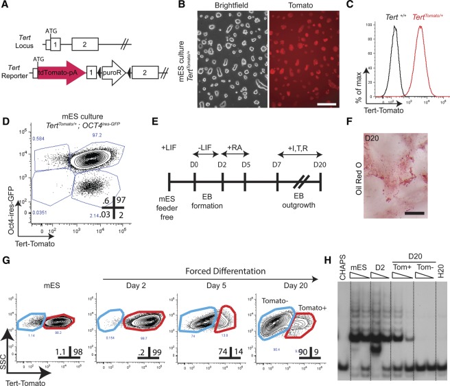
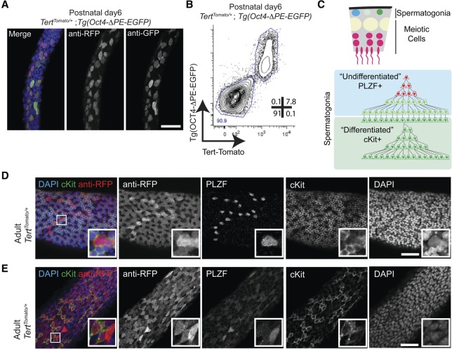
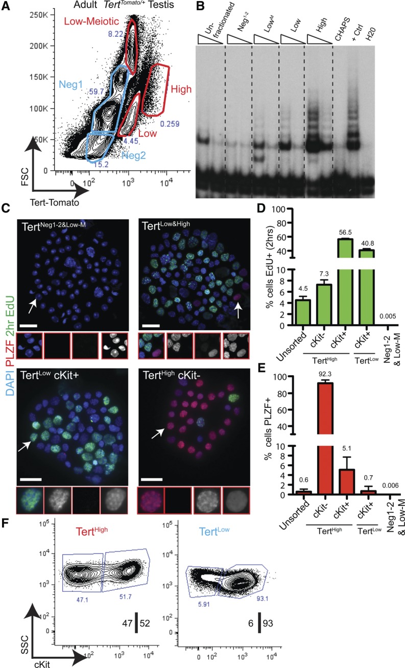
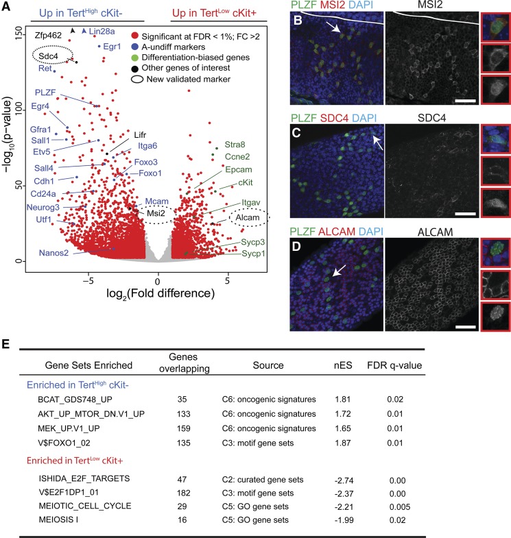
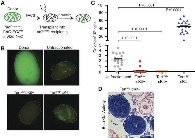
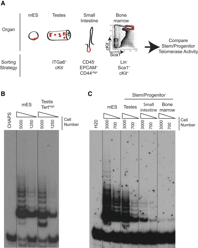
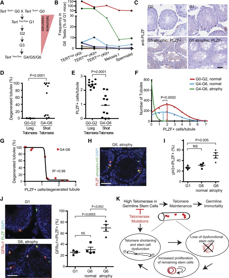
Telomerase inactivation causes loss of the male germline in worms, fish, and mice, indicating a conserved dependence on telomere maintenance in this cell lineage. Here, using telomerase reverse transcriptase (Tert) reporter mice, we found that very high telomerase expression is a hallmark of undifferentiated spermatogonia, the mitotic population where germline stem cells reside. We exploited these high telomerase levels as a basis for purifying undifferentiated spermatogonia using fluorescence-activated cell sorting. Telomerase levels in undifferentiated spermatogonia and embryonic stem cells are comparable and much greater than in somatic progenitor compartments. Within the germline, we uncovered an unanticipated gradient of telomerase activity that also enables isolation of more mature populations. Transcriptomic comparisons of Tert(High) undifferentiated spermatogonia and Tert(Low) differentiated spermatogonia by RNA sequencing reveals marked differences in cell cycle and key molecular features of each compartment. Transplantation studies show that germline stem cell activity is confined to the Tert(High) cKit(-) population. Telomere shortening in telomerase knockout strains causes depletion of undifferentiated spermatogonia and eventual loss of all germ cells after undifferentiated spermatogonia drop below a critical threshold. These data reveal that high telomerase expression is a fundamental characteristic of germline stem cells, thus explaining the broad dependence on telomerase for germline immortality in metazoans.

Keywords: aging; germline stem cells; spermatogenesis; telomerase; telomeres.


Figures

Figure 1.
Figure 1.
A Tert promoter knock-in reporter accurately reflects telomerase activity in both pluripotent and differentiating ES cells. (A) Generation of a knock-in transcriptional Tert reporter. TdTomato was inserted at the initiating methionine of Tert, with a floxed resistance cassette placed into the first intron. (B) Phase contrast and epifluorescent microscopy of Tert-Tomato reporter in TertTomato/+ mES cells grown in LIF/2i conditions. Bar, 200 μm. (C) Expression of Tert-Tomato reporter in TertTomato/+mES cells grown in LIF/2i, quantified by FACS. Untargeted parental mES cells are shown in black. Results were consistent across independently targeted clones. (D) Expression of fluorescent reporter genes by FACS in doubly targeted TertTomato/+ Oct4ires-EGFP/+ mES cells grown in LIF/2i conditions. Gates were drawn relative to parental and singly targeted mES cells. (E) Schema for in vitro differentiation of mES cells to an adipogenic fate. (EB) Embryoid body; (RA) retinoic acid; (I,T,R) insulin, triiodothyronine, and rosiglitazone. (F) Oil-Red-O staining of mES cells at day 20 of differentiation. Bar, 500 μm. (G) Fluorescent reporter expression by FACS in TertTomato/+ mES cells during adipogenic differentiation. Gates were drawn based on the background fluorescence of untargeted parental mES cells. The percent of events in each gate is shown. (SSC) Side scatter. (H) Telomerase activity measured by telomere repeat amplification protocol (TRAP) in populations shown in G. CHAPS and H20 represent buffer-only negative controls. TRAP reactions were programmed with 3000 or 1000 FACS-sorted cell equivalents.
Figure 2.
Figure 2.
High telomerase levels are a hallmark of undifferentiated spermatogonia. (A) Whole-mount analysis of seminiferous tubules from postnatal day 6 TertTomato/+ Tg(Oct4-ΔPE-GFP) mice, immunostained with anti-RFP and anti-GFP antibodies. Bar, 50 μm. (B) FACS analysis of dissociated tubules from postnatal day 6 TertTomato/+ Tg(Oct4-ΔPE-GFP) mice. Cells were gated by scatter and DAPI exclusion. Gates were drawn based on the fluorescence properties of wild-type and single-heterozygous mice. (C) Model of adult spermatogenesis. Mitotic spermatogonia are found along the basement membrane of the seminiferous tubules and are generally divided into undifferentiated PLZF+ (promyelocytic leukemia zinc finger-positive) and differentiated cKit+ populations. (D,E) Whole-mount analysis of adult seminiferous tubules immunostained for PLZF, cKit, and RFP in TertTomato/+ seminiferous tubules. Bar, 50 μm. Of the PLZF+ cells, 99.7% ± 0.1% were Tert-Tomato+ (n = 2270 cells; n = 6 mice). Of the cKit+ cells, 100% ± 0% were Tert-Tomato+ (n = 4 mice; n = 2600 cells).
Figure 3.
Figure 3.
Isolation and characterization of the TertHigh undifferentiated spermatogonial pool. (A) FACS analysis of the TertTomato/+ testis in the adult mouse; Tomato fluorescence versus forward scatter (FSC) in live, single cells. Populations colored in blue expressed below background levels of Tert-Tomato (see Supplemental Fig. S5). Representative results from at least n = 50 mice are shown. (B) Telomerase activity by TRAP assay using FACS-isolated cell populations shown in A. TRAP reactions were programmed with 3000 or 750 cell equivalents. Results representative of three independent experiments are shown. (C) 5′-ethynyl-2′-deoxyuridine (EdU) and PLZF staining in FACS-isolated populations after cytospin. Mice received a 2-h EdU pulse prior to analysis. Bar, 25 μm. Regions highlighted by arrows are shown in higher magnification. (D) Quantification of EdU incorporation from C (n ≥ 5 mice; n = 900–21,000 cells). The error bar shows SEM. (E) Quantification of anti-PLZF immunofluorescence from C (n ≥ 5 mice; n = 3000–21,000 cells). The error bar shows SEM. (F) Sorting strategy for isolating undifferentiated and differentiated spermatogonia. TertHigh and TertLow fractions were discernible when TERT-Tomato expression was plotted against cell side scatter (SSC) in live singlet cells. The TertHigh fraction was an equal mixture of cKit+ and cKit cells, whereas the TertLow fraction was a >90% population of cKit+ cells. Representative results from at least 50 mice are shown.
Figure 4.
Figure 4.
Transcriptional landscape of adult spermatogonial subtypes. (A) Volcano plot of expression profiles comparing TertHigh cKit with TertLow cKit+ cells. Genes whose expression significantly differs between the populations are labeled in red (FDR threshold 1%, twofold change threshold). Genes in blue are undifferentiated (A-undiff) markers from the literature. Genes in green are differentiated-biased markers from the literature. The entire list of differentially expressed genes is reported in Supplemental Table S1. (B) Whole-mount analysis of adult seminiferous tubules stained for MSI2 and PLZF. Bar, 50 μm. (C) Whole-mount analysis of adult seminiferous tubules stained for SDC4 and PLZF. Bar, 50 μm. (D) Whole-mount analysis of adult seminiferous tubules stained for ALCAM and PLZF. Bar, 50 μm. BD represent multiple independent mice. (E) Gene set enrichment analysis (GSEA) of genes up-regulated in TertHigh cKit or TertLow cKit+ cells against a database of curated gene sets. Selected significant signatures are shown. The entire GSEA is reported in Supplemental Table S1.
Figure 5.
Figure 5.
TertHigh undifferentiated spermatogonia comprise the functional stem cell pool. (A) Outline of transplant experiments. TertTomato/+ mice were intercrossed with a strain expressing either GFP or LacZ ubiquitously. Permanently labeled Tert-Tomato cell populations were FACS-isolated and transplanted into sterile cKitW/Wv recipients. Colonies were counted 2 mo after injection. (B) Representative EGFP epifluorescence in recipient cKitW/Wv mice 2 mo after transplantation of cells shown in A. White lines represent the boundary of the testis. “Donor” shows the ubiquitous GFP fluorescence present in the donor testes. “Unfractionated” represents the transplantation of FACS-sorted DAPI cells not fractionated by Tert-Tomato expression or immunophenotype. (C) Quantification of the transplant results shown in B. Colony counts were normalized to 105 cells. Mean and SEM are shown. P-values are from unpaired t-test. n = 19–20 recipient testes per condition, except for TertHigh cKit+ transplant (n = 8). (D) Representative histological cross-sections from transplant recipients. R26-LacZ+ donor cells of the indicated immunophenotype were detected by LacZ staining.
Figure 6.
Figure 6.
Marked down-regulation of telomerase activity in somatic progenitor cells compared with germline progenitor cells. (A) Experimental overview. Telomerase activity was compared between identical numbers of progenitor cells of the indicated immunophenotypes isolated from different tissues. (B) Telomerase assays by TRAP using identical numbers of TertTomato/+ mES cells and TertHigh testis cells from TertTomato/+ mice. Dilutions represent 5000 and 1250 cell equivalents. Results representative of four independent experiments are shown. (C) Telomerase assays by TRAP using FACS-purified cells of the indicated immunophenotypes from wild-type mice compared with wild-type ES cells. Dilutions represent 3000 and 700 cell equivalents, respectively. Representative results of three biological replicates are shown.
Figure 7.
Figure 7.
Telomere dysfunction depletes undifferentiated spermatogonia and induces tubular degeneration. (A) Overview of generational mating strategy used to generate Tert-Tomato reporter mice with telomere dysfunction. (B) Flow cytometry-based quantification of the indicated germ cell populations within the G1 and G6 testes. The total number of events for each G6 mouse was normalized to a G1 control mouse. Data points arising from the same mice are graphed in the same color. (C) PLZF staining on testes cross-sections from Tert knockout mice of the indicated generation. Bar, 50 μm. (D,E) Quantification of the extent of tubular degeneration (D) and the average number of PLZF+ cells per tubule (E) in generation 0–2 and generation 4–6 Tert knockout mice. The P-value is from the Mann-Whitney test. n = 11–12 mice per condition. (F) Histogram of PLZF+ cells per normal tubule and per dysfunctional tubule in generation 0–2 and generation 4–6 Tert knockout mice. The line shows the nonlinear regression curve fitting the data. Mean is indicated with a dashed line. The P-value is from the Mann-Whitney test. (G) Relationship between PLZF+ cell number per tubule cross-section and the percent of tubules showing degeneration in each G4–G6 Tert knockout mouse. The line indicates a sigmoidal curve with a variable slope fitting the data. n = 11 mice. (H) Phosphorylated histone 3 (pH3) expression in G6 atrophic tubules. Bar, 50 μm. (I) Quantification of proliferative status from H. n = 4 mice per condition. The P-value is from the unpaired two-tailed t-test. n = 4 mice per condition. (J) GFRα1 staining in early-generation and late-generation mice. Quantification of GFRα1+ cells per PLZF+ cell. n = 4 mice per condition. The P-value is from the unpaired two-tailed t-test. (K) Model for how telomerase expression in GSCs promotes germline immortality and how telomerase mutations interrupt GSC maintenance.

Similar articles

Cited by

References

    1. Allsopp RC, Vaziri H, Patterson C, Goldstein S, Younglai EV, Futcher AB, Greider CW, Harley CB. 1992. Telomere length predicts replicative capacity of human fibroblasts. Proc Natl Acad Sci 89: 10114–10118. - PMC - PubMed
    1. Allsopp RC, Morin GB, DePinho R, Harley CB, Weissman IL. 2003. Telomerase is required to slow telomere shortening and extend replicative lifespan of HSCs during serial transplantation. Blood 102: 517–520. - PubMed
    1. Aloisio GM, Nakada Y, Saatcioglu HD, Pena CG, Baker MD, Tarnawa ED, Mukherjee J, Manjunath H, Bugde A, Sengupta AL, et al. 2014. PAX7 expression defines germline stem cells in the adult testis. J Clin Invest 124: 3929–3944. - PMC - PubMed
    1. Armanios M, Blackburn EH. 2012. The telomere syndromes. Nat Rev Genet 13: 693–704. - PMC - PubMed
    1. Armanios M, Chen JL, Chang YP, Brodsky RA, Hawkins A, Griffin CA, Eshleman JR, Cohen AR, Chakravarti A, Hamosh A, et al. 2005. Haploinsufficiency of telomerase reverse transcriptase leads to anticipation in autosomal dominant dyskeratosis congenita. Proc Natl Acad Sci 102: 15960–15964. - PMC - PubMed

Publication types

Associated data

LinkOut - more resources