. 2016 Feb 1:126:60-71.
doi: 10.1016/j.neuroimage.2015.11.022. Epub 2015 Nov 14.

Real-time measurement and correction of both B0 changes and subject motion in diffusion tensor imaging using a double volumetric navigated (DvNav) sequence

Affiliations

Real-time measurement and correction of both B0 changes and subject motion in diffusion tensor imaging using a double volumetric navigated (DvNav) sequence

A Alhamud et al. Neuroimage. .

Abstract

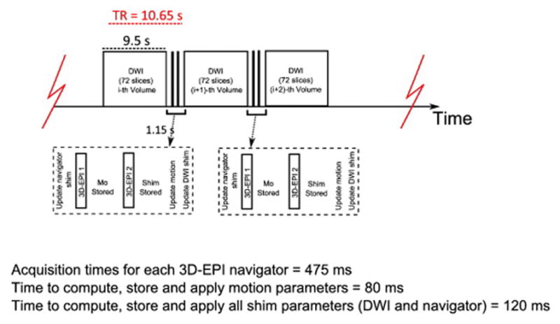
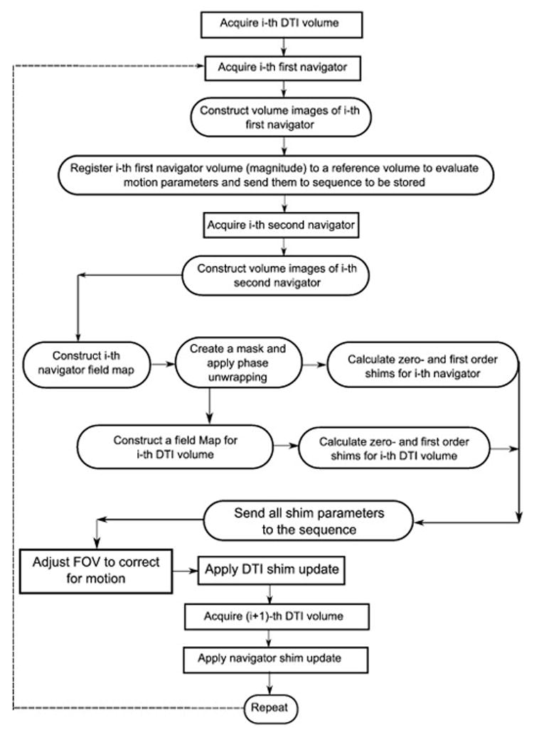
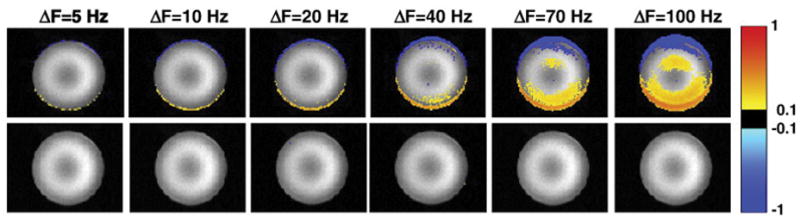
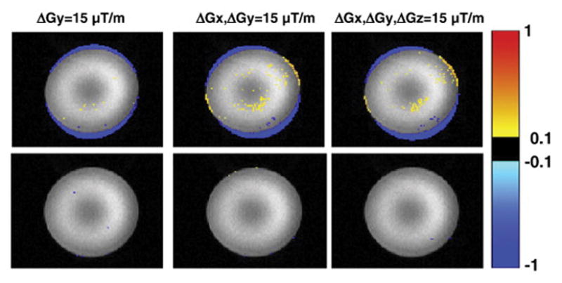
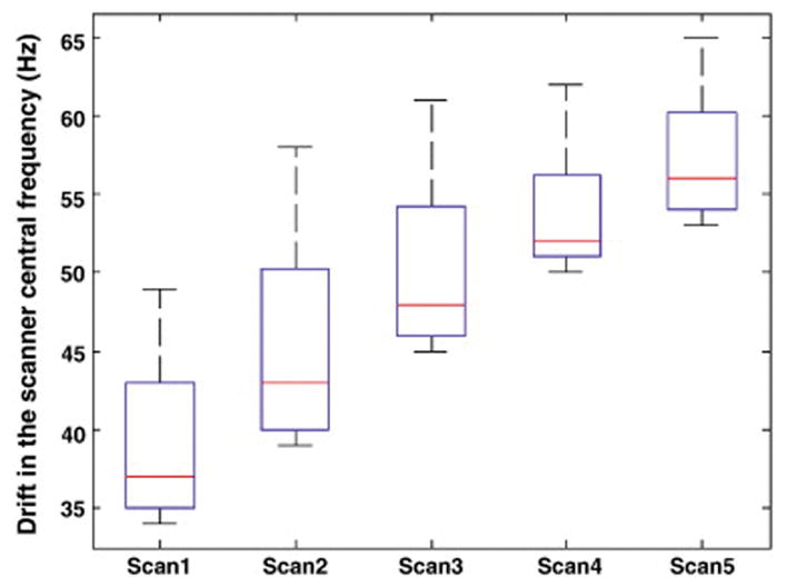
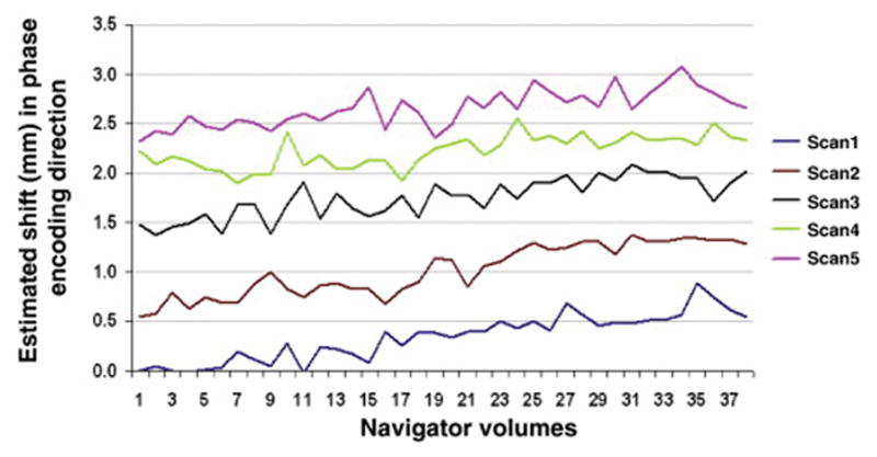
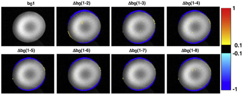
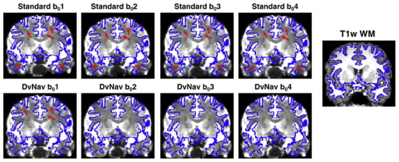
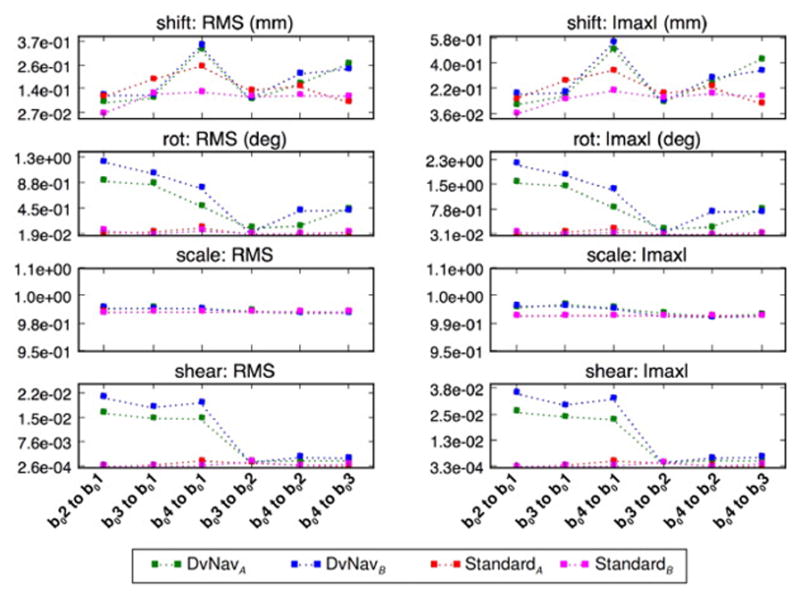
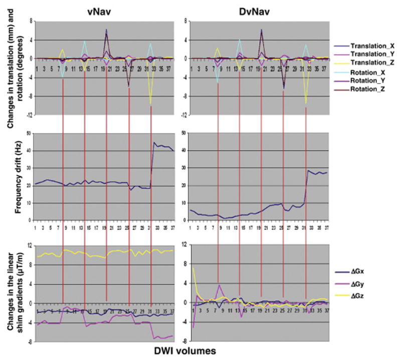
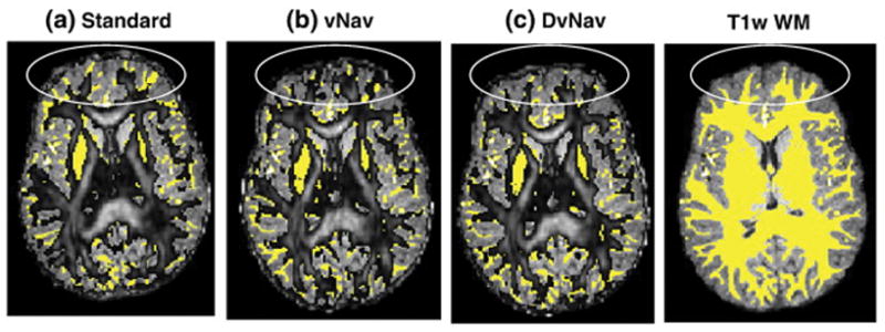
Diffusion tensor imaging (DTI) requires a set of diffusion weighted measurements in order to acquire enough information to characterize local structure. The MRI scanner automatically performs a shimming process by acquiring a field map before the start of a DTI scan. Changes in B0, which can occur throughout the DTI acquisition due to several factors (including heating of the iron shim coils or subject motion), cause significant signal distortions that result in warped diffusion tensor (DT) parameter estimates. In this work we introduce a novel technique to simultaneously measure, report and correct in real time subject motion and changes in B0 field homogeneity, both in and through the imaging plane. This is achieved using double volumetric navigators (DvNav), i.e. a pair of 3D EPI acquisitions, interleaved with the DTI pulse sequence. Changes in the B0 field are evaluated in terms of zero-order (frequency) and first-order (linear gradients) shim. The ability of the DvNav to accurately estimate the shim parameters was first validated in a water phantom. Two healthy subjects were scanned both in the presence and absence of motion using standard, motion corrected (single navigator, vNav), and DvNav DTI sequences. The difference in performance between the proposed 3D EPI field maps and the standard 3D gradient echo field maps of the MRI scanner was also evaluated in a phantom and two healthy subjects. The DvNav sequence was shown to accurately measure and correct changes in B0 following manual adjustments of the scanner's central frequency and the linear shim gradients. Compared to other methods, the DvNav produced DTI results that showed greater spatial overlap with anatomical references, particularly in scans with subject motion. This is largely due to the ability of the DvNav system to correct shim changes and subject motion between each volume acquisition, thus reducing shear distortion.

Keywords: B0 correction; Diffusion tensor imaging (DTI); Double volumetric navigators (DvNav); Navigated diffusion sequence (vNav); The first-order shim (linear gradients); Zero-order shim (frequency).


Figures

Fig. 1
Fig. 1
Sequence timing diagram of the standard twice-refocused, 2D diffusion pulse sequence (Reese et al., 2003) with double volumetric navigators inserted for simultaneous motion and shim correction. Acquisition times for ea ch 3D-EPI navigator = 475 ms Time to com pute, store and ap ply motion parameters = 80 ms Time to com pute, store and ap ply all shim parameters (DWI and navigator) = 120 ms
Fig. 2
Fig. 2
A flowchart showing the different steps performed to achieve real time motion and shim correction in the DvNav sequence. Events in rectangular boxes are performed in the sequence acquisition environment, while steps in the rounded rectangular boxes are performed in the image reconstruction environment.
Fig. 3
Fig. 3
Differences in normalized signal intensity between an undistorted reference scan (with zero frequency offset) and scans in which the system central frequency had been adjusted manually by ΔF=5, 10, 20, 40, 70 and 100 Hz, respectively. The top row shows, in a representative slice, the difference between the first volume of each distorted scan and the second volume of the reference scan. The bottom row shows the difference between the second volume of each distorted scan (after which frequency correction has been applied by the DvNav sequence) and the second volume of the reference scan (readout = left to right; phase encode = vertically upwards).
Fig. 4
Fig. 4
Differences in normalized signal intensity between a reference scan and scans in which the linear shim gradients had been adjusted manually (15 μT/m in each stated direction). The top row shows, in a representative slice, the difference between the first volume of the distorted acqusitions and the second volume of the reference scan. The bottom row shows the difference between the second volume of the distorted acquisitions (for which real-time shim correction had been applied) and the second volume of the reference scan. (readout = left to right; phase encode = vertically upwards).
Fig. 5
Fig. 5
Box-and-whisker plots showing the changes in the scanner central frequency estimated by the DvNav sequence for the 38 volumes of 5 consecutive DTI scans. The acquisition time for each scan was 6.7 mins. After the acquisition of the 5 scans (~ 33 mins), the water resonance frequency had drifted by roughly 30 Hz.
Fig. 6
Fig. 6
Translation (in mm) along the phase encode direction for each navigator volume for each successive scan. The shifts were estimated by registering successive navigators to the first navigator volume of the first scan.
Fig. 7
Fig. 7
Differences in normalized signal intensity between the first b0 volume (corrected only by the static shim of the MRI scanner) and the next seven b0 volumes (corrected by DvNav). For example, Δb0(1-2) = b01–b02. (readout = left to right; phase encode = vertically upwards).
Fig. 8
Fig. 8
A comparison of spatial distortions arising from standard and DvNav acquisitions. The outline of the T1w WM mask is overlaid in blue on each of the first four b0 volumes from the standard (top row) and DvNav (bottom row) acquisitions. Arrows highlight areas of significant distortion. (readout = left to right; phase encode = anterior to posterior).
Fig. 9
Fig. 9
A comparison of the magnitudes of the different affine transformation parameters for pairwise registration of the first four b0 volumes acquired using the standard (hot colors) and DvNav (cool colors) sequences in two subjects (A and B). In the DvNav acquisition correction is applied after the first volume. As such, registration of the corrected b0 volumes (b0(2-4)) to the uncorrected b01 volume quantifies the amount (and type) of distortion correction effected by the DvNav sequence.
Fig. 10
Fig. 10
Comparison of motion derivatives (1st row), zero (2nd row) and first order shim (3rd row) parameters for a scan with only prospective motion correction (left) and a scan with both real-time motion and shim correction (right).
Fig. 11
Fig. 11
FA maps (0.2

References

    1. Aksoy M, Forman C, Straka M, Skare S, Holdsworth S, Hornegger J, Bammer R. Real-time optical motion correction for diffusion tensor imaging. Magnetic resonance in medicine. 2011;66:366–378. - PMC - PubMed
    1. Alhamud A, Hess AT, Tisdall MD, Meintjes EM, van der Kouwe AJ. Implementation of real time motion correction in diffusion tensor imaging. Proceedings of the 19th Annual Meeting of ISMRM; May; Montreal, Canada. 2011.
    1. Alhamud A, Taylor PA, Laughton B, van der Kouwe AJ, Meintjes EM. Motion artifact reduction in pediatric diffusion tensor imaging using fast prospective correction. Journal of magnetic resonance imaging. 2015;41:1353–1364. - PMC - PubMed
    1. Alhamud A, Tisdall MD, Hess AT, Hasan KM, Meintjes EM, van der Kouwe AJ. Volumetric navigators for real-time motion correction in diffusion tensor imaging. Magnetic resonance in medicine. 2012;68:1097–1108. - PMC - PubMed
    1. Andersson JL, Skare S, Ashburner J. How to correct susceptibility distortions in spin-echo echo-planar images: application to diffusion tensor imaging. Neuroimage. 2003;20:870–888. - PubMed