

Chelator-Free Labeling of Layered Double Hydroxide Nanoparticles for in Vivo PET Imaging
- PMID: 26585551
- PMCID: PMC4653656
- DOI: 10.1038/srep16930
Chelator-Free Labeling of Layered Double Hydroxide Nanoparticles for in Vivo PET Imaging
Abstract
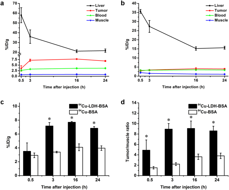
Layered double hydroxide (LDH) nanomaterial has emerged as a novel delivery agent for biomedical applications due to its unique structure and properties. However, in vivo positron emission tomography (PET) imaging with LDH nanoparticles has not been achieved. The aim of this study is to explore chelator-free labeling of LDH nanoparticles with radioisotopes for in vivo PET imaging. Bivalent cation (64)Cu(2+) and trivalent cation (44)Sc(3+) were found to readily label LDH nanoparticles with excellent labeling efficiency and stability, whereas tetravalent cation (89)Zr(4+) could not label LDH since it does not fit into the LDH crystal structure. PET imaging shows that prominent tumor uptake was achieved in 4T1 breast cancer with (64)Cu-LDH-BSA via passive targeting alone (7.7 ± 0.1%ID/g at 16 h post-injection; n = 3). These results support that LDH is a versatile platform that can be labeled with various bivalent and trivalent radiometals without comprising the native properties, highly desirable for PET image-guided drug delivery.
Figures


Similar articles
-
Facile Preparation of Multifunctional WS2 /WOx Nanodots for Chelator-Free 89 Zr-Labeling and In Vivo PET Imaging.Small. 2016 Nov;12(41):5750-5758. doi: 10.1002/smll.201601696. Epub 2016 Sep 4. Small. 2016. PMID: 27593416 Free PMC article.
-
Novel method to label solid lipid nanoparticles with 64cu for positron emission tomography imaging.Bioconjug Chem. 2011 Apr 20;22(4):808-18. doi: 10.1021/bc100478k. Epub 2011 Mar 9. Bioconjug Chem. 2011. PMID: 21388194 Free PMC article.
-
Comparison of the octadentate bifunctional chelator DFO*-pPhe-NCS and the clinically used hexadentate bifunctional chelator DFO-pPhe-NCS for 89Zr-immuno-PET.Eur J Nucl Med Mol Imaging. 2017 Feb;44(2):286-295. doi: 10.1007/s00259-016-3499-x. Epub 2016 Aug 30. Eur J Nucl Med Mol Imaging. 2017. PMID: 27573793 Free PMC article.
-
Radiolabeling Silica-Based Nanoparticles via Coordination Chemistry: Basic Principles, Strategies, and Applications.Acc Chem Res. 2018 Mar 20;51(3):778-788. doi: 10.1021/acs.accounts.7b00635. Epub 2018 Feb 28. Acc Chem Res. 2018. PMID: 29489335 Free PMC article. Review.
-
Recent Advances in Zirconium-89 Chelator Development.Molecules. 2018 Mar 12;23(3):638. doi: 10.3390/molecules23030638. Molecules. 2018. PMID: 29534538 Free PMC article. Review.
Cited by
-
Homogeneous Incorporation of Gallium into Layered Double Hydroxide Lattice for Potential Radiodiagnostics: Proof-of-Concept.Nanomaterials (Basel). 2020 Dec 26;11(1):44. doi: 10.3390/nano11010044. Nanomaterials (Basel). 2020. PMID: 33375387 Free PMC article.
-
Radiolabeled inorganic nanoparticles for positron emission tomography imaging of cancer: an overview.Q J Nucl Med Mol Imaging. 2017 Jun;61(2):181-204. doi: 10.23736/S1824-4785.17.02969-7. Epub 2017 Jan 26. Q J Nucl Med Mol Imaging. 2017. PMID: 28124549 Free PMC article. Review.
-
Intrinsic radiolabeling of Titanium-45 using mesoporous silica nanoparticles.Acta Pharmacol Sin. 2017 Jun;38(6):907-913. doi: 10.1038/aps.2017.1. Epub 2017 Apr 17. Acta Pharmacol Sin. 2017. PMID: 28414201 Free PMC article.
-
Biomedical Approach of Nanotechnology and Biological Risks: A Mini-Review.Int J Mol Sci. 2023 Nov 24;24(23):16719. doi: 10.3390/ijms242316719. Int J Mol Sci. 2023. PMID: 38069043 Free PMC article. Review.
-
Multifunctional Layered Double Hydroxides for Drug Delivery and Imaging.Nanomaterials (Basel). 2023 Mar 19;13(6):1102. doi: 10.3390/nano13061102. Nanomaterials (Basel). 2023. PMID: 36985996 Free PMC article. Review.
References
-
- Xu Z. P., Jin Y. G., Liu S. M., Hao Z. P. & Lu G. Q. Surface charging of layered double hydroxides during dynamic interactions of anions at the interfaces. J. Colloid Interf. Sci 326, 522–529 (2008). - PubMed
-
- Wang Q. & O’Ha D. Recent advances in the synthesis and application of layered double hydroxide (LDH) nanosheets. Chem. Rev. 112, 4124–4155 (2012). - PubMed
-
- Xu Z. P., Kurniawan N. D., Bartlett P. F. & Lu G. Q. Enhancement of relaxivity rates of Gd-DTPA complexes by intercalation into layered double hydroxide nanoparticles. Chemistry 13, 2824–2830 (2007). - PubMed
-
- Kim S. Y., Oh J. M., Lee J. S., Kim T. J. & Choy J. H. Gadolinium (III) diethylenetriamine pentaacetic acid/ layered double hydroxide nanohybrid as novel T1-magnetic resonant nanoparticles. J. Nanosci. Nanotechnol. 8, 5181–5184 (2008). - PubMed
Publication types
MeSH terms
Substances
Grants and funding
LinkOut - more resources
Full Text Sources
Other Literature Sources
