. 2016 Jan 1;351(6268):53-8.
doi: 10.1126/science.aad2087. Epub 2015 Nov 19.

Structural basis for leucine sensing by the Sestrin2-mTORC1 pathway

Affiliations

Structural basis for leucine sensing by the Sestrin2-mTORC1 pathway

Robert A Saxton et al. Science. .

Abstract

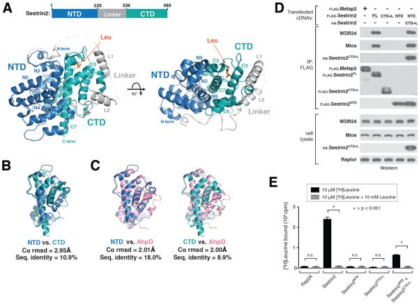
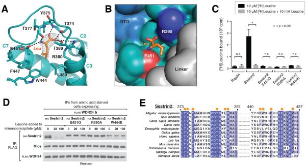
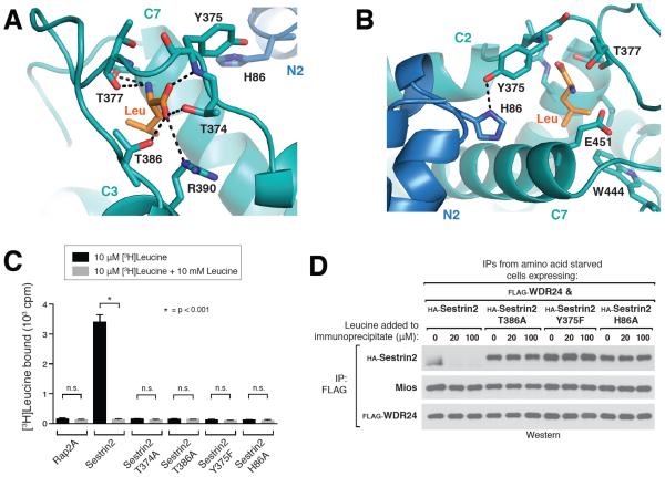
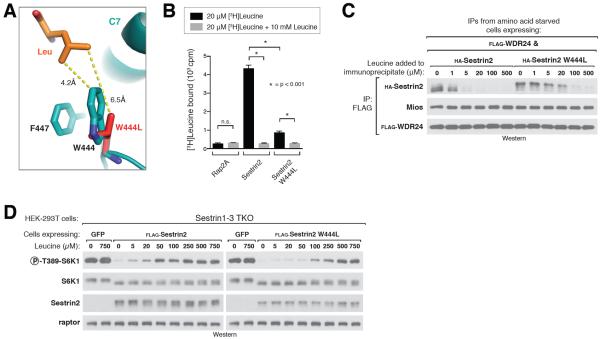
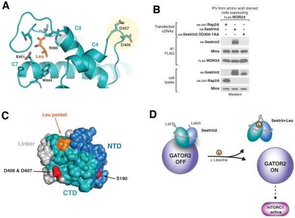
Eukaryotic cells coordinate growth with the availability of nutrients through the mechanistic target of rapamycin complex 1 (mTORC1), a master growth regulator. Leucine is of particular importance and activates mTORC1 via the Rag guanosine triphosphatases and their regulators GATOR1 and GATOR2. Sestrin2 interacts with GATOR2 and is a leucine sensor. Here we present the 2.7 angstrom crystal structure of Sestrin2 in complex with leucine. Leucine binds through a single pocket that coordinates its charged functional groups and confers specificity for the hydrophobic side chain. A loop encloses leucine and forms a lid-latch mechanism required for binding. A structure-guided mutation in Sestrin2 that decreases its affinity for leucine leads to a concomitant increase in the leucine concentration required for mTORC1 activation in cells. These results provide a structural mechanism of amino acid sensing by the mTORC1 pathway.


Figures

Figure 1
Figure 1. Structure of leucine-bound Sestrin2
A) Two views of human Sestrin2 are shown as ribbon diagrams, with the N-terminal (NTD), Linker, and C-terminal (CTD) domains colored in blue, gray, and teal respectively. The bound leucine molecule is shown in orange. Disordered regions not present in the crystal structure (1–65, 242–255, 272–280, 296–309) are shown as dashed lines. B) Structural superposition of Sestrin2 NTD (blue, residues 66–220) and CTD (teal, residues 339–480). C) Structural superposition on Sestrin2 NTD (blue) and CTD (teal) with a R. eutropha AhpD dimer (pink, PDB ID: 2PRR) D) Immunoprecipitation of N- and C- terminal fragments of Sestrin2. HEK-293T cells transiently transfected with FLAG-metap2, FLAG-Sestrin2 full length (FL), FLAG-Sestrin2-NTD (N-terminal domain, 1–220), FLAG-Sestrin2-CTD+L (C-terminal domain plus Linker, 220–480) or both Flag-Sestrin2-NTD and HA-Sestrin2-CTD+L were starved for amino acids for 50 minutes. FLAG-immunoprecipitates were prepared from cell lysates. Immunoprecipitates and lysates from one representative experiment were analyzed by immunoblotting for indicated proteins. WDR24 and Mios were used as representative GATOR2 components. E) [3H]Leucine binding assay using N- and C-terminal fragments of Sestrin2. FLAG-immunoprecipitates prepared from HEK-293T cells transiently expressing indicated proteins were used as described in the methods. Unlabeled leucine was used as a competitor where indicated. Values are Mean ± SD for 3 technical replicates from one representative experiment. Two-tailed t tests were used for comparison between two groups.
Figure 2
Figure 2. Recognition of leucine by Sestrin2
A) Close-up view of the leucine binding pocket in Sestrin2, focusing on the bound leucine (shown in orange) together with its 2Fo-Fc electron density map calculated and contoured at 1.5σ from an omit map lacking leucine and all pocket residues. Predicted hydrogen bonds or salt-bridges are shown as black dashed lines. Helix numbers are labeled as in 1A. B) Surface representation of leucine-bound Sestrin2, focusing on the leucine binding pocket. The bound leucine is represented as a stick model (orange). Residues 373–387 are omitted to allow visibility of the pocket. Residue Glu451, which contacts the amine of leucine is shown in red, and Arg390 which contacts the carboxyl of leucine is shown in blue. The domains of Sestrin2 are colored as in 1A. C) Binding of the E451Q, R390A and W444E mutants of Sestrin2 to leucine. HA-immunoprecipitates prepared from HEK-293T cells transiently expressing indicated HA-tagged proteins were used in binding assays with [3H]Leucine. Binding was analyzed as in 1E. D) Effect of leucine on the interactions of Sestrin2 E451Q, R390A or W444L with GATOR2. FLAG-immunoprecipitates were prepared from cells stably expressing FLAG-WDR24 and transiently expressing the indicated HA-tagged Sestrin2 constructs. The immunoprecipitates were treated with the indicated concentrations of leucine and analyzed by immunoblotting for the indicated proteins. E) Multiple Sequence Alignment of Sestrin2 homologs from various organisms. Positions of residues contacting leucine are indicated with orange dots. Positions are colored white to blue according to increasing sequence identity.
Figure 3
Figure 3. A lid-latch mechanism is required for leucine binding by Sestrin2
A) Top-down view of the leucine bound pocket, focusing on the “lid” residues Thr374, Thr377, and Thr386 which form hydrogen bonds with the amine and carboxyl groups of leucine (indicated by black dashed lines). Leucine is represented as a stick model (orange). Helix numbers are labeled as in 1A. B) Orthogonal view of the leucine binding pocket, focusing on the “latch” formed by the predicted hydrogen bond between Tyr375 and His86, which locks the lid in place over the bound leucine (orange). Helix numbers are labeled as in 1A. C) Binding of Sestrin2 T374A, T386A, Y375F and H86A mutants of Sestrin2 to leucine. Binding assays were performed and immunoprecipitates analyzed as in Figure 2C. D) Effect of leucine on the interactions of Sestrin2 T386A, Y375F or H86A with GATOR2 in vitro. FLAG immunoprecipitates were prepared from cells stably expressing FLAG-WDR24 and transiently expressing the indicated HA-tagged Sestrin2 constructs and analyzed as in 2D.
Figure 4
Figure 4. Altering the leucine sensitivity of the mTORC1 pathway in cells
A) Close-up view of Sestrin2-bound leucine (orange) and the pocket floor residues Phe447 and W444, with the W444L mutant (red) overlaid onto the wild-type protein (teal). Both residues are represented as stick models. Numbers indicate distance from leucine to residue 444 in Sestrin2 WT and Sestrin2 W444L. B) Leucine binding by Sestrin2 W444L. Binding assays were performed and immunoprecipitates analyzed as in Figure 2C. C) Higher concentrations of leucine are required to dissociate Sestrin2 W444L from GATOR2 compared to Sestrin2 WT. FLAG immunoprecipitates were prepared from cells stably expressing FLAG-WDR24 and transiently expressing the indicated HA-tagged Sestrin2 constructs and analyzed as in figure 2D. D) Sensitivity of the mTORC1 pathway to leucine in Sestrin1–3 triple-knock-out (TKO) cells expressing Sestrin2 wild type or W444L. HEK-293T cells generated with the CRISPR/Cas9 system expressing the indicated proteins via lentiviral transduction. Cells were starved of leucine for 50 minutes then re-stimulated with the indicated amount of leucine for 10 minutes. Cell lysates from one representative experiment were prepared and analyzed via immunoblotting.
Figure 5
Figure 5. Identification of GATOR2 binding site and model of leucine sensing by Sestrin2
A) View highlighting the conserved surface aspartates Asp406/407 and their position relative to the bound leucine (orange). B) Co-immunoprecipitation of GATOR2 with Sestrin2 wild type or DD406-7AA. FLAG immunoprecipitates were prepared from cells stably expressing FLAG-WDR24 and transiently expressing the indicated HA-tagged Sestrin2 constructs and analyzed as in 2D. C) Surface view of Sestrin2 highlighting the GATOR2 binding sites (red) and their position relative to the leucine-binding pocket (orange). Domains are colored as in 1A. D) Model of leucine sensing by Sestrin2. Binding of leucine (orange) causes closing of the lid-latch, resulting in a conformational change altering the position of the GATOR2 binding site in the CTD. This leads to dissociation of Sestrin2 from GATOR2, enabling GATOR2 to activate the mTORC1 pathway.

Comment in

References

    1. Zoncu R, Efeyan A, Sabatini DM. mTOR: from growth signal integration to cancer, diabetes and ageing. Nature reviews. Molecular cell biology. 2011;12:21. - PMC - PubMed
    1. Dibble CC, Manning BD. Signal integration by mTORC1 coordinates nutrient input with biosynthetic output. Nature Cell Biology. 2013;15:555–564. - PMC - PubMed
    1. Jewell JL, Russell RC, Guan KL. Amino acid signalling upstream of mTOR. Nature reviews. Molecular cell biology. 2013;14:133. - PMC - PubMed
    1. Potier M, Darcel N, Tomé D. Protein, amino acids and the control of food intake. Current Opinion in Clinical Nutrition and Metabolic Care. 2009;12:54–58. - PubMed
    1. Greiwe JS, Kwon G, McDaniel ML, Semenkovich CF. Leucine and insulin activate p70 S6 kinase through different pathways in human skeletal muscle. American journal of physiology. Endocrinology and metabolism. 2001;281:E466–471. - PubMed

Publication types

MeSH terms

Associated data