. 2016 Jan 15;409(2):473-88.
doi: 10.1016/j.ydbio.2015.09.023. Epub 2015 Nov 14.

Ibuprofen slows migration and inhibits bowel colonization by enteric nervous system precursors in zebrafish, chick and mouse

Affiliations

Ibuprofen slows migration and inhibits bowel colonization by enteric nervous system precursors in zebrafish, chick and mouse

Ellen Merrick Schill et al. Dev Biol. .

Abstract

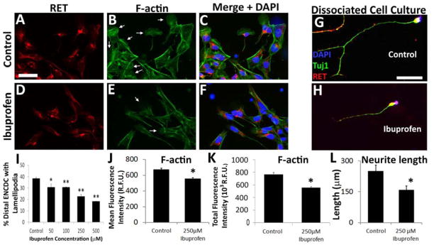
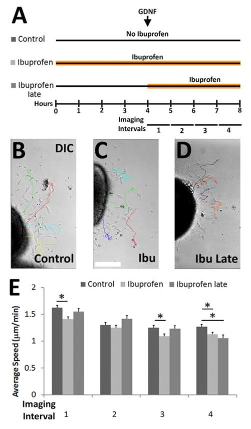
Hirschsprung Disease (HSCR) is a potentially deadly birth defect characterized by the absence of the enteric nervous system (ENS) in distal bowel. Although HSCR has clear genetic causes, no HSCR-associated mutation is 100% penetrant, suggesting gene-gene and gene-environment interactions determine HSCR occurrence. To test the hypothesis that certain medicines might alter HSCR risk we treated zebrafish with medications commonly used during early human pregnancy and discovered that ibuprofen caused HSCR-like absence of enteric neurons in distal bowel. Using fetal CF-1 mouse gut slice cultures, we found that ibuprofen treated enteric neural crest-derived cells (ENCDC) had reduced migration, fewer lamellipodia and lower levels of active RAC1/CDC42. Additionally, inhibiting ROCK, a RHOA effector and known RAC1 antagonist, reversed ibuprofen effects on migrating mouse ENCDC in culture. Ibuprofen also inhibited colonization of Ret+/- mouse bowel by ENCDC in vivo and dramatically reduced bowel colonization by chick ENCDC in culture. Interestingly, ibuprofen did not affect ENCDC migration until after at least three hours of exposure. Furthermore, mice deficient in Ptgs1 (COX 1) and Ptgs2 (COX 2) had normal bowel colonization by ENCDC and normal ENCDC migration in vitro suggesting COX-independent effects. Consistent with selective and strain specific effects on ENCDC, ibuprofen did not affect migration of gut mesenchymal cells, NIH3T3, or WT C57BL/6 ENCDC, and did not affect dorsal root ganglion cell precursor migration in zebrafish. Thus, ibuprofen inhibits ENCDC migration in vitro and bowel colonization by ENCDC in vivo in zebrafish, mouse and chick, but there are cell type and strain specific responses. These data raise concern that ibuprofen may increase Hirschsprung disease risk in some genetically susceptible children.

Keywords: Enteric nervous system development; Gene-environment interactions; Migration.


Conflict of interest statement

Conflict of Interest: The authors have declared that no conflict of interest exists.

Figures

Figure 1
Figure 1. Ibuprofen reduced ENCDC colonization of zebrafish bowel
(A–D) Zebrafish were treated with vehicle (1%DMSO) or ibuprofen (Ibu) from 34–96 hpf and then stained with HuC/D antibody. (A) White arrows indicate dorsal root ganglia. (C) White arrows highlight enteric neurons. Scale bar=500μm. (B, D) Higher magnification of fish midsection. Scale bar=100μm. (E) Length of bowel from most distal HuC/D+ cell (arrows in B, D) to bowel terminus (arrowheads in B, D). *P<0.05 (ANOVA on Ranks) (F) Length of whole zebrafish (1% DMSO n=4, 25 μM Ibuprofen n=6). *P=0.025 (t-test). (G) Distance between DRG and dorsal zebrafish edge (1% DMSO n=3, 25 μM ibuprofen n=4). *P>0.05 (t-test).
Figure 2
Figure 2. Ibuprofen inhibited hindgut colonization by chick ENCDC ex vivo and mouse ENCDC in vivo
(A, B) Ibuprofen (250 μM) almost completely blocked E6 chick colon colonization by N-cadherin+ (green) ENCDC during 48 hour culture. (Scale bar=200 μm, n=6 per group). Insets show the migration wavefront with EdU (red) and N-cadherin (green) immunohistochemistry. (Scale bar=35 μm). mg=midgut, hg=hindgut. (C–E) Ibuprofen feeding from E8.5 to E12.5 reduced Ret +/− mouse colon colonization by ENCDC visualized with TuJ1 antibody (red). Scale bars=400 μm. (E, F) Quantitative data show the proportion of the colon colonized by ENCDC (i.e., length of colon containing ENCDC divided by total colon length) (Control n=4, ibuprofen n=9). *P =0.042 (t-test). (F) Sox10 +/− colon was normally colonized by ENCDC when dams were fed ibuprofen from E8.5 to E12.5 (Control n = 19, ibuprofen n=13). P=0.79 (t-test).
Figure 3
Figure 3. Ibuprofen reduced murine ENCDC migration, but did not affect proliferation, caspase-3 activation, or neuronal differentiation
E12.5 CF-1 midgut slices cultured with or without ibuprofen for 16 hours after GDNF addition were stained for (A, B) RET (red), phalloidin (green), DAPI (blue) (scale bar=200μm), (C, D) RET (green), BrdU (red), DAPI (blue) (Scale bar=100μm), (E, F) RET (red), cleaved-caspase 3 (green), DAPI (blue) (Scale bar=100μm), or (G, H) RET (green), TuJ1 (red), DAPI (blue) (scale bar=100μm). (I) Ibuprofen reduced the distance ENCDC migrated from gut explants at 250 μM and 500 μM. Control n=275 slices, 29 biological replicates; 50 μM ibuprofen n = 48 slices, 3 biological replicates; 100 μM ibuprofen n = 55 slices, 3 biological replicates; 250 μM ibuprofen n=111 slices, 5 biological replicates; 500 μM ibuprofen n = 102 slices, 8 biological replicates) but not at lower ibuprofen concentrations. **P<0.001 (ANOVA). (J) Mean percentage BrdU+ ENCDC (RET+ cells) (Control n=21 slices, 7 biological replicates; ibuprofen n=9 slices, 3 biological replicates). P=0.24 (t-test). (K) Mean percentage cleaved-caspase 3+ ENCDC (RET+ cells) (Control n=15 slices, ibuprofen n=15 slices; both 3 biological replicates). P=0.36 (t-test). (L) Mean percentage TuJ1+ ENCDC (RET+ cells) (Control n=19 slices, ibuprofen n=23 slices; both 3 biological replicates). P=0.35 (t-test).
Figure 4
Figure 4. Ibuprofen did not affect migration or lamellipodia of NIH 3T3 or mouse gut mesenchymal cells
Confluent NIH 3T3 were pretreated with 250 μM ibuprofen (2 hours) before making a scratch to remove some cells. (A, B) NIH3T3 immediately after the scratch was made. (C, D) Two hours after the scratch. Cells were imaged every 5 minutes for 24 hours 250 μM ibuprofen did not affect (E) average speed of gap closure (Control n=5 assays, ibuprofen; n=7 assays), P=0.76 (t-test) or (F) percentage of NIH 3T3 with lamellipodia (Control n=8 assays, ibuprofen; n=9 assays). P=0.75 (t-test). (G, H) 250 μM ibuprofen did not affect distance CF-1 mesenchymal cells migrated from gut explants (Control n=19 slices, ibuprofen n=24 slices; 3 biological replicates). P=0.57 (t-test) or the percentage of mesenchymal cells with lamellipodia after 16 hours (Control=9 slices, ibuprofen=9 slices; 3 biological replicates). P=0.52 (t-test).
Figure 5
Figure 5. Ibuprofen reduced lamellipodia in migrating murine ENCDC and reduced neurite length in differentiating murine enteric neurons
(A–F) E12.5 CF-1 midgut slices were cultured with 250 μM ibuprofen or control media for 16 hours after GDNF addition and then stained for (A, D) RET, (B, E) Alexa488-phalloidin, and (C, F) DAPI (merged image). Arrows highlight well-formed lamellipodia. (G, H) Dissociated immunoselected E12.5 CF-1 ENCDC cultured 48 hours were stained for RET (red), TuJ1 (green) and DAPI (blue). Scale bars=50 μm. (I, J). (I) The percentage of migrating ENCDC with lamellipodia was reduced by 50 μM or higher ibuprofen concentrations. RET+ ENCDC most distant from the gut slices were analyzed. (Control n=61 slices, 13 biological replicates; 50 μM Ibuprofen n = 9 slices, 3 biological replicates; 100 μM ibuprofen n=9 slices, 3 biological replicates, 250 μM ibuprofen n=27 slices, 9 biological replicates; 500 μM ibuprofen n = 12 slices, 4 biological replicates). *P <0.05, **P <0.001, (ANOVA, Holm-Sidak method). (J) Ibuprofen reduced mean Alexa488-phalloidin fluorescence intensity for RET+ ENCDC (Control n=232 cells, ibuprofen n=236 cells; both 4 biological replicates). *P<0.001 (Mann-Whitney Rank Sum). (K) Ibuprofen reduced longest neurite in cultured differentiating ENCDC (Control n=50, ibuprofen n=76, both 3 biological replicates). *P=0.009 (Mann-Whitney Rank Sum).
Figure 6
Figure 6. E12.5 CF-1 mouse ENCDC had less immunoreactive PPARγ than E12.5 dorsal root ganglion (DRG) neurons
Sagittal sections were stained with antibodies to PPARγ (red) and neuron specific beta 3 tubulin (green, TuJ1). (A–D) PPARγ is readily detectable in DRG neurons. The boxed region in C contains DRG neurons that are immunoreactive for both PPARγ and TuJ1 antibodies (yellow cells) and is enlarged in D to show DRG neurons immunoreactive for PPARγ staining (red cells, white arrows). (E–H) Developing enteric neurons (TuJ1+ cells highlighted with arrows in F) have much lower levels of immunoreactive PPARγ (i.e., no yellow cells in the region of the ENS in G). The boxed region in G is enlarged in H to show the lack of PPARγ staining (red) in the ENCDC. Arrows highlight the region of the developing ENS. Images are from the same fetal mouse stained on the same slide. Scale bar = 100μm.
Figure 7
Figure 7. Ibuprofen had delayed effects on murine ENCDC migration
(A) Experimental paradigms. E12.5 CF-1 midgut slices were cultured on fibronectin. Ibuprofen was added four hours after plating (ibuprofen late) or at plating (ibuprofen). Time lapse images were then obtained every two minutes for four hours. (B–D) DIC images show final time points from time-lapse movies. Tracks show trajectories for single cells that migrated from explants. Immunohistochemistry after imaging confirmed tracked cells expressed RET. Scale bar = 100μm. (E) Mean ENCDC migration speed during each one hour interval. Ibuprofen slowed ENCDC speed, but not until 3–4 hours after ibuprofen exposure (Control n=130 cells, Ibuprofen n=180 cells, ibuprofen late n=92 cells; all 3 biological replicates). *P<0.05 (ANOVA on ranks).
Figure 8
Figure 8. Ibuprofen effects on murine ENCDC migration and lamellipodia do not appear to be cyclooxygenase dependent. Ptgs1−/− Ptgs2−/− mice have normal ENCDC migration in vitro and normal bowel colonization in vivo
(A, B) E12.5 WT C57BL/6 and Ptgs1−/− Ptgs2−/− midgut slices were cultured 16 hours after GDNF addition and then stained with RET antibody (red), phalloidin (green), and DAPI (blue). Scale bar=50 μm. (C, D) E12.5 bowel from WT and Ptgs1−/− Ptgs2−/− mice was stained with Tuj1 antibody. Scale bar=500 μm. Ptgs1−/− Ptgs2−/− mutations did not affect (E) the mean distance that ENCDC migrated from gut slices (WT n=57 slices, 5 embryos and Ptgs1−/− Ptgs2−/− n=48, slices, 7 embryos), P=0.55 (t-test), or (F) the mean percentage of ENCDC with a well-formed lamellipodia (WT n=11 slices, 4 embryos and Ptgs1−/− Ptgs2−/− n=10 slices, 5 embryos), P=0.38 (t-test), or (G) the extent of bowel colonization by ENCDC in vivo (WT n=6 and Ptgs1−/− Ptgs2−/− n=5), P=0.23 (t-test). Only ENCDC most distant from gut slices were evaluated for lamellipodia. (H–K) Stable Prostaglandin E2 (PGE2) and Prostaglandin F2 (PGF2) analogs did not rescue ibuprofen-induced ENCDC migration defects. E12.5 CF-1 midgut slices were cultured in media with GDNF with or without ibuprofen for 16 hours and then stained for RET, F-actin (phalloidin) and with DAPI. (H, I) Distance from the gut slice edge to the most distant RET+ ENCDC was measured. Neither 16, 16-dimethyl PGE2 (H) nor 16, 16-dimethyl PGF2 (I) restored ENCDC migration to control levels in the presence of ibuprofen, but 16,16-dimethyl PGE2 (H) did reduce ENCDC migration in the absence of ibuprofen. (For PGE2 studies: Control n = 165 slices, ibuprofen n = 112 slices, 16, 16-dimethyl PGE2 n = 137 slices, ibuprofen plus 16, 16-dimethyl PGE2 n = 53 slices; *P<0.05 compared to Control. #P<0.05 compared to Prostaglandin E2 (ANOVA). For PGF2 studies: Control n =103 slices, ibuprofen n = 96 slices, 16, 16-dimethyl PGF2 n = 127 slices, ibuprofen plus PGF2 n = 107 slices; *P<0.05 compared to Control. #P<0.05 compared to Prostaglandin F2 (ANOVA on Ranks)) (J, K) The percentage of ENCDC with well-formed lamellipodia was determined by examining phalloidin stained ENCDC that had migrated furthest from the gut slice edge. Neither 16, 16-dimethyl PGE2 nor 16, 16-dimethyl PGF2 rescued lamellipodia in the presence of ibuprofen (For PGE2 studies: Control n = 27 slices, ibuprofen n = 18 slices, PGE2 n = 27 slices, ibuprofen plus PGE2 n =18 slices) *P<0.05 compared to Control. #P<0.05 compared to Prostaglandin E2 (ANOVA on Ranks). For PGF2 studies: Control n = 105 slices, ibuprofen n = 83 slices, 16, 16-dimethyl PGF2 n = 105 slices, ibuprofen plus 16, 16-dimethyl PGF2 n =93 slices *P<0.05 compared to Control. #P<0.05 compared to Prostaglandin F2 (ANOVA)).
Figure 9
Figure 9. Ibuprofen reduced active RAC1/CDC42, but did not affect active RHOA in migrating murine ENCDC
E12.5 CF-1 midgut slices cultured 16 hours with GDNF were fixed, permeabilized and incubated with (A–H) PBD-GST that binds active GTP-RAC1 and GTP-CDC42, or (I–P) with RBD-GST that binds active GTP-RHOA. Cultures were then stained for (A, D, I, L) RET (green), (B, E, J, M) GST (red), and (C, F, K, N) DAPI (blue; merged image). Scale bar=50μm. (G, O) Mean fluorescence intensity for PBD-GST (G) and RBD-GST (O) for ENCDC that migrated furthest from gut slices. (G) 250 μM Ibuprofen treated ENCDC had less bound PBD-GST indicating reduced active RAC1/CDC42 (Control n=243 cells, ibuprofen n=205 cells, both 4 biological replicates); *P<0.001 (Mann-Whitney Rank Sum), but did not reduce (O) RBD-GST bound to active RHOA in actively migrating ENCDC (Control n=144 cells, ibuprofen n=136 cells, both 4 biological replicates). P=0.408 (Mann-Whitney Rank Sum). (H, P) Because ENCDC shape is altered by ibuprofen and this may change mean fluorescence intensity per pixel, total fluorescence intensity (mean fluorescence intensity/pixel x pixels/cell) was determine for (H) active RAC1/CDC42 (via PGD-GST binding) (Control n=243 cells, ibuprofen n=205 cells, both 4 biological replicates, and (P) active RHOA (via RBD-GST binding) (Control n =144 cells, ibuprofen n=136 cells, both 4 biological replicates) per cell. *P<0.001, Mann-Whitney Rank Sum. These data indicate that ibuprofen reduced total cellular levels of F-actin and active RAC1/CDC42 without affecting active RHOA levels. Only ENCDC furthest from the gut slice edge were analyzed.
Figure 10
Figure 10. ROCK inhibition rescued ibuprofen effects on murine ENCDC migration
(A–L) E12.5 CF-1 midgut slices were cultured with or without 250μM ibuprofen four hours before GDNF and 5 μM Y-27632 were added. Sixteen hours later cultures were fixed and stained for (A, D, G, J) RET (red), (B, E, H, K) phalloidin (green), and (C, F, I, L) DAPI (blue; merged image). Scale bar = 50 μm. (M) Distance from the gut slice edge to the most distant RET+ ENCDC was measured. Y-27632 increased migration of ibuprofen treated ENCDC, but not control ENCDC (Control n =43 slices, ibuprofen n=48 slices, Y-27632 n=54 slices, ibuprofen + Y-27632 n=59 slices, all from 7 biological replicates). *P<0.05 compared to control, #P<0.05 compared to ibuprofen + Y-27632 (ANOVA on ranks).
Figure 11
Figure 11. Model
RAC1 organizes the actin cytoskeleton to form lamellipodia and support migration. Ibuprofen reduced ENCDC RAC1 activation. ROCK is a RHOA effector that inhibits RAC1. Y-27632 inhibits ROCK to permit RAC1 activation, enhancing ENCDC migration.

References

    1. Airaksinen MS, Saarma M. The GDNF family: signalling, biological functions and therapeutic value. Nat Rev Neurosci. 2002;3:383–394. - PubMed
    1. Aluri JB, Stavchansky S. Determination of guaifenesin in human plasma by liquid chromatography in the presence of pseudoephedrine. J Pharm Biomed Anal. 1993;11:803–808. - PubMed
    1. Amiel J, Sproat-Emison E, Garcia-Barcelo M, Lantieri F, Burzynski G, Borrego S, Pelet A, Arnold S, Miao X, Griseri P, Brooks AS, Antinolo G, de Pontual L, Clement-Ziza M, Munnich A, Kashuk C, West K, Wong KK, Lyonnet S, Chakravarti A, Tam PK, Ceccherini I, Hofstra RM, Fernandez R. Hirschsprung disease, associated syndromes and genetics: a review. J Med Genet. 2008;45:1–14. - PubMed
    1. Anderson RB, Newgreen DF, Young HM. Neural crest and the development of the enteric nervous system. Adv Exp Med Biol. 2006;589:181–196. - PubMed
    1. Arnold S, Pelet A, Amiel J, Borrego S, Hofstra R, Tam P, Ceccherini I, Lyonnet S, Sherman S, Chakravarti A. Interaction between a chromosome 10 RET enhancer and chromosome 21 in the Down syndrome-Hirschsprung disease association. Hum Mutat. 2009;30:771–775. - PMC - PubMed

Publication types

MeSH terms