. 2016 Jan 1;143(1):24-34.
doi: 10.1242/dev.124602. Epub 2015 Nov 19.

Selection and dynamics of embryonic stem cell integration into early mouse embryos

Affiliations

Selection and dynamics of embryonic stem cell integration into early mouse embryos

Stoyana Alexandrova et al. Development. .

Abstract

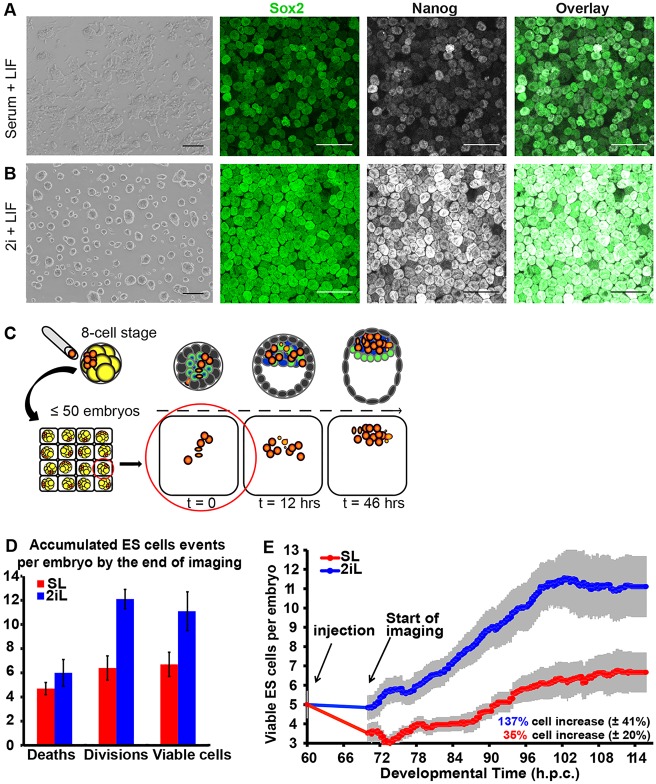
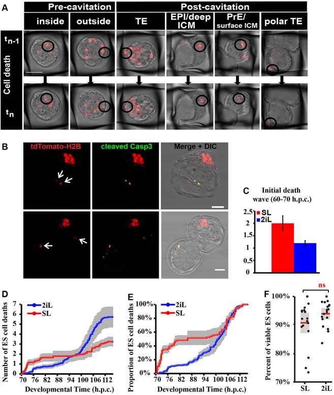
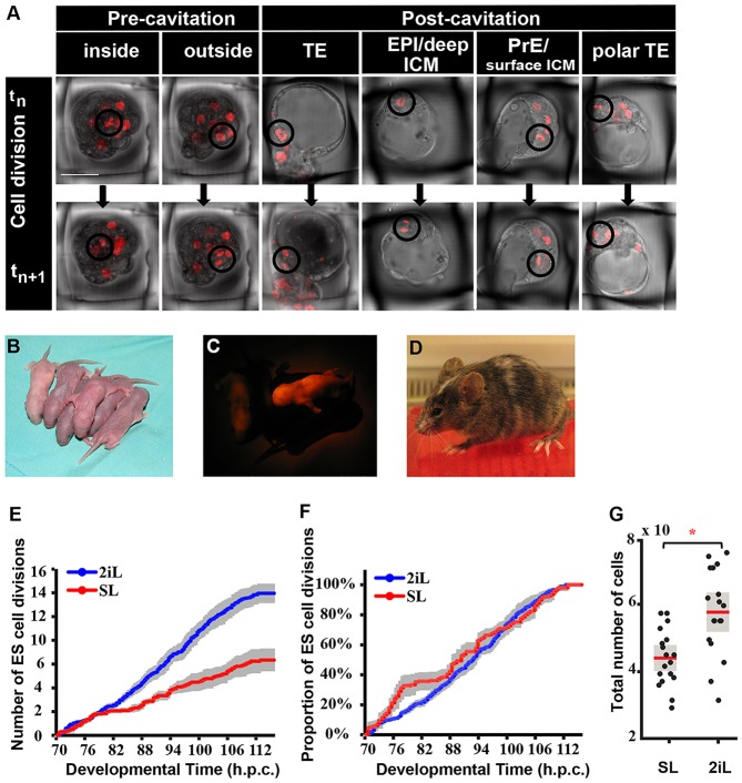
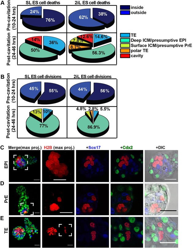
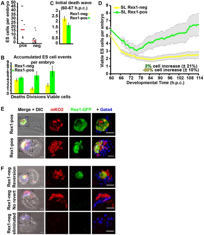
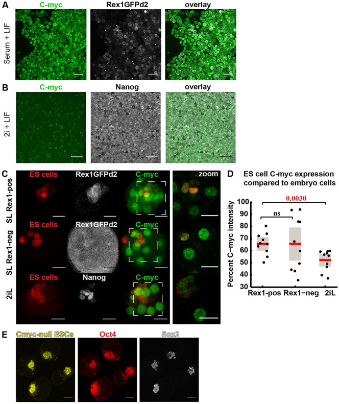
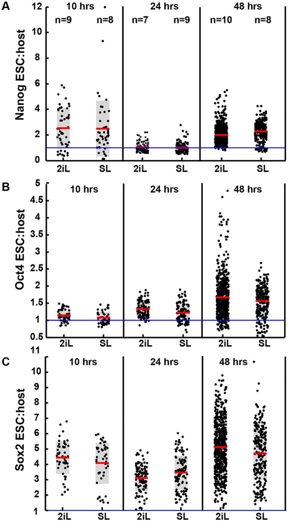
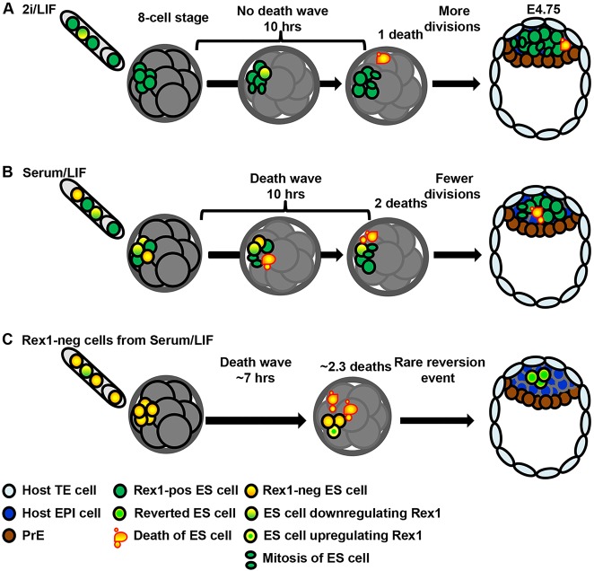
The process by which pluripotent cells incorporate into host embryos is of interest to investigate cell potency and cell fate decisions. Previous studies suggest that only a minority of the embryonic stem cell (ESC) inoculum contributes to the adult chimaera. How incoming cells are chosen for integration or elimination remains unclear. By comparing a heterogeneous mix of undifferentiated and differentiating ESCs (serum/LIF) with more homogeneous undifferentiated culture (2i/LIF), we examine the role of cellular heterogeneity in this process. Time-lapse ex vivo imaging revealed a drastic elimination of serum/LIF ESCs during early development in comparison with 2i/LIF ESCs. Using a fluorescent reporter for naive pluripotency (Rex1-GFP), we established that the acutely eliminated serum/LIF ESCs had started to differentiate. The rejected cells were apparently killed by apoptosis. We conclude that a selection process exists by which unwanted differentiating cells are eliminated from the embryo. However, occasional Rex1(-) cells were able to integrate. Upregulation of Rex1 occurred in a proportion of these cells, reflecting the potential of the embryonic environment to expedite diversion from differentiation priming to enhance the developing embryonic epiblast.

Keywords: Chimaera; Embryonic stem cell; Live imaging; Mouse blastocyst; Pluripotency.


Conflict of interest statement

Competing interests

The authors declare no competing or financial interests.

Figures

Fig. 1.
Fig. 1.
Comparison of ESCs cultured in conventional versus ground-state conditions. Morphology and immunohistochemistry of ESCs cultured for 2 days in (A) conventional, serum/LIF (SL) or (B) ground-state, 2i/LIF (2iL) conditions. Left panels: bright field; second and third panels: immunoreactivity to Sox2 (green) and Nanog (white), respectively; right panels: overlay of Sox2 and Nanog. (C) Scheme for the experimental strategy: 8-cell embryos were injected with fluorescently labelled ESCs and chimaeras transferred to an immobilising grid for live imaging for 2 days. (D) Bar plot of the average numbers of ESC deaths, divisions and resulting viable ESCs by the end of captured development. (E) Plot of the average numbers of viable ESCs per embryo over time (hpc). Grey bars reflect s.e.m. between the curves of the five embryo groups (profiles per embryo injected with 3-7 ESCs). See Table S1 for full data. Scale bars: 100 µm in A,B.
Fig. 2.
Fig. 2.
Differential ESC death dynamics during embryonic development. (A) Selected progressive snap shots of ESC death events, visualised by disintegration of nuclei during live imaging. (B) ESC nuclear fragments (red) colocalise with cleaved caspase 3 (Casp3, green) in two representative embryos injected with SL ESCs. (C) Bar plot of average numbers of ESC deaths accumulated in the first ∼10 h of development. See Table S2 for full data. (D) Accumulation in developmental time of average number of ESC deaths per embryo. (E) Cumulative distribution plot (CDF) of the ESC deaths per embryo, showing temporal distribution of accumulated ESC deaths from total ESC deaths per embryo. Grey bars reflect s.e.m. between the five curves, where each curve is the temporal profile per embryo injected with 3-7 ESCs. (F) Results of the in vitro control experiment for cell death: 300,000 ESCs plated per well in SL or 2iL (18 wells per condition) and counted 24 h later. Each black dot displays percentage viable ESCs of total cells counted per well. Each box plot is overlaid with the raw data, distributed along x-axis for clarity. Red line represents average value and grey box 1 s.d. P=0.1372 [non-significant (ns)]. Scale bars: 50 µm in A; 30 µm in B.
Fig. 3.
Fig. 3.
Differential ESC division dynamics during embryonic development. (A) Selected progressive images of ESC division events, visualised by condensation of fluorescently tagged chromatin and appearance of two smaller cells at the next time point during live imaging. (B) Pups born from injected embryos transferred to recipients after imaging for 40 h. (C) Red fluorescent image of chimaeric pups. (D) Adult chimaera from pup shown in C. (E) Accumulation in developmental time of average ESC divisions per embryo. (F) Cumulative distribution plot (CDF) of ESC divisions per embryo, exhibiting temporal distribution of accumulated ESC divisions from total per embryo. Grey bars reflect s.e.m. between curves, where each curve is the temporal profile per embryo injected with 3-7 ESCs. (G) Result of in vitro control experiment (shown in Fig. 2F) with respect to cell division; each black dot displays total number of ESCs in a single well in SL or 2iL 24 h after plating. Total cell number includes viable ESCs plus non-viable (apoptotic) cells. The total number of ESCs is significantly different in SL versus 2iL groups, *P<0.0001. Scale bar: 50 µm in A.
Fig. 4.
Fig. 4.
Localisation of ESCs during chimaera formation. (A) Pie charts displaying percentage of SL or 2iL ESC deaths recorded at different locations in embryos. Top panels depict ESC localisation during day 1 of development prior to cavitation; bottom panels display day 2. Percentages are exclusive of deaths or divisions not clearly assigned to an embryonic location because of restricted visibility (20-30% of events). (B) Pie charts displaying percentages of SL or 2iL ESC divisions recorded at different locations in embryos, as for cell death events in A. (C-E) Representative images of ESCs localising to epiblast (C, 30/30), primitive endoderm (D, 2/30) or trophectoderm (E, 3/30) in late blastocyst chimaeras. Two left panels are maximum projections; remaining images are single planes. tdTomato-H2B, red; Sox17, blue; Cdx2, green. Scale bars: 30 µm in C-E.
Fig. 5.
Fig. 5.
ESCs commencing differentiation are preferentially eliminated from host embryos. (A) Number of ESCs, sorted from SL cultures for presence (pos) or absence (neg) of Rex1-GFP, incorporated into injected embryos at the blastocyst stage (113 hpc). 8/22 Rex1 chimaeras and 10/12 Rex1+ chimaeras are displayed as black dots above baseline. A second experiment, conducted with mKO ESCs, is presented in Fig. S5. Each boxplot is overlaid with raw data, where each black dot represents data from a single embryo; red line shows mean value. (B) Bar plot for average numbers of ESC deaths, divisions and resulting viable ESCs by the end of culture (see Table S1). (C) Bar plot of average numbers of ESC deaths accumulated in first 7 h of development; for full details, see Table S2. (D) Plot of average numbers of viable ESCs per embryo; grey bars reflect s.e.m. between embryos. (E) Immunohistochemistry of embryos injected with mKO ESCs sorted for high (Rex1+) GFP expression after 2 days of culture (see Fig. S6 for details of sorting). 16/17 chimaeras exhibited a mixture of positive and negative Rex1-GFP cells, ranging from a single Rex1 cell in the epiblast (top embryo, single planes) to a significant proportion of the epiblast (bottom embryo, max projection). (F) Immunohistochemistry images (max projections) of three representative embryos for the three outcomes generated from Rex1 ESC injections. Top panels show Rex1+ epiblasts in embryos injected with Rex1 ESCs (4/19). Middle panels illustrate chimaeras from Rex1 ESCs not expressing GFP (7/19). Bottom panels show embryos that lost Rex1 ESCs during culture (8/19). Scale bars: 30 µm in E,F.
Fig. 6.
Fig. 6.
Expression of c-Myc in ESCs and chimaeras. (A) Immunohistochemistry of SL ESCs and (B) 2iL ESCs for c-Myc (green), Rex1-GFPd2 (white) or Nanog (white). SL ESCs are mKO2 Rex1-GFP, whereas 2iL ESCs are membrane-bound CFP Confetti. (C) Representative chimaeras from SL Rex1+ (top panels, n=10), SL Rex1 (middle panels, n=10) and 2iL (bottom panels, n=10) ESC injections. Left-hand images show maximum projection of whole embryos; right-most images show higher magnification of single planes. (D) Ratio of c-Myc expression in ESCs relative to host cells in chimaeras. Each black dot represents relative expressions levels in a single chimaera. Each box plot is overlaid with raw data distributed along x-axis for clarity; red lines indicate average values; grey box is s.e. Intensity of fluorescence for all ESCs and host cells were measured manually on ImageJ (see the Materials and Methods). (E) Representative chimaeras whose ES-derived epiblasts express naive pluripotency markers from 13/14 embryos injected with EYFP c-Myc null ESCs. Scale bars: 50 µm in A,B,E; 20 µm in C.
Fig. 7.
Fig. 7.
Ratio of marker expression in donor ESCs relative to embryonic cells. Intensity levels for expression of (A) Nanog, (B) Oct4 and (C) Sox2 in ESCs relative to average expression level for host embryo cells. Values at the top (A) indicate number of chimaeras inspected at different time points. Each black dot represents expressions levels for a single ESC relative to the host. Each box plot is overlaid with data distributed along x-axis for clarity; red lines indicate mean values, grey box is s.e. Intensity of fluorescence was detected using Volocity (see the Materials and Methods).
Fig. 8.
Fig. 8.
Model for incorporation of ESCs into early mammalian embryos. (A) ESCs cultured in stringent ground state conditions (2i/LIF) represent a nearly homogeneous culture of undifferentiated cells. Most injected cells incorporate and contribute to the epiblast. (B) ESCs cultured in conventional conditions (serum/LIF) represent a heterogeneous culture of undifferentiated and more developmentally advanced cells. In the embryonic environment, a wave of elimination is observed within a few hours. (C) Rex1 ESCs are acutely eliminated when placed into the embryonic environment. The outcome by E4.75 is either complete loss of ESC progeny or incorporation of Rex1 ESCs into the blastocyst (at variable frequencies), or in rare cases (depicted here), upregulation of Rex1 in one or more cells during the late blastocyst stage.

References

    1. Beddington R. S. and Robertson E. J. (1989). An assessment of the developmental potential of embryonic stem cells in the midgestation mouse embryo. Development 105, 733-737. - PubMed
    1. Boroviak T., Loos R., Bertone P., Smith A. and Nichols J. (2014). The ability of inner-cell-mass cells to self-renew as embryonic stem cells is acquired following epiblast specification. Nat. Cell Biol. 16, 516-528. 10.1038/ncb2965 - DOI - PMC - PubMed
    1. Bradley A., Evans M., Kaufman M. H. and Robertson E. (1984). Formation of germ-line chimaeras from embryo-derived teratocarcinoma cell lines. Nature 309, 255-256. 10.1038/309255a0 - DOI - PubMed
    1. Brook F. A. and Gardner R. L. (1997). The origin and efficient derivation of embryonic stem cells in the mouse. Proc. Natl. Acad. Sci. USA 94, 5709-5712. 10.1073/pnas.94.11.5709 - DOI - PMC - PubMed
    1. Chambers I., Silva J., Colby D., Nichols J., Nijmeijer B., Robertson M., Vrana J., Jones K., Grotewold L. and Smith A. (2007). Nanog safeguards pluripotency and mediates germline development. Nature 450, 1230-1234. 10.1038/nature06403 - DOI - PubMed

Publication types

LinkOut - more resources