. 2016 Jan;57(1):120-30.
doi: 10.1194/jlr.M063453. Epub 2015 Nov 18.

Characterization of circulating APOL1 protein complexes in African Americans

Affiliations

Characterization of circulating APOL1 protein complexes in African Americans

Allison Weckerle et al. J Lipid Res. 2016 Jan.

Abstract

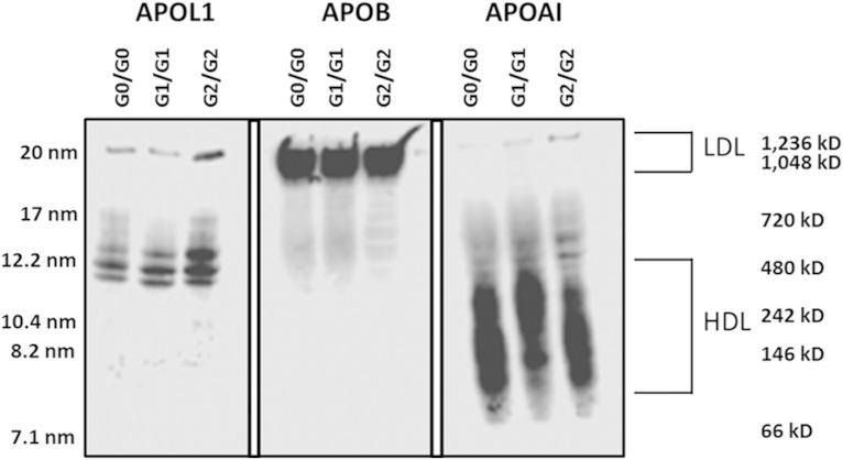
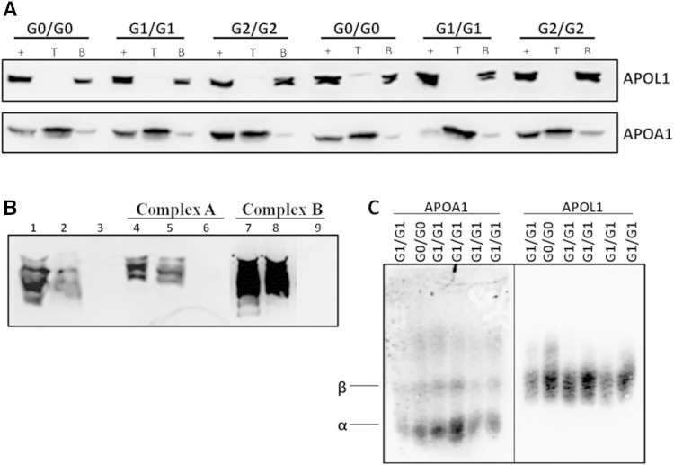
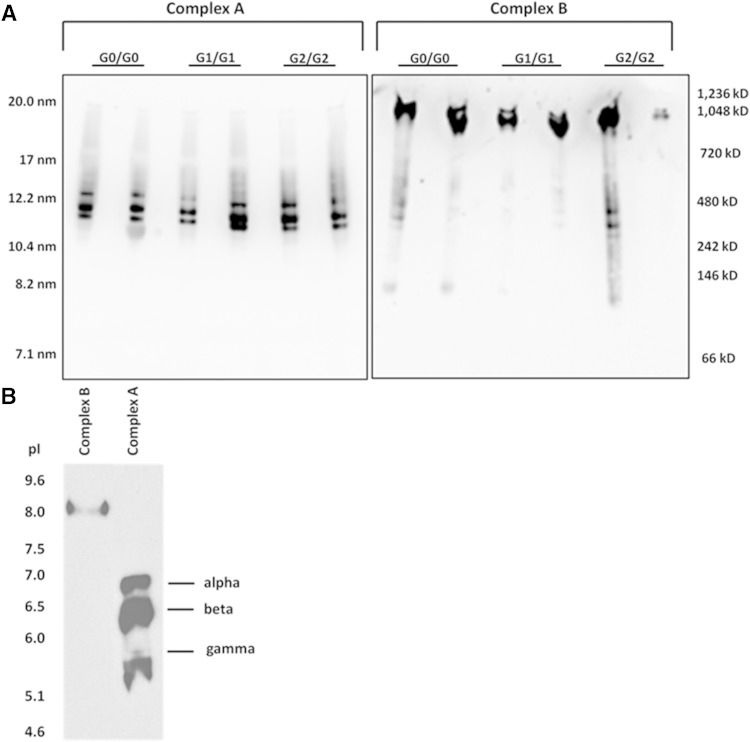
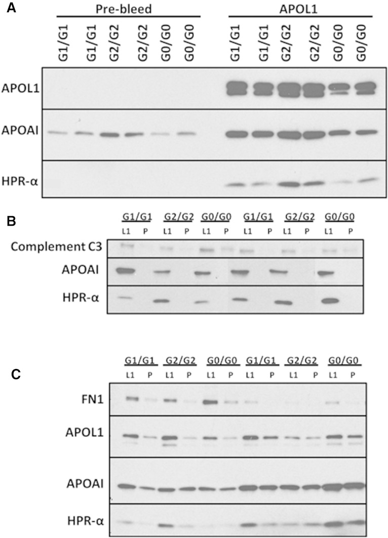
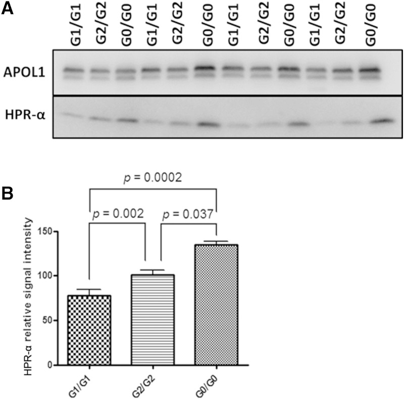
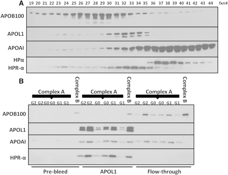
APOL1 gene renal-risk variants are associated with nephropathy and CVD in African Americans; however, little is known about the circulating APOL1 variant proteins which reportedly bind to HDL. We examined whether APOL1 G1 and G2 renal-risk variant serum concentrations or lipoprotein distributions differed from nonrisk G0 APOL1 in African Americans without nephropathy. Serum APOL1 protein concentrations were similar regardless of APOL1 genotype. In addition, serum APOL1 protein was bound to protein complexes in two nonoverlapping peaks, herein referred to as APOL1 complex A (12.2 nm diameter) and complex B (20.0 nm diameter). Neither of these protein complexes associated with HDL or LDL. Proteomic analysis revealed that complex A was composed of APOA1, haptoglobin-related protein (HPR), and complement C3, whereas complex B contained APOA1, HPR, IgM, and fibronectin. Serum HPR was less abundant on complex B in individuals with G1 and G2 renal-risk variant genotypes, relative to G0 (P = 0.0002-0.037). These circulating complexes may play roles in HDL metabolism and susceptibility to CVD.

Keywords: apolipoprotein L1; apolipoproteins; high density lipoprotein; kidney; proteomics; renal disease.


Figures

Fig. 1.
Fig. 1.
APOL1 serum concentration is variable and genotype independent. Representative immunoblots of serum samples from homozygous reference (G0/G0), homozygous G1 (G1/G1), homozygous G2 (G2/G2), heterozygous G1 (G1/G0), heterozygous G2 (G2/G0), and compound heterozygous (G1/G2) African Americans (n = 14 per genotype) compared with an APOL1-MBP fusion protein standard of known concentrations (left).
Fig. 2.
Fig. 2.
Apolipoprotein distribution among serum lipoprotein subfractions. One microliter of serum from homozygous APOL1 (G0, G1, and G2) individuals was frac­tionated on 4–20% nondenaturing gradient gels and subjected to immunoblot analysis for APOL1, APOB, and APOA1. The average hydrodynamic diameter of standard proteins is shown on the left side of the blot and the molecular mass range is shown on the right side along with the approximate size range for LDL and HDL.
Fig. 3.
Fig. 3.
APOL1 mainly elutes with particles similar in size to large HDL. Human serum was size-fractionated by FPLC and eluted fractions were monitored for TC) (A) and apolipoprotein distribution (B). A: TC distribution for representative serum sample for homozygous APOL1 (G0, G1, and G2) individuals. B: Individual FPLC fractions (fxn) were immunoblotted for APOB, APOL1, and APOA1. Two column regions enriched in APOL1 were identified; one containing most of the serum APOL1 (complex A; fractions 31–38) and a minor fraction (complex B; fractions 26–28).
Fig. 4.
Fig. 4.
APOL1 does not associate with HDL in human serum. A: African American serum was subjected to density ultracentrifugation at d = 1.225 g/ml. Top (T) and bottom (B) fractions were then analyzed by immunoblot to visualize APOL1 and APOA1. Immunoblots of bottom fractions indicate that most of the APOL1 in circulation does not associate with HDL, whereas APOA1 floats in the top fractions. The + represents serum that did not undergo density ultracentrifugation. Results are representative of homozygous APOL1 (G0, G1, and G2) individuals. B: Following ultracentrifugation, bottom fractions were size-fractionated by FPLC. Complex A, complex B, and the lipid-free fraction (fractions 45–48, Fig. 3) were pooled and separated by nondenaturing gradient gel electrophoresis to determine the elution position of APOL1. Lanes 1–3: serum was adjusted to d = 1.225 g/ml prior to FPLC fractionation (lane 1, starting serum; lanes 2 and 3, complex A and lipid-free fraction, respectively, from FPLC fractionation). Results demonstrate that complex A was not disrupted by 1.225 g/ml KBr. Lanes 4–6: bottom fraction (d > 1.225 g/ml) from ultracentrifugation of serum was size-fractionated by FPLC (lane 4, unfractionated bottom fraction; lanes 5 and 6, complex A and lipid-free fraction, respectively, from FPLC fractionation of bottom fraction). Lanes 7–9: bottom fraction (d > 1.225 g/ml) from ultracentrifugation of serum was size-fractionated by FPLC (lane 7, unfractionated bottom fraction; lanes 8 and 9, complex B and lipid-free fraction, respectively, from FPLC fractionation of bottom fraction). C: One microliter of human serum from African Americans homozygous for APOL1 (G0 and G1) was electrophoresed in 0.7% agarose and immunoblotted for APOA1 and APOL1 to examine apolipoprotein migration in agarose gels. APOL1 does not comigrate with α particles, which correspond to HDL.
Fig. 5.
Fig. 5.
Determining the composition of APOL1-containing complexes. Five FPLC fractions surrounding APOL1 complex A and complex B were pooled and concentrated. Samples were loaded onto 4–20% nondenaturing gradient gels (A) or IEF gels (B), followed by immunoblot analysis.
Fig. 6.
Fig. 6.
Confirmation of APOL1-associated protein components. A: Co-immunoprecipitation of APOL1 with protein components in whole serum using the N-terminal rabbit anti-APOL1 antibody (APOL1) and pre-bleed normal IgG (Pre-bleed). APOL1, APOA1, and HPR are confirmed to be present in co-immunoprecipitated APOL1 complexes. B: Co-immunoprecipitation of APOL1 in complex A FPLC peak fractions. APOA1, HPR, and complement C3 are confirmed to be present in co-immunoprecipitated APOL1 complex A. C: Co-immunoprecipitation of APOL1 in complex B FPLC peak fractions. Fibronectin, APOA1, and HPR are confirmed to be present in co-immunoprecipitated APOL1 complex. L1, anti-APOL1 antibody (Lampire); P, pre-bleed normal IgG from the same rabbit.
Fig. 7.
Fig. 7.
HPR-α is less abundant on complex B in APOL1 G1/G1 and G2/G2 individuals relative to G0/G0. A: APOL1 complex B peak fractions from AA individuals with different APOL1 genotypes (G1/G1, G2/G2, and G0/G0) were first quantified for APOL1 concentration by denatured immunoblot using Epitomics anti-APOL1 antibody and comparison with a series of APOL1 fusion proteins with known concentrations (see Fig. 1). Each well contains 10 ng APOL1. Samples were subjected to 4–20% SDS-PAGE and immunoblotting using specific antibody to APOL1 (Lampire anti-APOL1 polyclonal antibody) and HPR. The signal intensity of APOL1 was similar among complex B samples of different APOL1 genotypes. However, the relative amount of HPR-α residing on APOL1 complex B appeared to be less abundant for homozygous G1 carriers. B: Comparison of the relative signal intensity of HPR-α on serum APOL1 complex B containing the same amount of APOL1 (10 µg) of different genotypes (G1/G1, G2/G2, G0/G0), n = 3 each. P values were determined by t-test (2-sided).
Fig. 8.
Fig. 8.
HPR-α distribution is consistent with that of APOL1 following FPLC. A: FPLC fractions (fxn#) were subjected to 4–20% SDS-PAGE and probed with APOB, APOA1, APOL1, and HPR antibodies via immuno­blot analysis. APOA1 and APOB100 peak fractions represent the enrichment of HDL and LDL, respectively. The peak fractions for APOL1 and HPR-α are consistent, indicating the positions of complexes A and B. B: APOL1 complex A peak fractions from G1/G1, G2/G2, and G0/G0 individuals and one sample of peak APOL1 complex B from a G0/G0 subject were co-immunoprecipitated with rabbit anti-N-terminal APOL1 antibody (APOL1) or pre-bleed IgG from the same rabbit (Pre-bleed). APOL1 and HPR-α only appeared in co-immunoprecipitated APOL1, but not in the supernatant (Flow-through), indicating the coexistence of APOL1 and HPR in serum complexes A and B. The absence of APOL1 and HPR in the APOL1 supernatant suggests that anti-APOL1 IgG was sufficient to precipitate all APOL1 in the sample. The absence of APOL1, APOA1, and HPR in the pre-bleed rabbit IgG co-immunoprecipitation indicates the specificity of the antibodies. Because APOB is enriched in complex B fractions and APOB also appeared in the pre-bleed rabbit co-immunoprecipitation, it is undetermined whether APOB is specifically present on complex B.
Fig. 9.
Fig. 9.
Estimate of potential surface epitope conformational change of APOL1 protein caused by G1 and G2 variants. APOL1 complex B peak fractions containing 10 ng of APOL1 (quantified by Epitomics anti-APOL1 antibody shown in Fig. 7) were run on a 4–20% nondenaturing gradient gel at 150 V for 1 h at room temperature, transferred to a cellulose membrane, and probed with the same Epitomics anti-APOL1 antibody (immunogen within amino acids 300-330) via immunoblot. APOL1 signal intensity was significantly higher for complex B in G1/G1 and G2/G2 individuals compared with G0/G0, n = 3/genotype.

References

    1. Genovese G., Friedman D. J., Ross M. D., Lecordier L., Uzureau P., Freedman B. I., Bowden D. W., Langefeld C. D., Oleksyk T. K., Uscinski Knob A. L., et al. . 2010. Association of trypanolytic ApoL1 variants with kidney disease in African Americans. Science. 329: 841–845. - PMC - PubMed
    1. Tzur S., Rosset S., Shemer R., Yudkovsky G., Selig S., Tarekegn A., Bekele E., Bradman N., Wasser W. G., Behar D. M., et al. . 2010. Missense mutations in the APOL1 gene are highly associated with end stage kidney disease risk previously attributed to the MYH9 gene. Hum. Genet. 128: 345–350. - PMC - PubMed
    1. Freedman B. I., Kopp J. B., Langefeld C. D., Genovese G., Friedman D. J., Nelson G. W., Winkler C. A., Bowden D. W., and Pollak M. R.. 2010. The apolipoprotein L1 (APOL1) gene and nondiabetic nephropathy in African Americans. J. Am. Soc. Nephrol. 21: 1422–1426. - PMC - PubMed
    1. Madhavan S. M., O’Toole J. F., Konieczkowski M., Ganesan S., Bruggeman L. A., and Sedor J. R.. 2011. APOL1 localization in normal kidney and nondiabetic kidney disease. J. Am. Soc. Nephrol. 22: 2119–2128. - PMC - PubMed
    1. Ma L., Shelness G. S., Snipes J. A., Murea M., Antinozzi P. A., Cheng D., Saleem M. A., Satchell S. C., Banas B., Mathieson P. W., et al. . 2015. Localization of APOL1 protein and mRNA in the human kidney: nondiseased tissue, primary cells, and immortalized cell lines. J. Am. Soc. Nephrol. 26: 339–348. - PMC - PubMed

Publication types

MeSH terms