. 2016 Feb;139(Pt 2):547-62.
doi: 10.1093/brain/awv338. Epub 2015 Nov 19.

Cascading network failure across the Alzheimer's disease spectrum

Affiliations

Cascading network failure across the Alzheimer's disease spectrum

David T Jones et al. Brain. 2016 Feb.

Abstract

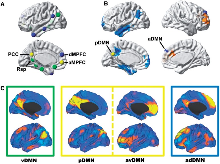
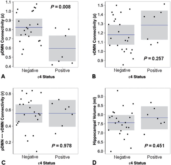
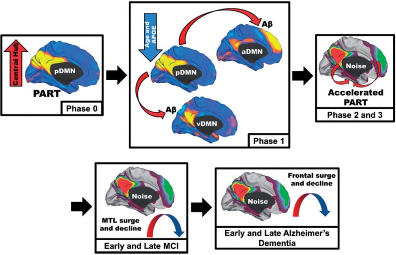
Complex biological systems are organized across various spatiotemporal scales with particular scientific disciplines dedicated to the study of each scale (e.g. genetics, molecular biology and cognitive neuroscience). When considering disease pathophysiology, one must contemplate the scale at which the disease process is being observed and how these processes impact other levels of organization. Historically Alzheimer's disease has been viewed as a disease of abnormally aggregated proteins by pathologists and molecular biologists and a disease of clinical symptoms by neurologists and psychologists. Bridging the divide between these scales has been elusive, but the study of brain networks appears to be a pivotal inroad to accomplish this task. In this study, we were guided by an emerging systems-based conceptualization of Alzheimer's disease and investigated changes in brain networks across the disease spectrum. The default mode network has distinct subsystems with unique functional-anatomic connectivity, cognitive associations, and responses to Alzheimer's pathophysiology. These distinctions provide a window into the systems-level pathophysiology of Alzheimer's disease. Using clinical phenotyping, metadata, and multimodal neuroimaging data from the Alzheimer's Disease Neuroimaging Initiative, we characterized the pattern of default mode network subsystem connectivity changes across the entire disease spectrum (n = 128). The two main findings of this paper are (i) the posterior default mode network fails before measurable amyloid plaques and appears to initiate a connectivity cascade that continues throughout the disease spectrum; and (ii) high connectivity between the posterior default mode network and hubs of high connectivity (many located in the frontal lobe) is associated with amyloid accumulation. These findings support a system model best characterized by a cascading network failure--analogous to cascading failures seen in power grids triggered by local overloads proliferating to downstream nodes eventually leading to widespread power outages, or systems failures. The failure begins in the posterior default mode network, which then shifts processing burden to other systems containing prominent connectivity hubs. This model predicts a connectivity 'overload' that precedes structural and functional declines and recasts the interpretation of high connectivity from that of a positive compensatory phenomenon to that of a load-shifting process transiently serving a compensatory role. It is unknown whether this systems-level pathophysiology is the inciting event driving downstream molecular events related to synaptic activity embedded in these systems. Possible interpretations include that the molecular-level events drive the network failure, a pathological interaction between the network-level and the molecular-level, or other upstream factors are driving both.

Keywords: Alzheimer’s disease; cascading failure; complex systems; default mode network; pathophysiology.


Figures

None
Subsystems within the default mode network are differentially affected in Alzheimer’s disease. Jones et al. present an in-depth analysis of changes within these subsystems and relate them to biomarker profiles across the Alzheimer’s disease spectrum. Results support a cascading network failure model of Alzheimer’s disease.
Figure 1
Figure 1
Subsystems of the DMN have distinct anatomical, functional, and pathophysiological profiles and can be isolated from one another in task-free functional MRI data. (A) Nodes within the DMN segregate into distinct subsystems, comprised of midline core regions (yellow), medial temporal lobe memory system (green), and a dorsal medial prefrontal cortex system (blue)—the regions and groupings were derived from Andrews-Hanna et al. (2010) and displayed on a cortical surface. (B) Seed-based analysis of posterior (left hemisphere) and anterior (right hemisphere) DMNs showing the divergent patterns of Alzheimer’s disease dementia-related changes in connectivity in these subsystems (decreased in blue and increased in orange)—modified from Jones et al. (2011). (C) These same subsystems were identified in a high-dimensional GICA of intrinsic connectivity networks in a large (n = 892) population-based sampling of cognitively normal elderly participants in the Mayo Clinic Study of Aging (Jones et al., 2012). The caret software package (Van Essen, 2005) was used to display these four independent components (i.e. ventral, posterior, anterior ventral, and anterior dorsal DMN) from this GICA analysis on brain surface renderings. Boxes around each of these renderings are colour coded to correspond to the subsystems nodes identified in A and B. aMPFC = anterior medial prefrontal cortex; dMPFC = dorsal medial prefrontal cortex; GICA = group independent component analysis; PCC = posterior cingulate cortex; Rsp = retrosplenial cingulate.
Figure 2
Figure 2
Connectivity measured during the ‘resting state’ is associated with out-of-scanner memory performance in cognitively normal participants (n = 43). The within-subsystem connectivity for each of the four DMN subsystems is plotted versus Rey Auditory Verbal Learning Test delayed recall (AVLT-DR) performance (A–D) and for ventral DMN hippocampal connectivity (Hc) versus AVLT-DR (E and F). Individual data points (black circles), regression lines (blue), and 95% confidence intervals (grey bands) are displayed for linear regressions (A–E) and for the second order natural spline non-linear regression (F).
Figure 3
Figure 3
DMN subsystem elements have distinct functional forms in their association with progression along the Alzheimer’s disease spectrum. The within-subsystem connectivity for the posterior DMN (A), ventral DMN (B), and anterior ventral DMN (C) is plotted versus raw score on the 13-item Alzheimer’s Disease Assessment Scale-cognitive subscale within generalized additive models controlling for motion, age, gender, and APOE ε4. The medial surface topography for within-subsystem connectivity is inset for A–C. Similar plots are made for significant between-subsystem elements (D and E) and ventral DMN to hippocampal connectivity (F). The deviance explained by the models and P-value for each element displayed is inset. Individual data points (black circles), regression lines (blue), and 95% CIs (grey bands) are displayed. Identical functional forms were obtained replacing 13-item Alzheimer’s Disease Assessment Scale-cognitive subscale with the Clinical Dementia Rating Scale-sum of boxes (data not shown).
Figure 4
Figure 4
Incorporating network changes into models of the molecular- and cellular-level changes across the Alzheimer’s disease spectrum. Amyloid-PET (A) and hippocampal volume (B) are plotted versus raw score on the 13-item Alzheimer’s Disease Assessment Scale-cognitive subscale within generalized additive models controlling for age, gender, APOE ε4, and total intracranial volume (hippocampal volume only). The deviance explained by the models and P-value for each element displayed is inset. Individual data points (black circles), regression lines (blue), and 95% CIs (grey bands) are displayed. The same analysis performed for network elements (blue, gold, and red solid lines for the posterior DMN, posterior to ventral DMN connection, and ventral DMN to hippocampal connection, respectively) are plotted with amyloid-PET (purple dotted line) and hippocampal volume (green dotted line) (C). For clarity, the 95% CIs are omitted, but can be inspected in the plots for each element individually (see Figs 3A, E, F, 4A and B).
Figure 5
Figure 5
The effect of APOE ε4 carriage in cognitively normal subjects without evidence of amyloid plaques. The response variable is plotted with mean and 95% CIs by APOE ε4 status holding all other variable constant including age, gender, motion (A–C only) and total intracranial volume (D only).
Figure 6
Figure 6
Schematic of the proposed cascading network failure model of Alzheimer’s disease. Phase 0: The posterior DMN (pDMN) serves as the central hub processing and integrating association cortices and is highly metabolically active. Independently, the medial temporal lobe (MTL) has accumulated age-related damage from neocortical processing of a different kind (pattern separation and completion) contributing to primary age-related tauopathy (PART) in these regions. Phase 1: The declining posterior DMN (Fig. 3A), more prominent with advancing age and APOE ε4 carriage, transfers information processing duties (or starts passing noisy signals) to the neocortical regions including the ventral DMN (Fig. 3D) and/or the anterior dorsal DMN (Fig. 3E). Aberrant between-neocortical network synaptic activity leads to dysregulated amyloid precursor protein (APP) processing promoting amyloid-β (Aβ) plaque formation in neocortical layers. Phase 2: Given that the hippocampus is continually processing information from these same regions, noise in these cortical systems is propagated down to the hippocampus. This increased burden on the hippocampus accelerates the pre-existing PART. Phase 3: Neurodegeneration expands to adjacent systems. This creates a detrimental positive feedback loop because degeneration lowers the noise handling capacity of the system leading to further degeneration. MCI Phase: Posterior brain regions supporting memory succumb to the degenerative feedback loop as hippocampal regions increase processing (Fig. 3F). Later the frontal brain regions begin to bear the high connectivity burden (Fig. 3C). Early Alzheimer’s disease phase: The high frontal connectivity firmly establishes the neurodegenerative feed-back loop in these systems before declining as Alzheimer’s disease progresses.

References

    1. Agosta F, Pievani M, Geroldi C, Copetti M, Frisoni GB, Filippi M. Resting state fMRI in Alzheimer's disease: beyond the default mode network. Neurobiol Aging 2012; 33: 1564–78. - PubMed
    1. Andrews-Hanna JR, Reidler JS, Sepulcre J, Poulin R, Buckner RL. Functional-anatomic fractionation of the brain's default network. Neuron 2010; 65: 550–62. - PMC - PubMed
    1. Ashburner J. A fast diffeomorphic image registration algorithm. Neuroimage 2007; 38: 95–113. - PubMed
    1. Behzadi Y, Restom K, Liau J, Liu TT. A component based noise correction method (CompCor) for BOLD and perfusion based fMRI. Neuroimage 2007; 37: 90–101. - PMC - PubMed
    1. Braak H, Braak E. Neuropathological stageing of Alzheimer-related changes. Acta Neuropathol 1991; 82: 239–59. - PubMed

Publication types