. 2016 Jan;15(1):48-59.
doi: 10.1158/1535-7163.MCT-15-0527. Epub 2015 Nov 19.

Identification of Selective Lead Compounds for Treatment of High-Ploidy Breast Cancer

Affiliations

Identification of Selective Lead Compounds for Treatment of High-Ploidy Breast Cancer

Alka Choudhary et al. Mol Cancer Ther. 2016 Jan.

Abstract

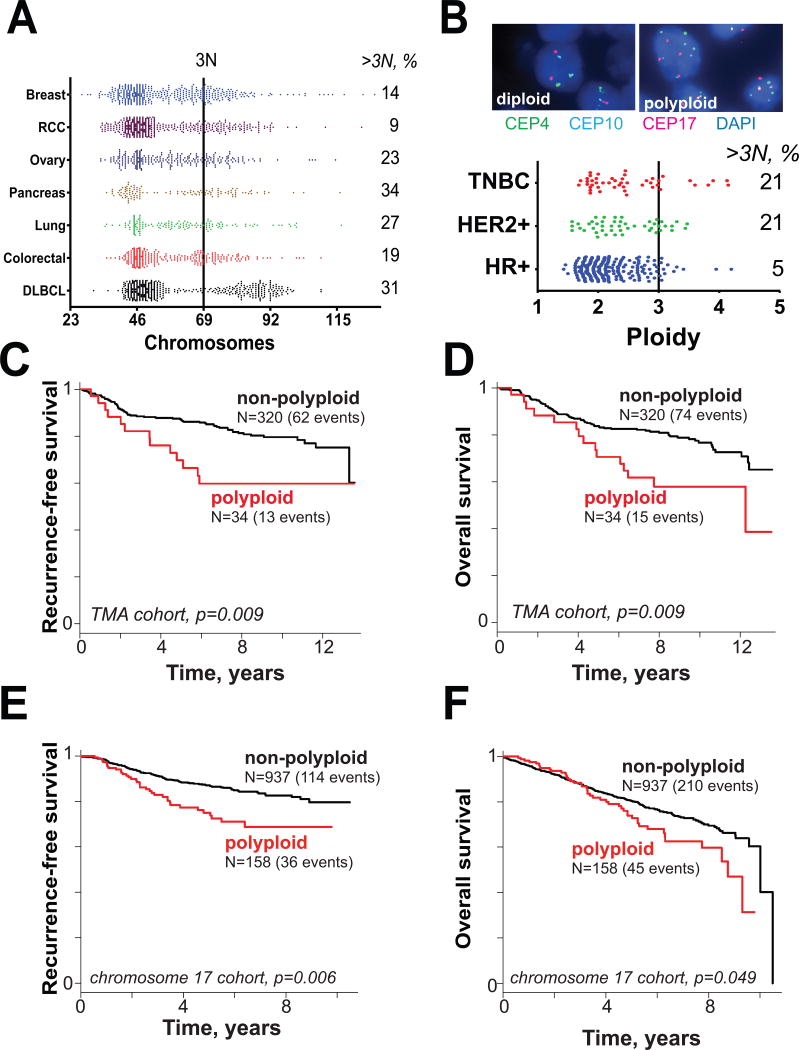
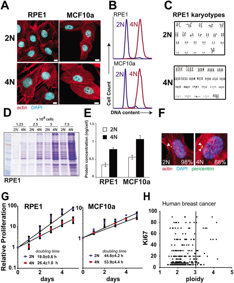
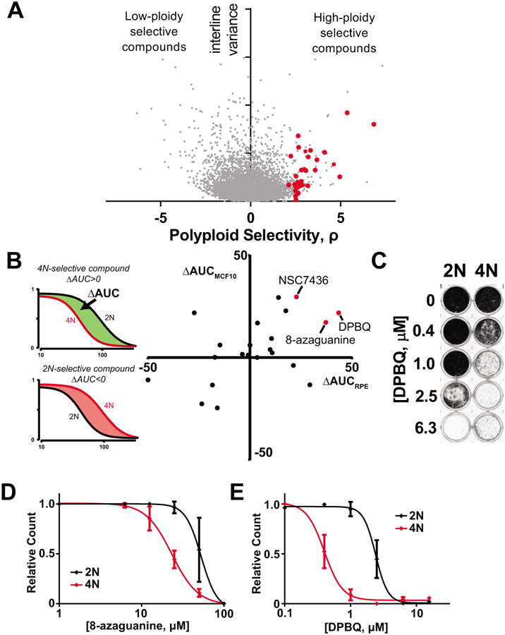
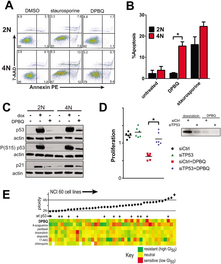
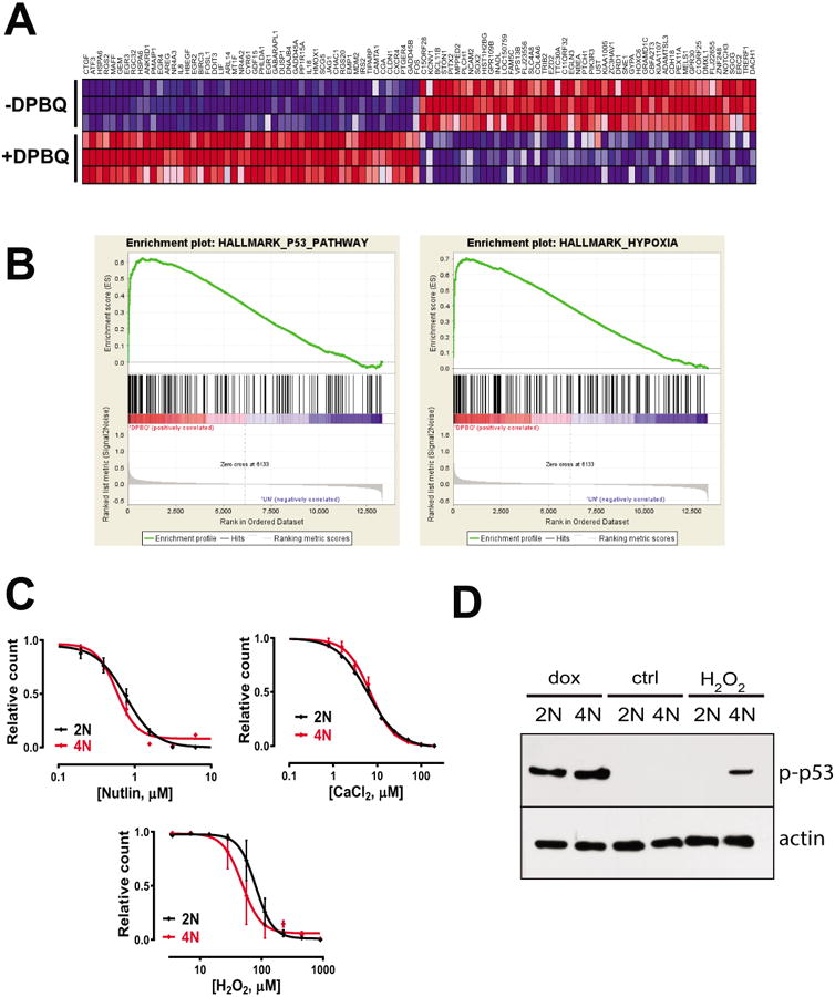
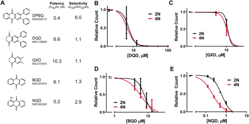
Increased ploidy is common in tumors but treatments for tumors with excess chromosome sets are not available. Here, we characterize high-ploidy breast cancers and identify potential anticancer compounds selective for the high-ploidy state. Among 354 human breast cancers, 10% have mean chromosome copy number exceeding 3, and this is most common in triple-negative and HER2-positive types. Women with high-ploidy breast cancers have higher risk of recurrence and death in two patient cohorts, demonstrating that it represents an important group for improved treatment. Because high-ploidy cancers are aneuploid, rather than triploid or tetraploid, we devised a two-step screen to identify selective compounds. The screen was designed to assure both external validity on diverse karyotypic backgrounds and specificity for high-ploidy cell types. This screen identified novel therapies specific to high-ploidy cells. First, we discovered 8-azaguanine, an antimetabolite that is activated by hypoxanthine phosphoribosyltransferase 1 (HPRT1), suggesting an elevated gene-dosage of HPRT1 in high-ploidy tumors can control sensitivity to this drug. Second, we discovered a novel compound, 2,3-diphenylbenzo[g]quinoxaline-5,10-dione (DPBQ). DPBQ activates p53 and triggers apoptosis in a polyploid-specific manner, but does not inhibit topoisomerase or bind DNA. Mechanistic analysis demonstrates that DPBQ elicits a hypoxia gene signature and its effect is replicated, in part, by enhancing oxidative stress. Structure-function analysis defines the core benzo[g]quinoxaline-5,10 dione as being necessary for the polyploid-specific effects of DPBQ. We conclude that polyploid breast cancers represent a high-risk subgroup and that DPBQ provides a functional core to develop polyploid-selective therapy. Mol Cancer Ther; 15(1); 48-59. ©2015 AACR.


Conflict of interest statement

Conflict of interests: The authors declare that they have no competing interests.

Figures

Figure 1
Figure 1
Polyploidy is a feature of many cancers. A. Chromosome number in multiple cancer types from Mitelman database. RCC = renal cell carcinoma; DLBCL = diffuse large B-cell lymphoma. B. (top) Representative 3-color centromeric FISH images from breast cancer tissue microarray analysis of 354 distinct breast cancers. A total of 6 chromosomes were sampled by centromeric probes (4, 10, 17 on one section and 3, 7, 9 on a separate section). (bottom) Mean ploidy status by 6-chromosome sampling of 354 breast cancers by subtype. C-F. Kaplan-Meier survival curves for polyploid versus non-polyploid cancers. C. Recurrence-free survival for tissue microarray (TMA) cohort where polyploid tumors are defined as those that have mean ≥3 centromere signals per cell among 6 centrosomes counted. D. Overall survival for TMA cohort. E. Relapse-free survival in single-chromosome FISH cohort where polyploid tumors are those defined as those that have mean ≥3 centromere 17 per cell. F. Overall survival for single-chromosome FISH cohort.
Figure 2
Figure 2
Matched pairs of diploid-polyploid cells were generated from epithelial breast cell types. A. Representative images of diploid (2N) and polyploid (4N) cells derived from RPE1 and MCF10a cell lines. Scale bar = 10 μm. B. Flow cytometry demonstrating double DNA content of tetraploid cells after staining with propidium iodide. C. Karyotype of diploid and polyploid matched RPE1 cells. D-E. Protein levels are higher in tetraploid cells relative to diploid as determined by coomassie staining of extracts (D) and Bradford Assay (E). F. Polyploid RPE1 cells have supernumerary centrosomes. Cells were synchronized using aphidicolin and stained. Pericentrin foci are marked with arrows and percentage of cells that harbor the indicated number of centrosomes is indicated (n>40). G. 4N cells proliferate more slowly than 2N cells. Cell number was quantified in proliferating 2N and 4N cells and doubling time was calculated. H. High ploidy in human breast cancer samples does not correlate strongly with the Ki67 proliferation marker.
Figure 3
Figure 3
Discovery of DPBQ and 8-azaguanine as polyploid-specific disruptors of proliferation. A. Correlation with drug sensitivity and cell ploidy from NCI-60 data of over 45,342 chemicals across ∼60 cell lines. Polyploid selectivity, ρ, is plotted against interline variance for each chemical. Red circles indicate chemicals obtained for secondary screens. B. Secondary screen to test selectivity of hits in matched pairs of diploid and polyploid RPE1 and MCF10a cells. Red circles indicate hits that are polyploid selective in both MCF10a and RPE1 cell types. C. DPBQ 8-day proliferation assay in RPE1 cells. Crystal violet staining marks viable cells remaining. D-E. Quantification of proliferation assay as in C. plotting absorbance for 8-azaguanine (D) and DPBQ (E) in RPE1. n=3, SD shown.
Figure 4
Figure 4
Mechanism of DPBQ. A-B. DPBQ elicits polyploid-specific apoptosis. A. Apoptosis by representative Annexin assay. B. Averaged apoptosis (early and late) for n=3 assays, SD shown. *p<0.05 by T-test. C. 1 μM DPBQ elicits 4N-specific p53 induction and activation; dox=doxorubicin. D. p53 is required for the DPBQ effect. 4N RPE1 cells were transfected with siRNA against p53 (siTP53) or control (siCtrl) and then exposed to DPBQ or vehicle. DPBQ restrained prolilferation only when p53 was present (red). Right: blot demonstrating suppression of phospho(S15)-p53 with knockdown. *p<0.05 by T-test. E. Among NCI-60 lines, DPBQ has its strongest effects against polyploid cell lines that express wildtype p53.
Figure 5
Figure 5
Evaluation of DPBQ mechanism by gene expression analysis. A. Top gene expression altered by DPBQ is shown, both upregulated (red) and downregulated (blue) in 3 technical replicates. B. Gene set enrichment analysis showing strong enrichment of hallmark pathways for p53 and hypoxia. C. Concentration-response curves for nutlin, CaCl2, and H2O2 on diploid and tetraploid cells. D. Expression of phospho-P53 (top) and actin after treatment with doxorubicin, control, or H2O2.
Figure 6
Figure 6
Structure-function analysis of DPBQ. A. DPBQ-like molecules were tested for potency and specificity for in paired diploid-polyploid RPE1 cells. B-E. Concentration response curves for DPBQ-like molecules for 8-day proliferation assays, as in Fig. 3C-E. n=2, SD shown.

References

    1. Storchova Z, Kuffer C. The consequences of tetraploidy and aneuploidy. J Cell Sci. 2008;121:3859–66. - PubMed
    1. Storchova Z, Pellman D. From polyploidy to aneuploidy, genome instability and cancer. Nat Rev Mol Cell Biol. 2004;5:45–54. - PubMed
    1. Janssen A, Kops GJ, Medema RH. Elevating the frequency of chromosome mis-segregation as a strategy to kill tumor cells. Proc Natl Acad Sci U S A. 2009;106:19108–13. - PMC - PubMed
    1. Lissa D, Senovilla L, Rello-Varona S, Vitale I, Michaud M, Pietrocola F, et al. Resveratrol and aspirin eliminate tetraploid cells for anticancer chemoprevention. Proc Natl Acad Sci U S A. 2014;111:3020–5. - PMC - PubMed
    1. Tang YC, Williams BR, Siegel JJ, Amon A. Identification of aneuploidy-selective antiproliferation compounds. Cell. 2011;144:499–512. - PMC - PubMed

Publication types

MeSH terms