. 2016 Jan 22;291(4):1763-1773.
doi: 10.1074/jbc.M115.688002. Epub 2015 Nov 19.

The Relay/Converter Interface Influences Hydrolysis of ATP by Skeletal Muscle Myosin II

Affiliations

The Relay/Converter Interface Influences Hydrolysis of ATP by Skeletal Muscle Myosin II

Marieke J Bloemink et al. J Biol Chem. .

Abstract

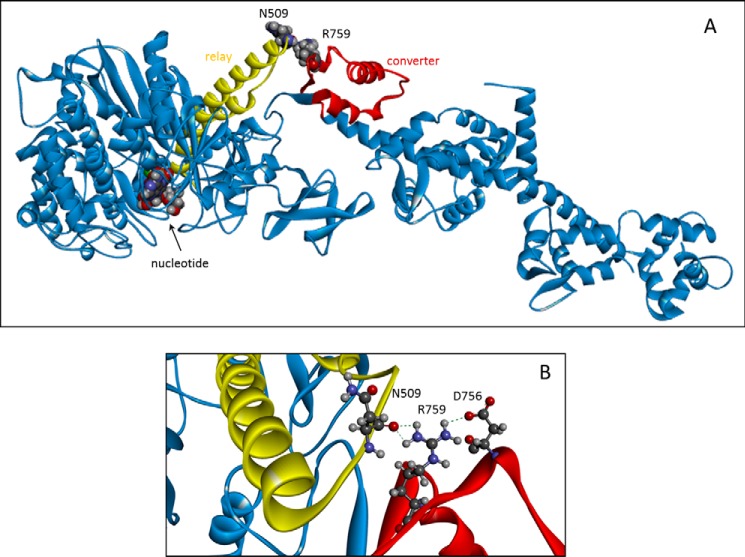
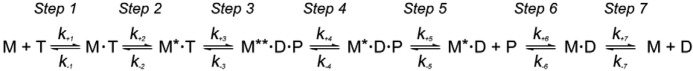
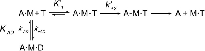
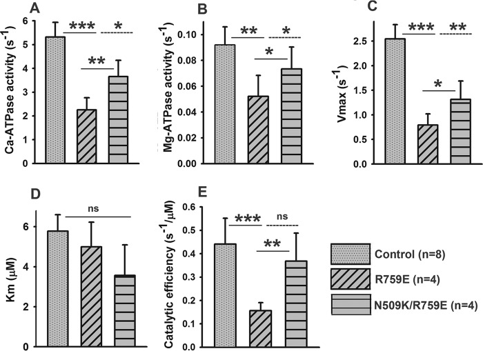
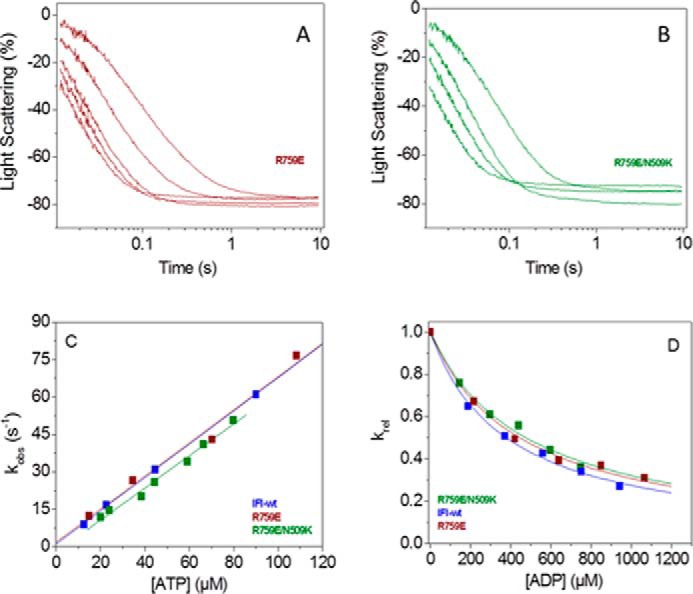
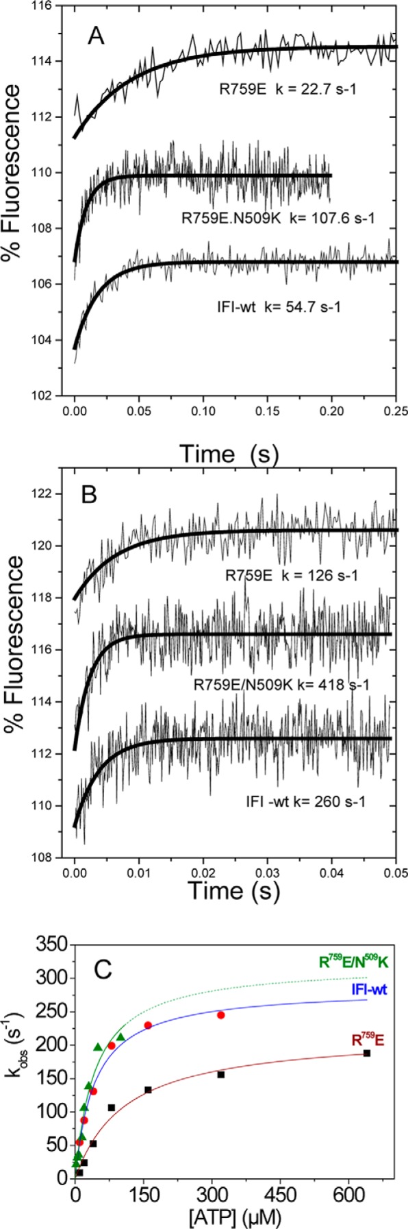
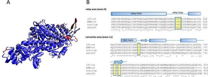
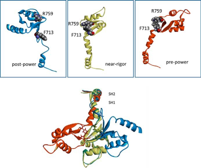
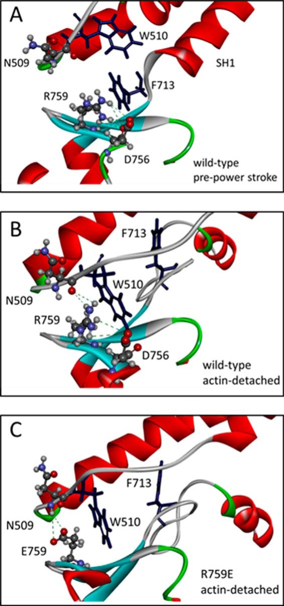
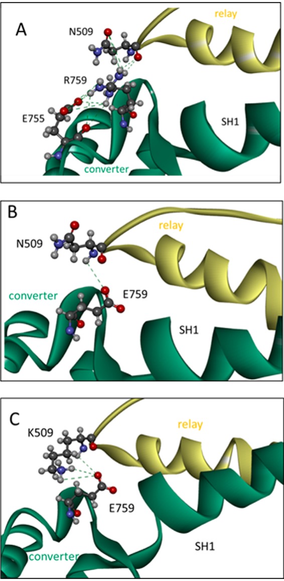
The interface between relay and converter domain of muscle myosin is critical for optimal myosin performance. Using Drosophila melanogaster indirect flight muscle S1, we performed a kinetic analysis of the effect of mutations in the converter and relay domain. Introduction of a mutation (R759E) in the converter domain inhibits the steady-state ATPase of myosin S1, whereas an additional mutation in the relay domain (N509K) is able to restore the ATPase toward wild-type values. The R759E S1 construct showed little effect on most steps of the actomyosin ATPase cycle. The exception was a 25-30% reduction in the rate constant of the hydrolysis step, the step coupled to the cross-bridge recovery stroke that involves a change in conformation at the relay/converter domain interface. Significantly, the double mutant restored the hydrolysis step to values similar to the wild-type myosin. Modeling the relay/converter interface suggests a possible interaction between converter residue 759 and relay residue 509 in the actin-detached conformation, which is lost in R759E but is restored in N509K/R759E. This detailed kinetic analysis of Drosophila myosin carrying the R759E mutation shows that the interface between the relay loop and converter domain is important for fine-tuning myosin kinetics, in particular ATP binding and hydrolysis.

Keywords: actin; fluorescence; homology modeling; kinetics; muscle; myosin; protein structure-function; sequence alignment.


Figures

FIGURE 1.
FIGURE 1.
Overview of myosin S1 with relay area (yellow) and converter domain (red) indicated. A, residues 509 (relay loop) and 759 (converter) are shown as space-filling models. The nucleotide is also shown at the opposite end of the relay helix. B, close-up of the interface between the relay loop and converter domain with converter residue Arg-759 interacting with variable relay loop residue Asn-509 and converter residue Asp-756. Homology model of the Drosophila IFI myosin was built using the coordinates of PDB 1KK8 (actin-detached post power-stroke state) as a template.
SCHEME 1.
SCHEME 1.
Interaction of S1 with ATP and ADP. S1, ATP, and ADP are represented as M, T, and D, respectively. * indicates the different levels of tryptophan fluorescence and represents different conformational states of the myosin.
SCHEME 2.
SCHEME 2.
Interaction of S1 with actin, ATP, and ADP. Myosin, actin, ATP, and ADP are represented as M, A, T, and D, respectively. Dashed interactions represent a weakly bound complex, and dotted interactions represent strongly bound states. Cross-bridge detachment from the rigor state (A·M) involves the complex binding ATP, governed by the association constant, K′1, followed by the rate-limiting isomerization, k′+2, after which actin-myosin affinity becomes weak, and the complex separates rapidly.
FIGURE 2.
FIGURE 2.
Steady-state ATPase activity of wild-type control, single mutant R759E, and double mutant R759E/N509K Drosophila myosin. Basal Ca2+-ATPase activity (A), basal Mg2+-ATPase activity (B), actin-stimulated Mg2+-ATPase activity Vmax (C), actin affinity relative to Mg2+-ATPase (Km) (D), and catalytic efficiency (E) were determined as described under “Experimental Procedures.” Notations above histograms indicate the level of statistically significant differences (*, p < 0.05; **, p < 0.01; ***, p < 0.001; ns, not statistically significant). Significant differences were assumed for p < 0.05.
FIGURE 3.
FIGURE 3.
Summary of transient kinetics measurements of converter mutant and suppressor. A and B, example of light scattering traces measured for R759E (A) and R759E/N509K (B) using flash photolysis and fitted to single exponentials, from which the rate constant kobs is determined. C, kobs as a function of [ATP] yields the actomyosin dissociation constant K1k+2, which is not significantly different for R759E and R759E/N509K compared with wild type. D, ATP-induced dissociation of acto-S1 with increasing [ADP] shows similar ADP affinity (KAD) for R759E and R759E/N509K compared with IFI-WT.
FIGURE 4.
FIGURE 4.
ATP binding to Drosophila S1. Intrinsic fluorescence transients observed for ATP binding to wild-type myosin S1 and converter mutants R759E and R759E/N509K S1 using stopped flow. 50 nm S1 is rapidly mixed with 10 μm ATP (A) or 320 μm ATP (IFI-WT and R759E) or 160 μm ATP (R759E/N509K) (B). Note that the amplitudes are small (2–3%) and evaluation of kobs values above 200 s−1 becomes unreliable. C, summary of stopped-flow data: kobs as a function of [ATP] yields kmax = 223 s−1 (R759E), 322 s−1 (R759E/N509K), and 286 s−1 (IFI-wt) for a single data set measured for wild-type S1, R759E, and R759E/N509K.
FIGURE 5.
FIGURE 5.
Myosin head domains of scallop and Drosophila are very similar. A, overlay of crystal structures of myosin head domains of scallop (1RS6) and Drosophila embryonic myosin (4QBD). The coloring represents the r.m.s.d. values between the two structures with similar structural elements shown in blue and deviating structural elements (large r.m.s.d. values) shown in red. The backbone of the two structures overlay very well with an average r.m.s.d. < 1.8 Å (shown in blue). Flexible loops that diverge somewhat are shown in red. B, alignments for Drosophila (IFI and EMB), scallop, and chicken smooth myosin. The conserved residues Arg-759 and Phe-713 in the converter area (bottom) are highlighted in yellow. Note that in regard to the variable residues surrounding the conserved tryptophan (yellow box in the relay loop) the Asn-509 residue (Drosophila IFI) is replaced by alanine (scallop) and glutamate (chicken smooth).
FIGURE 6.
FIGURE 6.
Interaction between converter residue Arg-759 and SH1 helix residue Phe-713. Top panels, homology models of IFI-WT indicate a strong interaction between Arg-759 and Phe-713 in near-rigor (yellow) and pre-power stroke state (red), whereas in the post-power stroke state (blue) this interaction is not seen, as SH1 has become disordered. Bottom panel, overlay of the models for IFI-WT myosin showing the SH1/SH2 and converter area for different myosin conformations. The SH2 area overlays very well but structures start to divert in SH1 area. Residue Arg-759 is indicated for each conformation, together with Phe-713. Homology models of the Drosophila IFI myosin isoform were built using the coordinates of scallop myosin PDB 1QVI (pre-power stroke state), PDB 1SR6 (near-rigor state), and PDB 1KK8 (actin-detached post power stroke state) as a templates.
FIGURE 7.
FIGURE 7.
Details of the relay-converter interaction. A, Arg-759 of wild-type IFI in the pre-power stroke state cannot make contacts with relay loop residues and makes contacts with converter residues (Pro-755/Asp-756) and SH1 residue Phe-713. B, in the post-power stroke state (actin-detached), Arg-759 contacts relay residue Asn-509 and Trp-510, in addition to converter residues (Pro-755/Asp-756). Phe-713 has moved away after SH1 helix structure is lost. C, R759E mutant residue is able to contact relay loop residue Asn-509 as well in the post-power stroke/actin-detached state. However, additional contacts with converter residues Pro-755/Asp-756 are missing. Homology models were built using scallop myosin PDB 1QVI (pre-power stroke) and PDB 1KK8 (actin-detached, post power stroke state) as a templates.
FIGURE 8.
FIGURE 8.
Side chain interactions between converter residue 759 and relay loop residue 509 for wild-type IFI (A), R759E (B) and N509K/R759E (C). A, close-up of the interface between the relay loop and converter domain with converter residue Arg-759 interacting with the side chain of residue Asn-509, via H-bonds, and converter residue Glu-755 via a salt bridge. B, close-up of the interface between the relay loop and converter domain of R759E S1 mutant. The converter residue Glu-759 only forms H-bonds with backbone NH of Asn-509. C, close-up of the interface between the relay loop and converter domain of N509K/R759E S1 mutant. The interaction between the side chains of residues 759 and 509 is restored as the side chains are close enough to form hydrogen bonds or a salt bridge. (Homology model was built using the coordinates of chicken smooth muscle myosin in the pre-power stroke state (PDB 3J04) as a template.)

References

    1. Odronitz F., and Kollmar M. (2007) Drawing the tree of eukaryotic life based on the analysis of 2,269 manually annotated myosins from 328 species. Genome Biol. 8, R196. - PMC - PubMed
    1. O'Connell C. B., Tyska M. J., and Mooseker M. S. (2007) Myosin at work: motor adaptations for a variety of cellular functions. Biochim. Biophys. Acta 1773, 615–630 - PubMed
    1. Reggiani C., Bottinelli R., and Stienen G. J. (2000) Sarcomeric myosin isoforms: fine tuning of a molecular motor. News Physiol. Sci. 15, 26–33 - PubMed
    1. Bloemink M. J., and Geeves M. A. (2011) Shaking the myosin family tree: biochemical kinetics defines four types of myosin motor. Semin. Cell Dev. Biol. 22, 961–967 - PMC - PubMed
    1. Geeves M. A., Fedorov R., and Manstein D. J. (2005) Molecular mechanism of actomyosin-based motility. Cell. Mol. Life Sci. 62, 1462–1477 - PMC - PubMed

Publication types

MeSH terms

LinkOut - more resources