. 2015 Nov;22(6):760-72.
doi: 10.1016/j.sjbs.2015.03.005. Epub 2015 Mar 22.

Regulation of ceramide channel formation and disassembly: Insights on the initiation of apoptosis

Affiliations

Regulation of ceramide channel formation and disassembly: Insights on the initiation of apoptosis

Majdouline Abou-Ghali et al. Saudi J Biol Sci. 2015 Nov.

Abstract

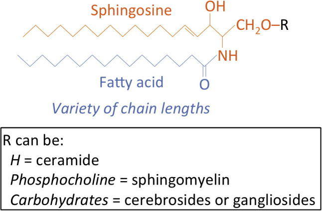
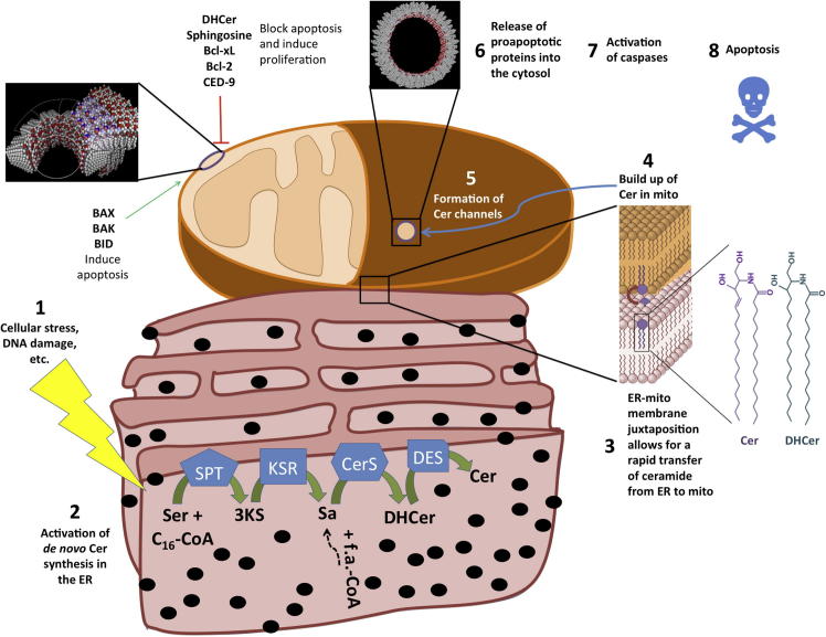
Sphingolipid research has surged in the past two decades and has produced a wide variety of evidence supporting the role of this class of molecules in mediating cellular growth, differentiation, senescence, and apoptosis. Ceramides are a subgroup of sphingolipids (SLs) that are directly involved in the process of initiation of apoptosis. We, and others, have recently shown that ceramides are capable of the formation of protein-permeable channels in mitochondrial outer membranes under physiological conditions. These pores are indeed good candidates for the pathway of release of pro-apoptotic proteins from the mitochondrial intermembrane space (IMS) into the cytosol to initiate intrinsic apoptosis. Here, we review recent findings on the regulation of ceramide channel formation and disassembly, highlighting possible implications on the initiation of the intrinsic apoptotic pathway.

Keywords: Apoptosis; Assembly and disassembly; Bcl-2 family proteins; Bcl-2, B cell CLL/lymphoma-2; Cer, ceramide; CerS, ceramide synthase; Ceramide channels; Chain length; DES, dihydroceramide desaturase; DHCer, dihydroceramide; ER, endoplasmic reticulum; IMS, intermembrane space; KSR, 3-ketosphinganine reductase; MOMP, mitochondrial outer membrane permeability; Mitochondria; SLs, sphingolipids; SM, sphingomyelin; SPT, serine palmitoyl transferase; So, sphingosine; Sphingolipids; de novo synthesis.


Figures

Figure 1
Figure 1
The basic structure of sphingolipids. SLs have a So backbone (orange) that is N-acylated with fatty acids of a variety of chain lengths (blue). The C-1 hydroxyl can be as simple as a hydrogen atom (in ceramides) or as complex as multiple carbohydrate subunits in cerebrosides and gangliosides. The trans double bond at C-4 is characteristic of Cer and when it is saturated the molecule is DHCer.
Figure 2
Figure 2
Ceramide is the hub of sphingolipid metabolism. In the cell three different pathways produce Cer: the de novo synthesis from serine and palmitoyl-CoA (top), SM hydrolysis (left) and the salvage pathway (right). Cer can be used to produce various simple and complex sphingolipids (bottom). The enzymes used in those pathways are boxed. SPT: serine palmitoyl transferase; KSR: 3-ketosphinganine reductase; CerS1-6: ceramide synthases 1–6; DES: dihydroceramide desaturase; SMS: SM synthase; SMase: sphingomyelinase.
Figure 3
Figure 3
An overview of mechanisms of apoptosis. Apoptosis can be activated extrinsically or intrinsically. In extrinsic apoptosis, a macrophage carrying a Fas ligand (FasL) binds to Fas receptor (FasR) on the plasma membrane of the cell inducing the trimerization of the receptor. This induces the recruitment of Fas activated death domain (FADD) which recruits and activates procaspase-8 which ultimately activates the executioner caspase-3 that is responsible for the activities that lead to apoptosis. On the other hand, intrinsic apoptosis is centered around mitochondrial outer membrane permeabilization. Different pro-apoptotic proteins reside in the IMS of mitochondria and when the integrity of the outer membrane is compromised they leak out and cause the activation of apoptosomes, the condensation of chromatin, membrane blebbing and nuclear envelope destruction. Different proteins affect various steps in both pathways positively (green arrows) or negatively (red blunt-ended lines). IAP: inhibitor of apoptosis protein; FLIP: (FADD-like IL-1β-converting enzyme)-inhibitory protein; Smac/DIABLO: second mitochondria-derived activator of caspases/Direct IAP-binding protein with low pI; HSP 27: heat shock protein 27; Apaf-1: apoptotic protease activating factor 1; Bid and tBid: BH3 interacting domain death agonist and truncated Bid; Bax: Bcl-2-associated X protein; Bcl-xL: Bcl-2-extra large; EndoG: endonuclease G; Omi/HtrA2: high temperature requirement A serine endoprotease; AIF: apoptosis-inducing factor; mPTP: mitochondrial permeability transition pore. Note that the release of all pro-apoptotic proteins from the IMS can be through any one of the shown channels.
Figure 4
Figure 4
Cytology of apoptosis. The different stages of apoptotic cell death start by cellular shrinkage and chromatin condensation, concomitant with formation of membrane blebs. Organelles and nucleus fragment and the blebs begin formation of apoptotic bodies which are eventually engulfed by macrophages or neighboring cells by endocytosis/phagocytosis. The lack of release of cellular components to the extracellular fluid results in the absence of inflammation.
Figure 5
Figure 5
A mechanistic view of the action at the mitochondrial outer membrane following the activation of intrinsic apoptosis. The sequence of events leading to the death of the cell is summarized as follows: (1) cellular stresses induce (2) the biosynthesis of Cer in the ER via the de novo synthesis pathway. (3) Cer synthesized in the ER is then readily imported into mitochondria via ER-mitochondria contact sites termed MAM leading to (4) a buildup of Cer in mitochondria. (5) The increased local Cer concentration allows those lipids to self-assemble into a barrel-stave channel through which (6) pro-apoptotic proteins are released into the cytosol. Those channels are inhibited by DHCer, So, anti-apoptotic Bcl-2 family proteins; and they are potentiated by pro-apoptotic Bcl-2 proteins (see the text for details). The release of proteins into the cytosol (7) activates caspases which ultimately (8) induce apoptosis. Note: the structures in black of Cer channels were obtained from Prof. Marco Colombini at the University of Maryland College Park.

References

    1. Adan-Gokbulut A., Kartal-Yandim M., Iskender G., Baran Y. Novel agents targeting bioactive sphingolipids for the treatment of cancer. Curr. Med. Chem. 2013;20:108–122. - PubMed
    1. Allouche M., Bettaieb A., Vindis C., Rousse A., Grignon C., Laurent G. Influence of Bcl-2 overexpression on the ceramide pathway in daunorubicin-induced apoptosis of leukemic cells. Oncogene. 1997;14:1837–1845. - PubMed
    1. Anishkin A., Sukharev S., Colombini M. Searching for the molecular arrangement of transmembrane ceramide channels. Biophys. J. 2006;90:2414–2426. - PMC - PubMed
    1. Antonsson B. Bax and other pro-apoptotic Bcl-2 family “killer-proteins” and their victim the mitochondrion. Cell Tissue Res. 2001;306:347–361. - PubMed
    1. Antonsson B., Montessuit S., Lauper S., Eskes R., Martinou J.C. Bax oligomerization is required for channel-forming activity in liposomes and to trigger cytochrome c release from mitochondria. Biochem. J. 2000;345(Pt 2):271–278. - PMC - PubMed

LinkOut - more resources