. 2015 Nov 20;10(11):e0143174.
doi: 10.1371/journal.pone.0143174. eCollection 2015.

Gelsolin-Like Domain 3 Plays Vital Roles in Regulating the Activities of the Lily Villin/Gelsolin/Fragmin Superfamily

Affiliations

Gelsolin-Like Domain 3 Plays Vital Roles in Regulating the Activities of the Lily Villin/Gelsolin/Fragmin Superfamily

Dong Qian et al. PLoS One. .

Abstract

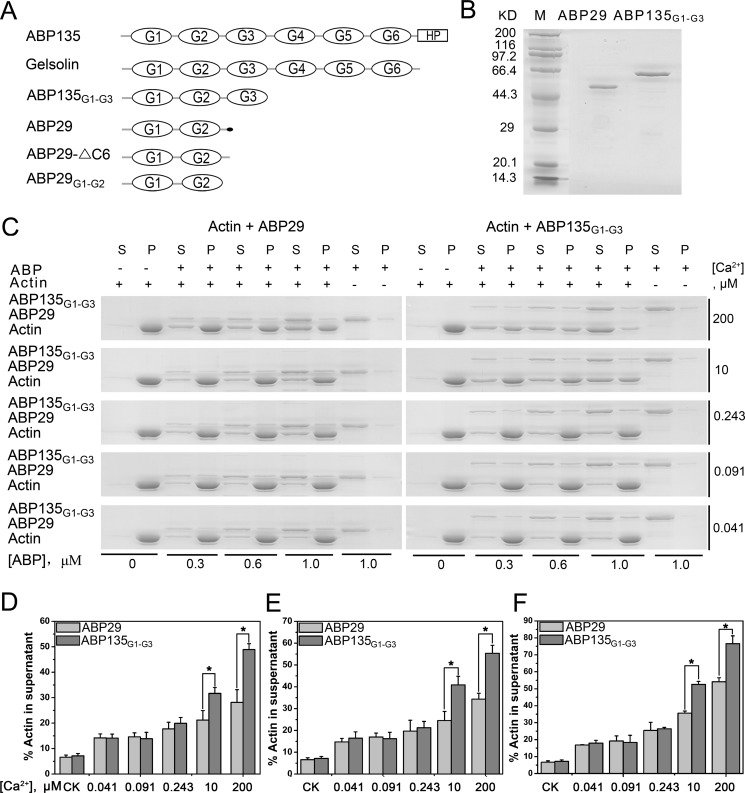
The villin/gelsolin/fragmin superfamily is a major group of Ca2+-dependent actin-binding proteins (ABPs) involved in various cellular processes. Members of this superfamily typically possess three or six tandem gelsolin-like (G) domains, and each domain plays a distinct role in actin filament dynamics. Although the activities of most G domains have been characterized, the biochemical function of the G3 domain remains poorly understood. In this study, we carefully compared the detailed biochemical activities of ABP29 (a new member of this family that contains the G1-G2 domains of lily ABP135) and ABP135G1-G3 (which contains the G1-G3 domains of lily ABP135). In the presence of high Ca2+ levels in vitro (200 and 10 μM), ABP135G1-G3 exhibited greater actin severing and/or depolymerization and nucleating activities than ABP29, and these proteins had similar actin capping activities. However, in the presence of low levels of Ca2+ (41 nM), ABP135G1-G3 had a weaker capping activity than ABP29. In addition, ABP29 inhibited F-actin depolymerization, as shown by dilution-mediated depolymerization assay, differing from the typical superfamily proteins. In contrast, ABP135G1-G3 accelerated F-actin depolymerization. All of these results demonstrate that the G3 domain plays specific roles in regulating the activities of the lily villin/gelsolin/fragmin superfamily proteins.


Conflict of interest statement

Competing Interests: The authors have declared that no competing interests exist.

Figures

Fig 1
Fig 1. Recombinant ABP29 and ABP135G1-G3 fusion proteins sever and/or depolymerize actin filaments in a calcium-dependent manner.
(A) Domain structures of gelsolin superfamily members studied in the paper. The lily ABP135,with six G domains and a headpiece (HP) domain at the C-terminus, is designated as ABP135. Full-length human gelsolin, containing six G-domains, is designated as Gelsolin. ABP135G1-G3 containing the G1-G3 domains is part of ABP135. Lilium ABP29 is a splicing product of ABP135, and the small black dot at the end of ABP29 represents the six amino acids that differ from ABP135. ABP29-△C6 has six fewer amino acids than ABP29 at the C-terminus. ABP29G1-G2 contains only the G1-G2 domains of ABP29. (B) SDS-PAGE analysis of purified recombinant ABP29 and ABP135G1-G3. Molecular mass markers (M) in kiloDaltons are shown to the left of the gel. (C) Detailed comparison of ABP29 and ABP135G1-G3 severing and/or depolymerization activities in the presence of different concentrations of Ca2+ with high-speed co-sedimentation assays; "(s)" indicates supernatant, "(p)" indicates pellets, and "ABP" indicates ABP29 or ABP135G1-G3. (D-F) Statistical analysis results from (C). The graph shows the percentage of actin in the supernatant. The concentrations of the recombinant proteins are 0.3, 0.6 and 1.0 μM, respectively in (D-F). The CK is the percentage of actin in the supernatant in the absence of ABP. The error bars represent ± SE (n = 3); *P < 0.05 (Student’s t test).
Fig 2
Fig 2. Direct visualization of actin filament severing and/or depolymerization by recombinant ABP29 and ABP135G1-G3.
(A) Direct observation of the severing and/or depolymerization activities of ABP29 and ABP135G1-G3 at different concentrations (100, 200 nM) in the presence of 200 μM free Ca2+. The scale bar is 10 μm. (B) Direct observation of the severing and/or depolymerization activities of 200 nM ABP29 and ABP135G1-G3 in the presence of various concentrations (200, 10, 0.243, 0.091 and 0.041 μM) of free Ca2+. The scale bar is 10 μm. (C) Quantification of actin filament length from the micrographs from (B). The lengths of actin filaments were measured by Image J. Thirty pictures for each condition were analyzed (25–70 actin filaments in every picture), and quantification of 10 pictures represents a single result. Error bars represent ± SE (n = 3). *P < 0.05, **P < 0.01 (Student’s t test).
Fig 3
Fig 3. Effects of ABP135G1-G3 on actin filament severing.
A total of 2 μM preformed CapG capped pyrene actin filaments was diluted to 100 nM in S2 buffer with various concentrations of ABP135G1-G3, and immediately, the decrease in pyrene fluorescence intensity that accompanied actin depolymerization was monitored for 300 s.
Fig 4
Fig 4. Recombinant ABP29 and ABP135G1-G3 nucleate actin polymerization.
(A) Time course of the change in pyrene fluorescence accompanying actin polymerization in the presence of ABP29 or ABP135G1-G3. Increasing amounts of ABP29 or ABP135G1-G3 were added to 5 μM G-actin (5% pyrene-labeled) in the presence of 200 μM free Ca2+. Different combinations of reactions are indicated in the upper left. (B) and (C) In the presence of 10 μM free Ca2+ and 0.041 μM free Ca2+ respectively, time course of the change in pyrene fluorescence accompanying actin polymerization in the presence of ABP29 or ABP135G1-G3. Different combinations of reactions are indicated in the upper left.
Fig 5
Fig 5. Recombinant ABP29 and ABP135G1-G3 inhibit actin filament elongation.
(A) Kinetics of actin filament barbed end elongation with various concentrations of ABP29 or ABP135G1-G3 in the presence of 200 μM free Ca2+. Different combinations of reactions are indicated in the upper left. (B) and (C) Kinetics of actin filament barbed end elongation with 20 nM and 60 nM ABP29 or ABP135G1-G3, respectively, in the presence of different concentrations of free Ca2+. Different combinations of reactions are indicated in the upper left.
Fig 6
Fig 6. Recombinant ABP29 and ABP135G1-G3 exert opposing effects on the actin dynamics of depolymerization.
(A) Kinetics of actin depolymerization in the presence of various concentrations of ABP29 or ABP135G1-G3 in the presence of 200 μM free Ca2+. The change in pyrene fluorescence accompanying actin depolymerization was monitored. Different combinations of reactions are indicated in the upper right. (B) and (C) Fixed concentrations of ABP29 or ABP135G1-G3 were incubated with 5 μM F-actin (50% pyrene-labeled) in the presence of free Ca2+ chelated by EGTA. The change in pyrene fluorescence accompanying actin depolymerization was monitored. Different combinations of reactions are indicated in the upper right.
Fig 7
Fig 7. ABP29 and ABP135G1-G3 compete with each other in actin depolymerization.
(A) Competition experiments between ABP29 and ABP135G1-G3 were performed during the F-actin depolymerization process. A fixed amount of ABP135G1-G3 (1000 nM) and different amounts of ABP29 (0, 200 nM and 300 nM) were simultaneously incubated with 5 μM F-actin (50% pyrene-labeled). The kinetics of actin depolymerization were monitored by measuring the decrease in pyrene fluorescence intensity. Different combinations of reactions are indicated in the upper right. (B) A fixed amount of ABP29 (300 nM) and different amounts of ABP135G1-G3 (0, 1000 nM and 2000 nM) were simultaneously incubated with 5 μM F-actin (50% pyrene-labeled). Different combinations of reactions are indicated in the upper right.
Fig 8
Fig 8. Recombinant ABP29-△C6 and ABP29G1-G2 sever and/or depolymerize F-actin and stabilize F-actin in vitro.
(A) The severing and/or depolymerization activity of ABP29-△C6 exerted on actin filaments was determined using high-speed co-sedimentation assays in the presence of 200 μM and 0.041 μM free Ca2+; "(s)" indicates supernatant, and "(p)" indicates pellets. (B) The severing and/or depolymerization activity of ABP29G1-G2 exerted on actin filaments was determined using high-speed co-sedimentation assays in the presence of various concentrations of free Ca2+ (200, 0.243 and 0.041 μM); "(s)" indicates supernatant, and "(p)" indicates pellets. (C) and (E) Statistical analysis results from (A) and (B), respectively. The error bars represent ± SE (n = 3); **P < 0.01 (Student’s t test). (E) and (F) Kinetics of actin depolymerization in the presence of various concentrations of ABP29G1-G2 and ABP29-△C6 in the presence of 200 μM free Ca2+. The change in pyrene fluorescence accompanying actin depolymerization was monitored. Different combinations of the reactions are indicated in the upper right. (G) and (H) 300 nM ABP29G1-G2 or ABP29-△C6, respectively, was incubated with 5 μM F-actin (50% pyrene-labeled) for 5 min in the presence of various concentrations of free Ca2+ chelated by EGTA. The kinetics of actin depolymerization were monitored by measuring the change in fluorescence intensity. Different combinations of reactions are indicated in the upper right.

Similar articles

Cited by

References

    1. Sun H, Yamamoto M, Mejillano M, Yin HL (1999) Gelsolin, a multifunctional actin regulatory protein. J Biol Chem 274: 33179–33182. - PubMed
    1. Silacci P, Mazzolai L, Gauci C, Stergiopulos N, Yin HL, Hayozb D (2004) Gelsolin superfamily proteins: Key regulation of cellular functions. Cell Mol Life Sci 61: 2614–2623. - PMC - PubMed
    1. Mcgough AM, Staiger CJ, Min JK, Simonetti KD (2003) The gelsolin family of actin regulatory proteins: Modular structures, versatile functions. FEBS Letters 552: 75–81. - PubMed
    1. Kwiatkowski DJ, Stossel TP, Orkin SH, Mole JE, Colten HR, Yin HL (1986) Plasma and cytoplasmic gelsolins are encoded by a single gene and contain a duplicated actin-binding domain. Nature 323: 455–458. - PubMed
    1. Burtnick LD, Koepf EK, Grimes J, Jones EY, Stuart DI, McLaughlin PJ, et al. (1997) The crystal structure of plasma gelsolin: Implications for actin severing, capping, and nucleation. Cell 90: 661–670. - PubMed

Publication types