. 2015 Nov 17;43(5):959-73.
doi: 10.1016/j.immuni.2015.10.014.

Identification of Common Features in Prototype Broadly Neutralizing Antibodies to HIV Envelope V2 Apex to Facilitate Vaccine Design

Affiliations

Identification of Common Features in Prototype Broadly Neutralizing Antibodies to HIV Envelope V2 Apex to Facilitate Vaccine Design

Raiees Andrabi et al. Immunity. .

Abstract

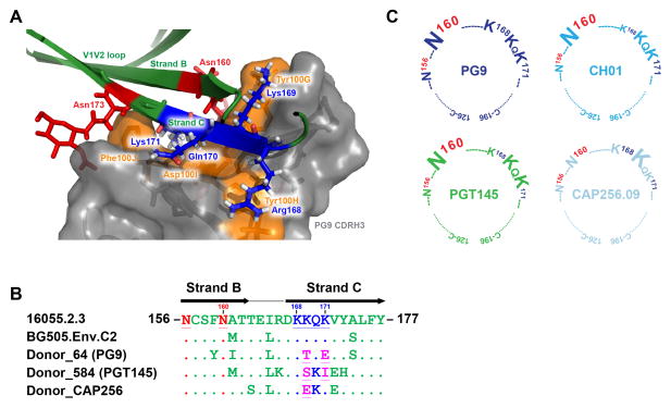
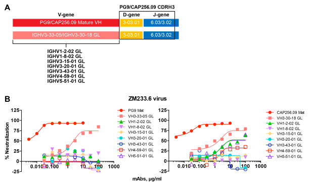
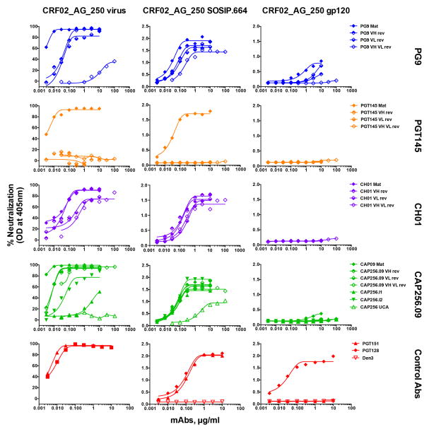
Broadly neutralizing antibodies (bnAbs) directed to the V2 apex of the HIV envelope (Env) trimer isolated from individual HIV-infected donors potently neutralize diverse HIV strains, but strategies for designing immunogens to elicit bnAbs have not been identified. Here, we compared four prototypes (PG9, CH01, PGT145, and CAP256.VRC26.09) of V2 apex bnAbs and showed that all recognized a core epitope of basic V2 residues and the glycan-N160. Two prototype bnAbs were derived from VH-germlines that were 99% identical and used a common germline D-gene encoded YYD-motif to interact with the V2-epitope. We identified isolates that were neutralized by inferred germline (iGL) versions of three of the prototype bnAbs. Soluble Env derived from one of these isolates was shown to form a well-ordered Env trimer that could serve as an immunogen to initiate a V2-apex bnAb response. These studies illustrate a strategy to transition from panels of bnAbs to vaccine candidates.


Figures

Figure 1
Figure 1. Epitope recognition by V2 apex bnAbs (See also Figure S1 and Table S1)
A. PG9 CDRH3 interaction with glycans and strand B and C of the V1V2 domain presented on a scaffold shown as a ribbon representation (modified from (McLellan et al., 2011)). PG9 CDRH3-interacting amino acid residues are shown in orange with side chains as sticks. The V2 glycans N160 and N173 are depicted in red, and the four key residues from the lysine-rich strand C are shown in blue with side chains as sticks. B. Amino acid sequence alignment of strands B and C of isolate 16055.2.3, BG505.Env.C2, IAVI donor 64 PG9 antibody escape virus, IAVI donor 584 PGT145 antibody escape virus and donor CAP256 CAP256.09 antibody escape virus (HXB2 numbering: 156–177). The sequences were aligned with clustalX in the BioEdit program. The N-linked glycan sites N156 and N160 are shown in red, the four amino acids in the lysine rich region on strand C are shown in blue and the escape mutations at position 169 and 171 in the donor escape viruses are highlighted in pink. C. Graphic representation of the epitopes for four (PG9, CH01, PGT145 and CAP256.09) V2 apex bnAb prototypes. Each cartoon represents the cysteine-linked V1V2 loops (HXB2: 126–196), highlighting the core epitope for all the V2 apex bnAb prototypes. The epitope includes the glycans N156, N160 and four strand C amino acid residues. The size of each amino acid letter is proportional to the dependence on that residue for neutralization.
Figure 2
Figure 2. Glycan specificity of V2 apex bnAbs (See also Tables S2-S3)
A. V2 apex bnAb neutralization of Env variants displaying differing glycan composition in the context of the 16055.2.3 isolate. Two Abs from each V2 apex bnAb prototype were tested for neutralization against 16055.2.3 wild type (WT), N156K, N160K glycan variants, and viruses produced in the presence of glycosidase inhibitors kifunensine (Kif) and swainsonine (Swain). B. The four V2 apex bnAb prototypes were tested in ELISA binding to Gallanthus Nivalis Lectin (GNL)-purified BG505.Env.C2.SOSIP.664 soluble trimer glycovariants. The gp120-gp41 glycan dependent Ab PGT151, the CD4bs Ab VRC01 and a Dengue Ab (DEN3) were used as controls. C. Reactivity of V2 apex bnAbs with glycans on a glycan microarray. The V2 apex bnAbs were tested for reactivity on a glycan array. The data are shown as Relative Fluorescence Units (RFU) and the error bars represent the average percentage error for all data points reported. The symbol for each monosaccharide is indicated. The glycan dependent antibody PGT151 was used as control.
Figure 3
Figure 3. Variable (V), Diversity (D) and Joining (J) gene families encoding V2 apex bnAbs and conservation of the YYD motif in PG9 and CAP256.09 prototype Abs (See also Figure S2 and Table S4)
A. Representation based on the amino acid sequence alignment of the inferred germline-encoding heavy chain variable regions (VH). The V, D and J gene usage for various V2 apex bnAb prototypes (denoted on the right) is listed in the boxes. The V-gene sequence alignment shows that PG9 and CAP256.09 Ab prototypes, derived from different gene families share 99% sequence similarity at amino acid level (shown in red) and use same germline D-gene. Different gene families encode the J-genes for each of the Ab prototypes. Abs possess (N)-nucleotide insertions of variable lengths at V-D and D-J junctions in the CDRH3. B. Sequence alignment of CDRH3 sequences of V2 apex bnAbs shows conservation of the YYD motif (underlined bold) in PG9, PG16 and CAP256.09 antibody prototypes. IMGT numbering is shown. C. The effects of substitutions in the YYD motif amino acid residues in PG9 and PG16 Abs on neutralization of viruses. The IC50 neutralization titers of mature (WT) PG9 and PG16 Abs and variant antibodies against a 4-virus panel are represented as bars. D. Binding of WT PG9 and PG16 Abs and their variants to recombinant GNL-purified gp120 proteins by ELISA. Antibodies to the high mannose patch (PGT128), CD4bs (b6, b12 and PGV04) and Dengue (DEN3) were used as control mAbs.
Figure 4
Figure 4. Neutralization breadth and potency of mature, chimeric and inferred germline reverted V2 apex bnAbs (See also Figures S3-S4 and Table S5-S6)
A. A schematic representation of chimeric and inferred germline (iGL) reverted antibodies. The mature antibody, referred to as ‘Mat’ shows constant region (grey), the variable heavy (VH) chain (red) and variable light (VL) chain (green). The third Complementarity Determining Regions of VH (CDRH3) and VL (CDRL3) chains are shown at the end of each variable region. The VH and VL genes are further divided into framework regions (FR1-3) and other CDRs (CDR1-2) shown with ImMunoGeneTics (IMGT) numbering. To generate the VH or VL regions in iGL Abs were replaced in with their closest inferred germline sequence predicted by in the IMGT human IgG germline database (Brochet et al., 2008). The CDRH3 and CDRL3 from the Mat antibodies were retained in the reverted iGL Abs. B. Association of IC50 neutralization titers with number of somatic mutations in VH and VL for the four prototype Abs, PG9, CH01, CAP256.09 and PGT145. C. Neutralization breadth of Mat, chimeric and iGL reverted V2 apex bnAbs. The number of viruses in an 18-virus panel neutralized with an IC50 <10 μg/ml is shown for Mat and chimeric Abs. For the less potent iGL Abs, neutralization is scored as positive with IC50 <100 μg/ml.
Figure 5
Figure 5. Neutralization of ZM233M.PB6 by PG9 and CAP256.09 heavy chain inferred germline antibody variants (See also Table S7)
A. Schematic representation of PG9 and CAP256.09 inferred germline-VH Ab variants. The PG9 or CAP256.09 VH-germline gene was replaced by germline-VH encoding sequences from diverse HIV-1 specific Abs. The selected germline genes are used by envelope specific bnAbs that include IGHV1-2-02 (CD4bs), IGHV1-8-02, IGHV3-15-01, IGHV3-20-01, IGHV3-43-01 (V2 apex), IGHV4-59-01 (N332-V3), IGHV5-51-01 (V3). B. Neutralization of ZM233M.PB6 virus by PG9 and CAP256.09 iGL variant antibodies in which the VH germline has been substituted as in (A) above.
Figure 6
Figure 6. Comparison of mutations in PG9 and CAP256.09 contributing to broad neutralization (See also Figure S5)
A–B. Sequence alignment of mature heavy (A) and light (B) chain sequences of PG9, PG16 and CAP256.09 antibodies with their corresponding inferred germline (iGL) encoding sequences. Each VH and VL gene sequence is divided into FR1-3 and CDR1-2 with junctions underlined. A. Common positions at which somatic mutations occur in the mature HC of PG9, PG16 and CAP256.09 Abs are highlighted in cyan, common mutations shown in red. B. Common somatic mutations at 8 positions in mature PG9 and PG16 VL are highlighted in cyan. E37 in PG9, which is D in PG16, essential for glycan recognition (McLellan et al., 2011; Pancera et al., 2013) is included. C. The VH or VH-VL iGL-modified PG9 Ab variants were tested against the 18-virus panel and the median IC50 neutralization titers are shown for each. D. Ribbon representation of PG9 antibody in complex with a ZM109F V1V2 loop (green) scaffold (modified from (McLellan et al., 2011)). The glycans at N160 and N173 positions (red; represented as sticks) and strand C residues (blue) in V2 domain are labeled. The PG9 variable heavy (cyan) and light (aqua) chains illustrate common positions (shown as surface rendering) at which somatic mutations occur in PG9, PG16 and CAP256.09 HC (denoted by residues in black) and PG9 and PG16 LC (denoted by residues in red).
Figure 7
Figure 7. The CRF02_AG_250 virus is neutralized by 3 iGL prototype bnAbs and forms well-ordered recombinant Env trimers. (See also Figure S6-S7)
A. V2 apex bnAb neutralization of CRF02_AG_250 virus and binding to the corresponding SOSIP.664 and gp120 proteins. (A. Left column) the mature (Mat), VH or VL reverted chimeric and VH-VL iGL antibodies from each Ab prototype, an inferred unmutated common ancestor (CAP256 UCA) and two intermediate (CAP256.I1 and CAP256.I2) Abs derived from the CAP256 donor, were tested for neutralization against the CRF02_AG_250 virus. The neutralization curves show the activity of Abs, Mat (filled diamond), VH or VL reverted chimeras (half-filled diamond), VH-VL iGL (empty diamond). (A. Middle column) Abs were assessed for binding to the PGT145 antibody affinity purified soluble trimeric Env form (SOSIP.664) of CRF02_AG_250 by ELISA. (A. Right column) Abs were further tested for binding to the GNL-affinity purified monomeric gp120 protein corresponding to CRF02_AG_250 envelope, by ELISA. Antibodies, PGT128, PGT151 and DEN3 were used as controls.

Comment in

References

    1. Barbian HJ, Decker JM, Bibollet-Ruche F, Galimidi RP, West AP, Jr, Learn GH, Parrish NF, Iyer SS, Li Y, Pace CS, et al. Neutralization properties of simian immunodeficiency viruses infecting chimpanzees and gorillas. mBio. 2015;6 - PMC - PubMed
    1. Bonsignori M, Hwang KK, Chen X, Tsao CY, Morris L, Gray E, Marshall DJ, Crump JA, Kapiga SH, Sam NE, et al. Analysis of a clonal lineage of HIV-1 envelope V2/V3 conformational epitope-specific broadly neutralizing antibodies and their inferred unmutated common ancestors. Journal of virology. 2011;85:9998–10009. - PMC - PubMed
    1. Braibant M, Gong EY, Plantier JC, Moreau T, Alessandri E, Simon F, Barin F. Cross-group neutralization of HIV-1 and evidence for conservation of the PG9/PG16 epitopes within divergent groups. Aids. 2013;27:1239–1244. - PubMed
    1. Brochet X, Lefranc MP, Giudicelli V. IMGT/V-QUEST: the highly customized and integrated system for IG and TR standardized V-J and V-D-J sequence analysis. Nucleic acids research. 2008;36:W503–508. - PMC - PubMed
    1. Burton DR, Mascola JR. Antibody responses to envelope glycoproteins in HIV-1 infection. Nature immunology. 2015;16:571–576. - PMC - PubMed

Publication types

MeSH terms

Associated data