. 2015 Nov 17;43(5):1011-21.
doi: 10.1016/j.immuni.2015.10.016.

A Wave of Regulatory T Cells into Neonatal Skin Mediates Tolerance to Commensal Microbes

Affiliations

A Wave of Regulatory T Cells into Neonatal Skin Mediates Tolerance to Commensal Microbes

Tiffany C Scharschmidt et al. Immunity. .

Abstract

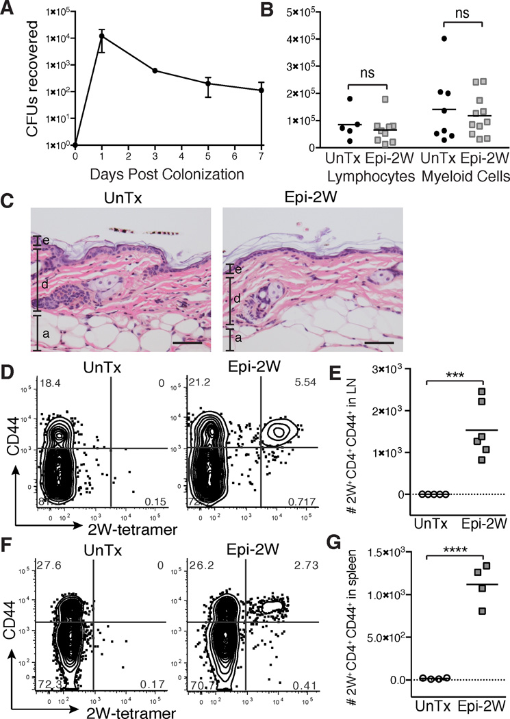
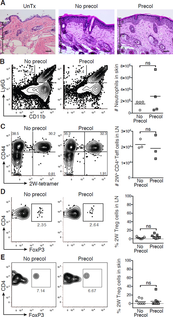
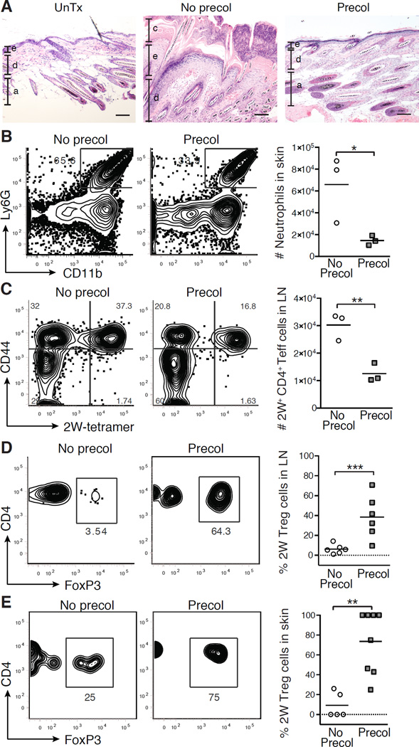
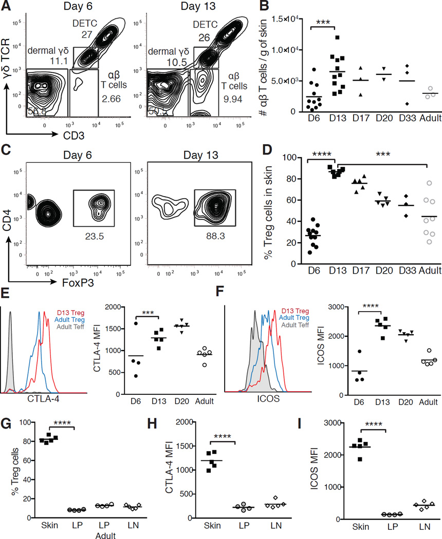
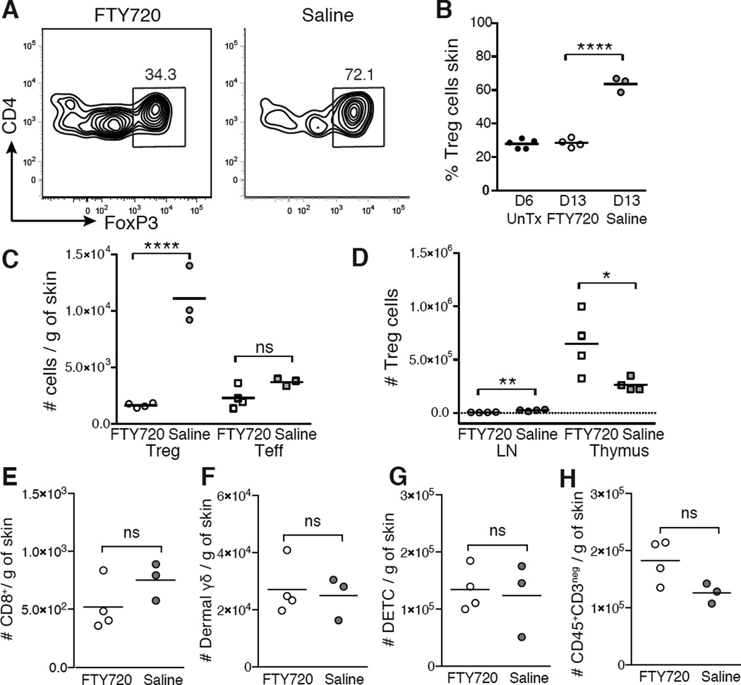
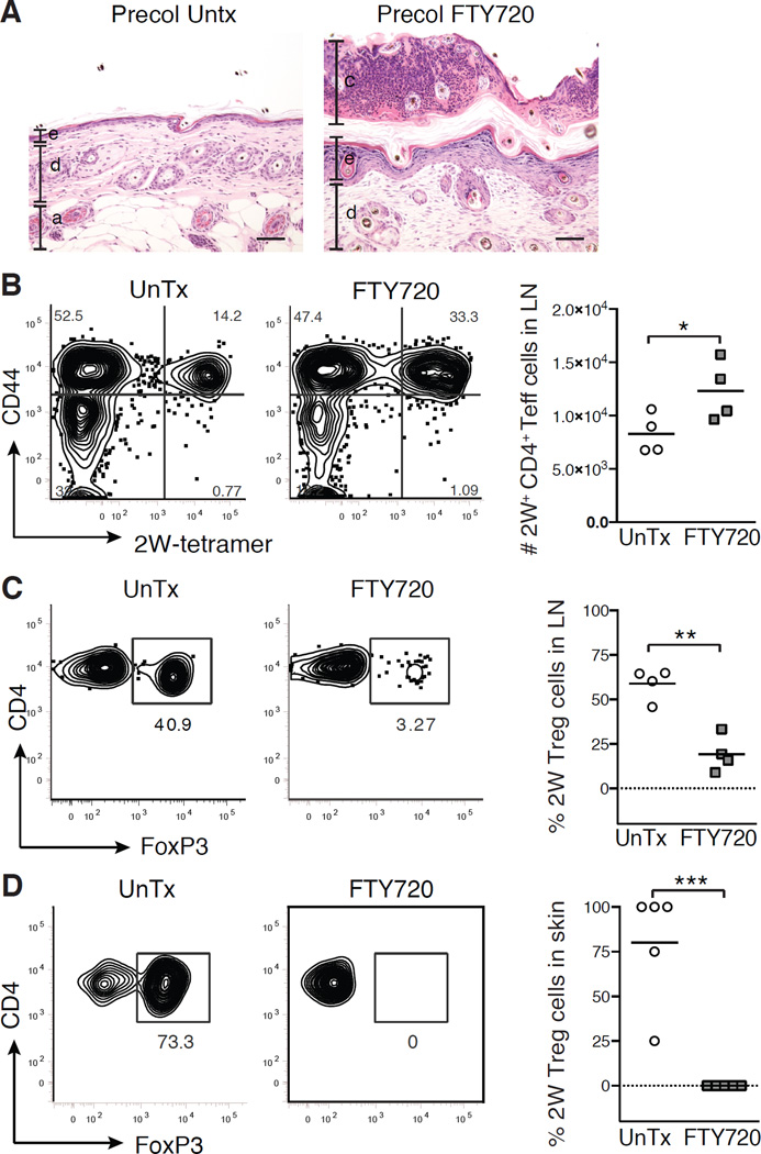
The skin is a site of constant dialog between the immune system and commensal bacteria. However, the molecular mechanisms that allow us to tolerate the presence of skin commensals without eliciting destructive inflammation are unknown. Using a model system to study the antigen-specific response to S. epidermidis, we demonstrated that skin colonization during a defined period of neonatal life was required for establishing immune tolerance to commensal microbes. This crucial window was characterized by an abrupt influx of highly activated regulatory T (Treg) cells into neonatal skin. Selective inhibition of this Treg cell wave completely abrogated tolerance. Thus, the host-commensal relationship in the skin relied on a unique Treg cell population that mediated tolerance to bacterial antigens during a defined developmental window. This suggests that the cutaneous microbiome composition in neonatal life is crucial in shaping adaptive immune responses to commensals, and disrupting these interactions might have enduring health implications.


Figures

Figure 1
Figure 1. Antigen-specific recognition of commensal microbes across an intact skin barrier
(A) Number of Epi-2W colony forming units (CFU) recovered via skin swab from mice colonized with Epi-2W once on day 0. Each data point represents average of 3 mice. Adult mice were colonized with Epi-2W or untreated (UnTx) every three days for three applications and then skin, skin-draining lymph nodes (SDLNs) and spleen were harvested on day 10. (B) Absolute number of lymphocytes (live CD45+CD3+) and myeloid cells (live CD45+CD3neg) per gram of skin and (C) skin histology. Scale bars represent 50 µm. e = epidermis, d = dermis, a = adipose. (D) Flow cytometry plots of CD4+ T cells (gated on live CD45+CD3+CD4+ from tetramer-enriched fraction) and (E) absolute numbers of CD44+CD4+2W+ cells in SDLNs and (F and G) spleen on day 10. Results in B-E representative of three independent experiments and in A, F and G from two independent experiments. See also Figure S1.
Figure 2
Figure 2. Colonization with commensal bacteria in adult mice does not establish immune tolerance
Adult mice were not colonized (No precol) or colonized with Epi-2W (Precol) every three days for one week and then challenged 3–4 weeks later with Epi-2W and superficial skin abrasion. (A) Representative skin histology 10 days post-challenge versus healthy age-matched skin. Scale bars represent 50 µm. e = epidermis, d = dermis, a = adipose. (B) Flow cytometry plots and absolute numbers of neutrophils in skin. Gated on live CD45+CD3neg population. (C) Flow cytometry and absolute numbers of CD44+CD4+2W+FoxP3neg cells in SDLNs. Gated on live DumpnegCD45+CD3+CD4+FoxP3neg population in tetramer-enriched fraction. (D) Flow cytometry and percent 2W-specific Treg cells in SDLNs and (E) skin. Gated on live DumpnegCD45+CD3+CD4+CD44+2W+ population in tetramer-enriched fraction for SDLNs and total unenriched fraction for skin. Each point represents pooled data from 2 mice. Representative data from three independent experiments with ≥6 mice per group. See also Figure S2.
Figure 3
Figure 3. Colonization of neonatal mice with commensal bacteria results in antigen-specific immune tolerance
Neonatal mice were not colonized (No Precol) or colonized (Precol) with Epi-2W on postnatal day 7, then challenged 3–4 weeks later with Epi-2W and superficial skin abrasion. (A) Representative skin histology 10 days post-challenge as compared to untreated age-matched skin. Scale bars represent 50 µm. e = epidermis, d = dermis, a = adipose, c = crust. (B) Flow cytometry and numbers of skin neutrophils. Gated on live CD45+CD3neg population. (C) Flow cytometry and absolute numbers of CD44+CD4+2W+FoxP3neg cells in SDLNs. Gated on live DumpnegCD45+CD3+CD4+FoxP3neg population in tetramer-enriched fraction. (D) Flow cytometry and percent of 2W-specific Treg cells in SDLNs and (E) skin. Gated on live DumpnegCD45+CD3+CD4+CD44+2W+ population in tetramer-enriched fraction for SDLNs and total unenriched fraction for skin. Each point represents pooled data from two mice. Representative data from three independent experiments with ≥6 mice per group. See also Figure S2.
Figure 4
Figure 4. Activated Treg cells abruptly accumulate in neonatal skin
(A) Representative flow cytometric plots outlining T cell subsets in murine skin at postnatal D6 and D13. Gated on live CD45+ cells. (B) Absolute numbers of skin αβ T cells by age. (C) Flow cytometry of skin CD4+ cells at D6 and D13. (D) Percent Treg cells in skin by age. Expression of (E) total CTLA-4 and (F) ICOS by flow cytometry and mean fluorescent intensity (MFI) on skin Treg cells by age. Teff cells = FoxP3negCD4+ T cells. (G) Percentage of Treg cells, (H) MFI of CTLA-4 and (I) MFI of ICOS in D13 Treg cells from skin, lamina propria (LP) and SDLNs. Each point represents data from an individual mouse. Data in A-D are representative of three independent experiments with ≥3 mice per group. Data in E-G is representative of two independent experiments with ≥3 mice per group. See also Figure S3.
Figure 5
Figure 5. FTY720 treatment preferentially blocks migration of Treg cells into neonatal skin
FTY720 or saline was administered every 48 hours between postnatal D5 and D11 and skin, thymus and SDLNs were harvested on D13. (A) Flow cytometry and (B) percent Treg cells in skin on D13 vs. untreated D6 neonates. Gated on live CD45+CD3+CD4+ cells. (C) Absolute numbers skin of Treg cells and CD4+FoxP3neg (Teff) cells in skin on D13. (D) Absolute numbers of Treg cells in thymus and SDLNs on D13. Absolute numbers of (E) CD8+ T cells, (F) dermal γδ T cells, (G) dendritic epidermal T cells (DETC) and (H) CD45+CD3neg myeloid cells in skin on D13. Data is representative of two independent experiments with ≥3 mice per group.
Figure 6
Figure 6. Inhibiting Treg cell migration to the skin in neonatal life prevents tolerance to commensal bacteria
Neonatal mice were colonized for one week with Epi-2W beginning on D7 and FTY720 or saline (UnTx) was administered on postnatal D5 and D7. Mice were then challenged 3–4 weeks later with Epi-2W and superficial skin abrasion. (A) Representative skin histology 10 days post-challenge. Scale bars represent 50 µm. e = epidermis, d = dermis, a = adipose, c = crust. (B) Flow cytometry and absolute numbers of CD44+CD4+2W+FoxP3neg cells in SDLNs. Gated on live DumpnegCD45+CD3+CD4+FoxP3neg population in tetramer-enriched fraction. (C) Flow cytometry and percent of 2W-specific Treg cells in SDLNs and (D) skin. Gated on live DumpnegCD45+CD3+CD4+CD44+2W+ population in tetramer-enriched fraction for SDLNs and total unenriched fraction for skin. Each point represents data pooled from two mice. Data are representative of three independent experiments with ≥6 mice per group. See also Figure S4.

Comment in

References

    1. Adkins B, Leclerc C, Marshall-Clarke S. Neonatal adaptive immunity comes of age. Nat Rev Immunol. 2004;4:553–564. - PubMed
    1. Allgaier H, Jung G, Werner RG, Schneider U, Zähner H. Epidermin: sequencing of a heterodetic tetracyclic 21-peptide amide antibiotic. Eur. J. Biochem. 1986;160:9–22. - PubMed
    1. Atarashi K, Tanoue T, Shima T, Imaoka A, Kuwahara T, Momose Y, Cheng G, Yamasaki S, Saito T, Ohba Y, et al. Induction of Colonic Regulatory T Cells by Indigenous Clostridium Species. Science. 2011;331:337–341. - PMC - PubMed
    1. Augustin J, Gotz F. Transformation of Staphylococcus epidermidis and other staphylococcal species with plasmid DNA by electroporation. FEMS Microbiology Letters. 1990;54:203–207. - PubMed
    1. Belkaid Y, Hand TW. Role of the Microbiota in Immunity and Inflammation. Cell. 2014;157:121–141. - PMC - PubMed

Publication types

MeSH terms

Substances

Associated data