. 2015 Nov 20:15:44.
doi: 10.1186/s12861-015-0094-5.

The developmental basis of mesenchymal stem/stromal cells (MSCs)

Affiliations

The developmental basis of mesenchymal stem/stromal cells (MSCs)

Guojun Sheng. BMC Dev Biol. .

Abstract

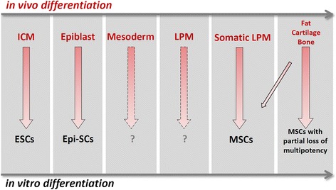
Background: Mesenchymal Stem/Stromal Cells (MSCs) define a population of progenitor cells capable of giving rises to at least three mesodermal lineages in vitro, the chondrocytes, osteoblasts and adipocytes. The validity of MSCs in vivo has been questioned because their existence, either as a homogeneous progenitor cell population or as a stem cell lineage, has been difficult to prove. The wide use of primary MSCs in regenerative and therapeutic applications raises ethical and regulatory concerns in many countries. In contrast to hematopoietic stem cells, a parallel concept which carries an embryological emphasis from its outset, MSCs have attracted little interest among developmental biologists and the embryological basis for their existence, or lack thereof, has not been carefully evaluated.

Methods: This article provides a brief, embryological overview of these three mesoderm cell lineages and offers a framework of ontological rationales for the potential existence of MSCs in vivo.

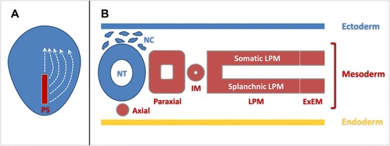
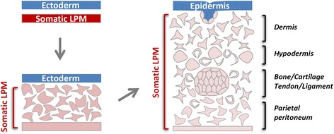
Results: Emphasis is given to the common somatic lateral plate mesoderm origin of the majority of body's adipose and skeletal tissues and of the major sources used for MSC derivation clinically. Support for the MSC hypothesis also comes from a large body of molecular and lineage analysis data in vivo.

Conclusions: It is concluded that despite the lack of a definitive proof, the MSC concept has a firm embryological basis and that advances in MSC research can be facilitated by achieving a better integration with developmental biology.


Figures

Fig. 1
Fig. 1
Schematic diagram of mesoderm formation and patterning during vertebrate early development. a Mesoderm precursors located in the primitive streak (PS) undergo epithelial-to-mesenchymal transition (EMT) and migrate between the ectoderm and endoderm germ layers to their final destinations in a spatially and temporally coordinated manner (white stippled lines). b Major mesoderm lineages (axial, paraxial, intermediate, lateral plate and extraembryonic) are laid out along the medio-lateral axis of the early embryo. IM: intermediate mesoderm; LPM: lateral plate mesoderm; ExEM: extraembryonic mesoderm; NT: neural tube; NC: neural crest. White stippled line represents the boundary between the LPM and ExEM
Fig. 2
Fig. 2
Schematic diagram of somatic LPM morphogenesis. Epithelial-to-mesenchymal transition (EMT) of the epithelial somatic LPM produces a homogenous mesenchymal cell population located between the ectoderm and the remaining LPM epithelium. These mesenchymal cells differentiate into many mesoderm lineageslineages, including the adipocytes, chondrocytes and osteoblasts. MSCs are hypothesized to exist both in the naïve somatic LPM population and in a more differentiated LPM tissue environment
Fig. 3
Fig. 3
Comparison of MSC-related phenomena in vivo and in vitro. In vivo, progenitor populations that will give rise to the adipocyte, chondrocyte and osteoblast lineages pass through developmental phases that progressively restrict their fate choices. This is correlated with progressively limited differentiation potentials of corresponding stem cell populations cultured in vitro. ICM: inner cell mass; ESCs: embryonic stem cells; Epi-SCs: epiblast stem cells; LPM: latral plate mesoderm; MSCs: mesenchymal stem cells. Corresponding stem cell populations for the mesoderm (pan-mesoderm stem cells) and LPM (pan-lateral plate mesoderm stem cells) have not yet been reported

References

    1. Dominici M, Le Blanc K, Mueller I, Slaper-Cortenbach I, Marini FC, Krause DS, et al. Minimal criteria for defining multipotent mesenchymal stromal cells. The international society for cellular therapy position statement. Cytotherapy. 2006;8(4):315–7. doi: 10.1080/14653240600855905. - DOI - PubMed
    1. Caplan AI. Mesenchymal stem cells. J Orthop Res. 1991;9(5):641–50. doi: 10.1002/jor.1100090504. - DOI - PubMed
    1. Bianco P. “Mesenchymal” stem cells. Annu Rev Cell Dev Biol. 2014;30(2014):677–704. doi: 10.1146/annurev-cellbio-100913-013132. - DOI - PubMed
    1. Cawthorn WP, Scheller EL, MacDougald OA. Adipose tissue stem cells meet preadipocyte commitment: going back to the future. J Lipid Res. 2012;53(2):227–46. doi: 10.1194/jlr.R021089. - DOI - PMC - PubMed
    1. Friedenstein AJ, Piatetzky S, II, Petrakova KV. Osteogenesis in transplants of bone marrow cells. J Embryol Exp Morphol. 1966;16(3):381–90. - PubMed

Publication types