. 2016 Jan;131(1):115-35.
doi: 10.1007/s00401-015-1506-0. Epub 2015 Nov 20.

SNTF immunostaining reveals previously undetected axonal pathology in traumatic brain injury

Affiliations

SNTF immunostaining reveals previously undetected axonal pathology in traumatic brain injury

Victoria E Johnson et al. Acta Neuropathol. 2016 Jan.

Abstract

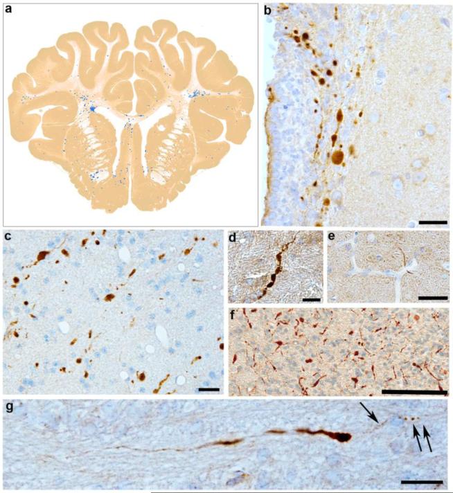
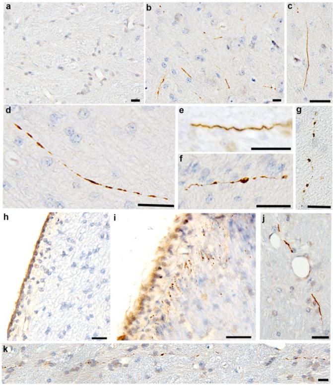
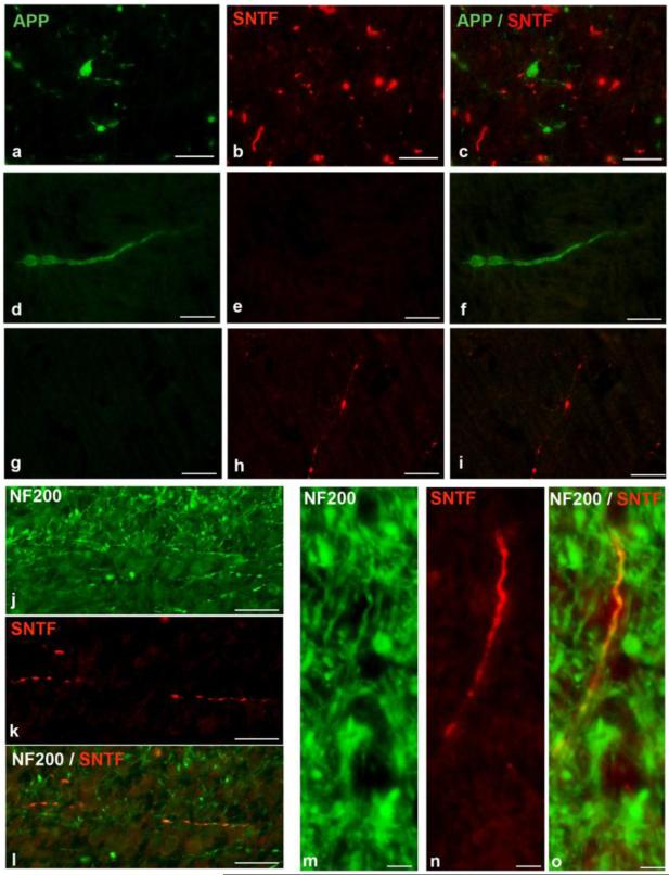
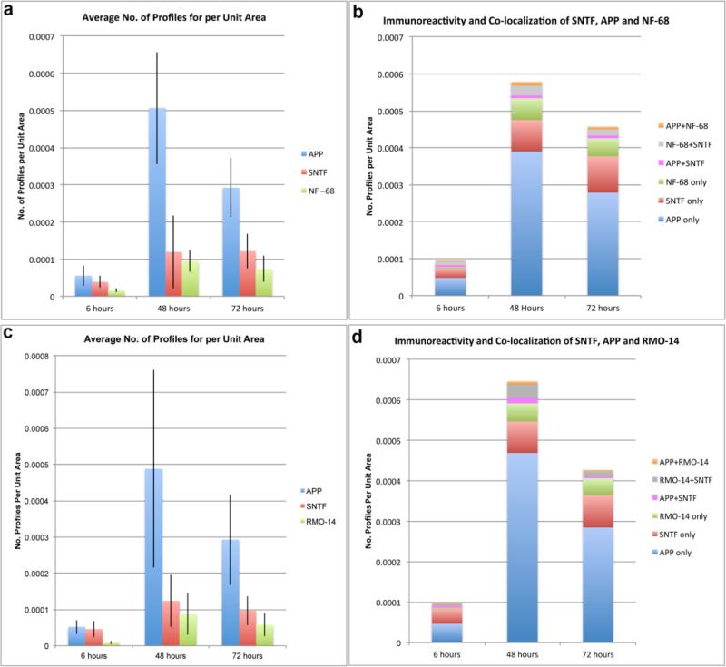
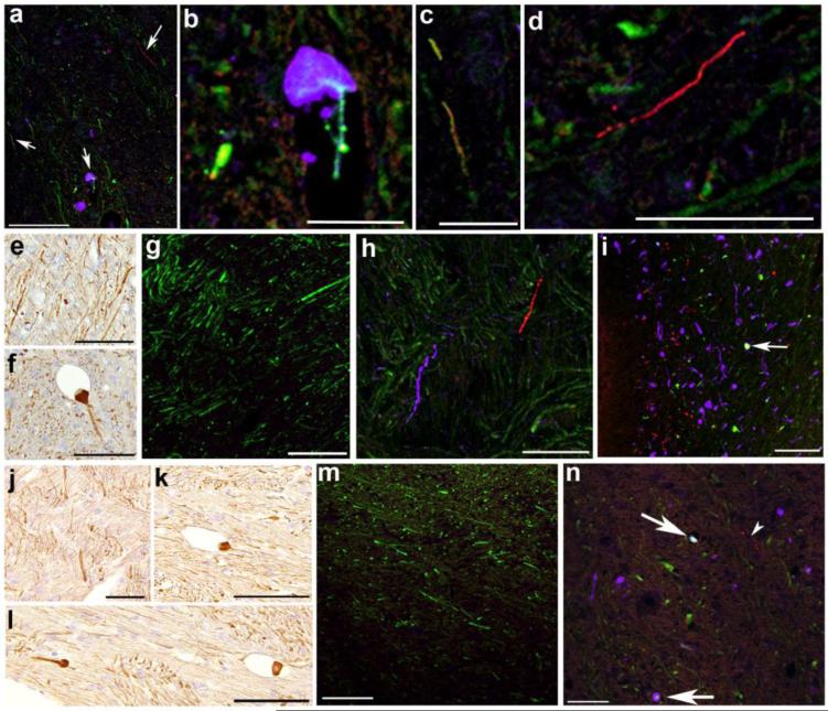
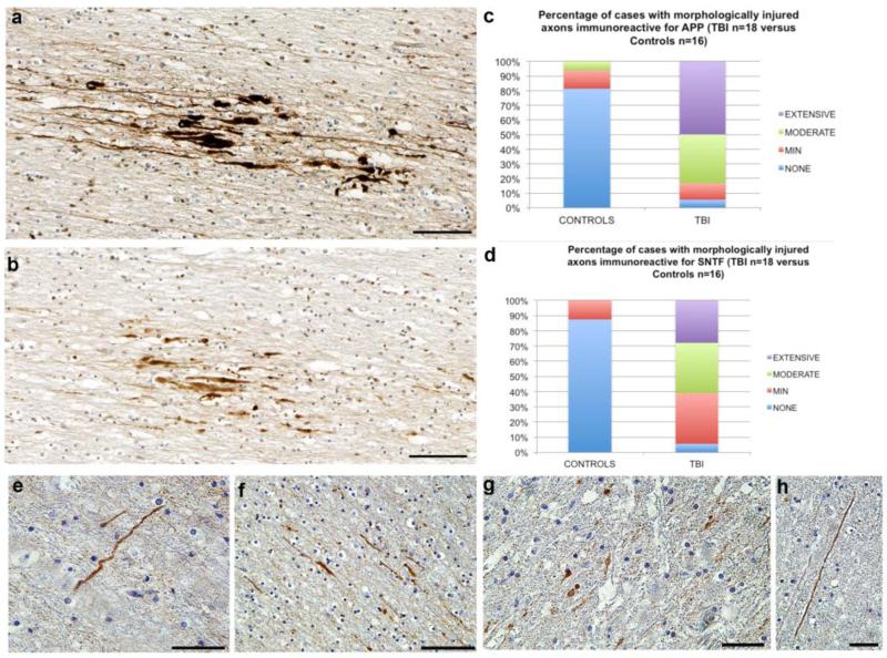
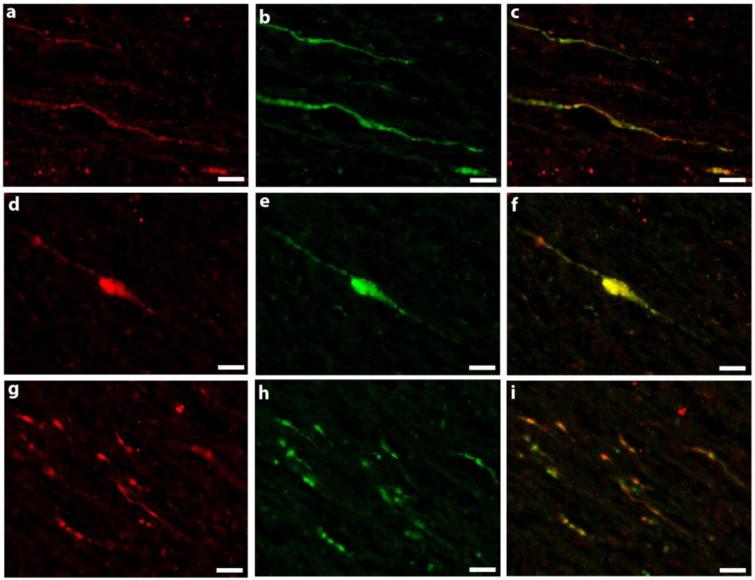
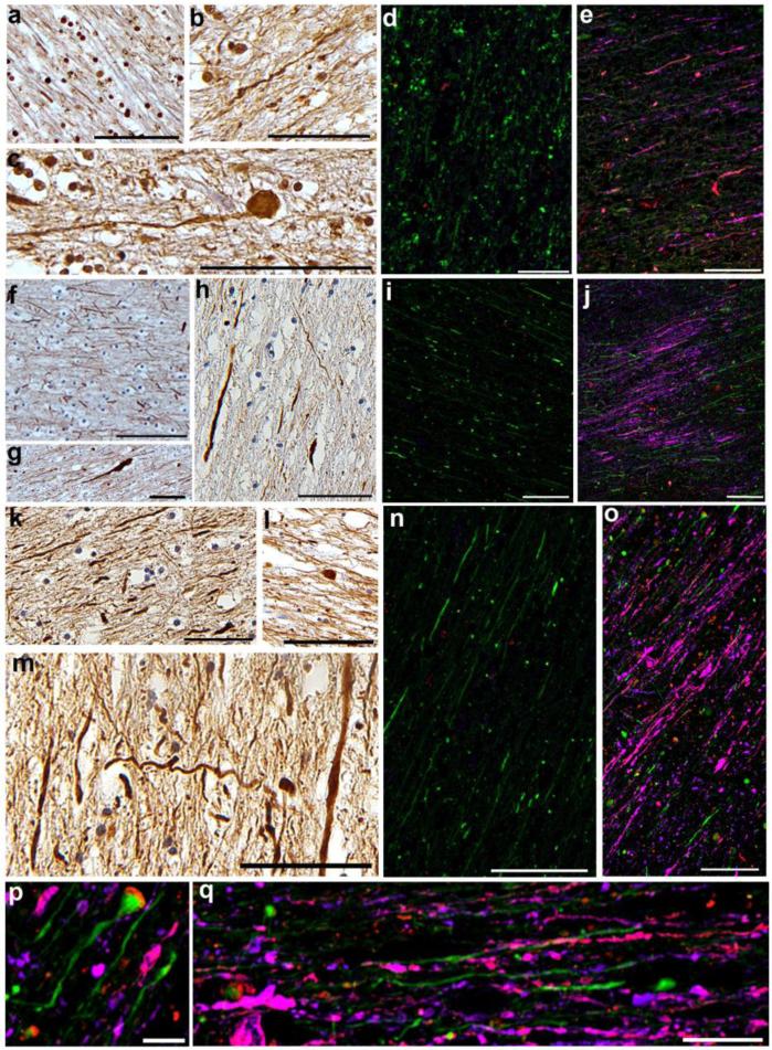
Diffuse axonal injury (DAI) is a common feature of severe traumatic brain injury (TBI) and may also be a predominant pathology in mild TBI or "concussion". The rapid deformation of white matter at the instant of trauma can lead to mechanical failure and calcium-dependent proteolysis of the axonal cytoskeleton in association with axonal transport interruption. Recently, a proteolytic fragment of alpha-II spectrin, "SNTF", was detected in serum acutely following mild TBI in patients and was prognostic for poor clinical outcome. However, direct evidence that this fragment is a marker of DAI has yet to be demonstrated in either humans following TBI or in models of mild TBI. Here, we used immunohistochemistry (IHC) to examine for SNTF in brain tissue following both severe and mild TBI. Human severe TBI cases (survival <7d; n = 18) were compared to age-matched controls (n = 16) from the Glasgow TBI archive. We also examined brains from an established model of mild TBI at 6, 48 and 72 h post-injury versus shams. IHC specific for SNTF was compared to that of amyloid precursor protein (APP), the current standard for DAI diagnosis, and other known markers of axonal pathology including non-phosphorylated neurofilament-H (SMI-32), neurofilament-68 (NF-68) and compacted neurofilament-medium (RMO-14) using double and triple immunofluorescent labeling. Supporting its use as a biomarker of DAI, SNTF immunoreactive axons were observed at all time points following both human severe TBI and in the model of mild TBI. Interestingly, SNTF revealed a subpopulation of degenerating axons, undetected by the gold-standard marker of transport interruption, APP. While there was greater axonal co-localization between SNTF and APP after severe TBI in humans, a subset of SNTF positive axons displayed no APP accumulation. Notably, some co-localization was observed between SNTF and the less abundant neurofilament subtype markers. Other SNTF positive axons, however, did not co-localize with any other markers. Similarly, RMO-14 and NF-68 positive axonal pathology existed independent of SNTF and APP. These data demonstrate that multiple pathological axonal phenotypes exist post-TBI and provide insight into a more comprehensive approach to the neuropathological assessment of DAI.

Keywords: Amyloid precursor protein; Axonal pathology; Concussion; Diffuse axonal injury; Mild TBI; Neurofilaments; SNTF; Spectrin breakdown; TBI; Traumatic brain injury.


Figures

Fig. 1
Fig. 1
APP (antibody 22C11) accumulation in injured axons following rotational acceleration-deceleration injury in swine. (a) Whole brain coronal map of APP accumulating axonal pathology 48 hours following injury. (b) Extensive APP positive axonal bulbs and swellings in the periventricular white matter 6 hours following injury. Note the associated disruption of the ependymal cell layer. (c) APP accumulating axons 48 hours following injury displaying a variety of injured morphologies including terminal swellings, beading and intact fusiform profiles with multiple points of transport interruption. (d) A highly fusiform APP positive axon 48 hours post injury indicating multiple points of cytoskeletal damage and transport interruption. (e) A single APP positive degenerating axon underlying a vessel at 48 hours post-injury, a common site of axonal pathology likely reflecting the impingement of axons on the comparatively more rigid vessel during dynamic motion. (f) Extensive APP positive axonal pathology in the periventricular white matter 72 hours following injury. (g) A single APP positive injured axon 6 hours post injury with APP accumulating along its length and giving a fusiform appearance. Note the largest point of swelling is followed by a section of the axon with a much narrower diameter and beaded appearance likely indicative of complete transport interruption with distal Wallerian-like degeneration (arrows). Scale bars (b-e,g) 25 μm, (f) 100 μm
Fig. 2
Fig. 2
IHC specific for SNTF (Ab2233) showing the range of axonal pathologies following mTBI in swine. (a) An absence of SNTF immunoreactivity in the subcortical white matter of a sham animal. (b) Multiple SNTF positive profiles in the subcortical white matter of the left hemisphere 6 hours post injury. Note that the axons are of comparatively smaller diameter to those reactive for APP with limited evidence of extensive swelling. (c). A single axon reactive for SNTF 6 hours post-injury in the periventricular region which remains intact along the immunoreactive portion without overt swellings. (d) A single axon reactive for SNTF 6 hours post-injury in the subcortical white matter with a more patchy immunoreactivity extending approximately 100μm. (e) A single SNTF positive axon 6 hours post-injury in the left subcortical white matter showing an undulating morphology indicative of cytoskeletal damage with an absence of large swellings. (f) 48 hours post-injury, a single SNTF positive axon with some small swellings and a more degenerative morphology. (g) A single SNTF positive axon at 72 hours post-injury in the subcortical white matter showing a highly beaded and degenerative morphology. (h) An absence of SNTF positive profiles in the periventricular region of a sham animal. (i) The same region as (h) showing multiple small accumulations of SNTF 48 hours post-injury. (j) An axon positive for SNTF close to a vessel, a known site of mechanical vulnerability. (k) What appears to be a single SNTF positive axon with multiple foci of SNTF immunoreactivity extending for several hundred microns through the subcortical white matter 72 hours post-injury in a region of known mechanical vulnerability. All scale bars 25μm
Fig. 3
Fig. 3
Double label IHC following rapid rotational acceleration / deceleration in swine using APP (22C11) and SNTF (Ab2233). (a-c) A region of the subcortical white matter showing APP and SNTF reactive profiles with notably little overlap at 72 hours post-injury. (d-f) An APP immunoreactive axonal swelling that is SNTF negative and (g-i) an SNTF immunoreactive axonal profile that is APP negative at 48 hours post-injury. Note the significantly reduced diameter of the SNTF positive axon when compared to that of the APP immunoreactive axon. SNTF reactive profiles were also immunoreactive for pan-NF-H (Sigma) confirming that SNTF reactive profiles are axons (j-l, m-o). All scale bars 25μm
Fig. 4
Fig. 4
Graphs demonstrating the average number of profiles per unit area (μm) following mTBI in swine for (a) SNTF, APP and NF-68 and (c) SNTF, APP and RMO-14. In addition, graphs (b) and (d) demonstrate the relative co-localization or absence of co-localization of each marker in identifying pathology at 6h, 48h and 72h post-mTBI identified using triple labeling for SNTF, APP and NF-68 and SNTF, APP and RMO-14 respectively.
Fig. 5
Fig. 5
A representative example of triple immunofluorescent labeling 48h following mTBI in swine showing SNTF (2233) (red), SMI-32 (green) and APP (purple) (a-d). Regions of interest from (a) identified with arrows are shown at high magnification (b-d). Note that while there is colocalization between SNTF and SMI-32 in some axons, they appeared to have a normal morphology without notably swelling. In addition, APP positive swellings did not co-localize with SMI-32 (b). Notably, many SNTF axons did not co-localize with either SMI-32 or APP (d). NF-68 immunoreactivity in a sham animal (e) and following mTBI in swine (f). Note the clear baseline staining in normal axons in shams (e). In contrast, large swollen axonal bulbs could be seen post-mTBI (f,i). Representative examples of triple immunofluorescent labeling in swine with SNTF (2233) (red), NF-68 (green) and APP (purple) (g-i). Sham animals displayed an absence of APP or SNTF immunoreactivity, but clear baseline NF-68 in axons was visible (g). At 6 hours post-mTBI there are very few NF-68 positive swellings, although APP and SNTF accumulating profiles can be observed without co-localization (h). In contrast, by 48 hours, clear NF-68 positive swellings can be observed (i), few of which co-localized with APP and or SNTF (i,arrow). RMO-14 staining in sham animal (j) and 48 hours following mTBI in swine (k-l). Note the clear baseline staining in shams (j). In contrast, large swollen axonal bulbs could be seen post-mTBI (k-l). Representative example of triple immunofluorescent labeling in swine with SNTF (2233) (red), RMO-14 (green) and APP (purple) (m-n). Sham animals displayed an absence of APP or SNTF immunoreactivity, yet baseline RM0-14 immunoreactivity in axons was observed (m). At 48 hours post-mTBI, RMO-14 positive swellings can be observed, with (arrows) and without co-localization with APP and SNTF (n). However, many injured axons displayed an absence of co-localization between any markers. Note the more subtle beaded profiles positive for SNTF alone (n, arrowhead). Scale bars: (a,e-g,j-m) 100μm, (b) 25μm, (c) 12.5 μm, (d,h,i) 50 μm
Fig. 6
Fig. 6
SNTF (Ab2233) versus APP (22C11) immunoreactivity acutely following severe TBI in humans. APP (a) and SNTF (b) immunoreactive profiles including fusiform swellings and terminal axonal bulbs in serial sections of the white matter adjacent to the cingulate gyrus in an 18 year old male who died 10 hours following severe TBI. Note the more extensive pathology revealed by APP IHC. Graphs showing semi-quantitative scores for the percentage of cases with APP (c) and SNTF (d) immunoreactive axonal profiles. The range of SNTF (Ab2233) immunoreactive profiles in white matter following acute severe TBI in humans shown in (e) SNTF immunoreactive axon with an undulating morphology in the parasagittal white matter of a 59 year old male, 4 days following a TBI caused by a fall. (f) Swollen and fusiform SNTF immunoreactive profiles in the corpus callosum of an 18 year old male who died 10 hours following TBI caused by an assault. (g) SNTF immunoreactive axonal bulbs and (h) a linear SNTF positive, swollen axonal profile observed in the corpus callosum of a 17 year old male who died 2 weeks following a road traffic accident. Scale bars (a-b,f) 100μm, (e,g-h) 50μm
Fig. 7
Fig. 7
SNTF (Ab2233) and APP (22C11) double labeling acutely following severe TBI in humans where SNTF is red and APP is green. (a-c) Shows patchy variation in the extent of APP and SNTF immunoreactivity along the length of a fusiform axon. (d-f) shows almost complete overlap in SNTF and APP immunoreactivity in a large axonal swelling which remains connected. (g-i) Shows axons that are both reactive for either APP or SNTF only, in addition to those with co-localization. All scale bars (d-l) 25μm
Fig. 8
Fig. 8
Baseline SMI-32 staining in a human tissue from a control case (a) versus following acute severe TBI (b-c). TBI cases demonstrate occasional scattered axons with altered morphology, including greatly swollen terminal bulbs (b-c). Representative examples of triple immunofluorescent labeling in human white matter with SNTF (2233) (red), SMI-32 (green) and APP (purple) (d-e). Typically, controls displayed no APP or SNTF positive staining, but clear baseline SMI-32 immunoreactivity was visible in normal axons (d). Following TBI, SMI-32 swollen profiles were few in comparison to that identified by APP or SNTF and can be difficult to interpret given the baseline staining of normal axons. Complex patterns of co-localization can be observed (e). SNTF frequently co-localizes with APP and more occasionally SMI-32. However, SNTF and APP profiles can be observed without co-localization with other markers (e). NF-68 staining in human cases demonstrated baseline staining in normal control cases (f) when compared to acute severe TBI cases that displayed altered morphology or occasional swollen terminal bulbs (g-h). Representative examples of triple immunofluorescent labeling in humans with SNTF (2233) (red), NF-68 (green) and APP (purple) (i-j). Controls displayed no APP or SNTF positive staining, but baseline NF-68 immunoreactivity was observed in normal axons (i). Following TBI (j), NF-68 swollen profiles were few in comparison to that identified by APP or SNTF. SNTF frequently co-localized with APP and only occasional co-localization with NF-68 could be observed. However, again, SNTF and APP profiles could be observed without co-localization. RMO-14 immunoreactivity demonstrating normal white matter staining in a normal control case (k) when compared to human cases of acute TBI which displayed altered axonal morphologies including undulations and terminal axonal bulbs (l-m). Representative examples of triple immunofluorescent labeling in humans showing SNTF (2233) (red), RMO-14 (green) and APP (purple) (n-q). Controls displayed no APP or SNTF positive staining, but clear baseline RMO-14 immunoreactivity was observed in normal axons (n). Following TBI (o-q), RMO-14 swollen profiles were observed. Notably, SNTF often co-localized with RMO-14, although they appeared to occupy different compartments within terminal axonal bulbs (o-p). There was also little overlap with between RMO-14 and APP. However, SNTF and APP profiles were observed without co-localization with any other markers (o,q). p,q show higher magnification regions from o. Scale bars (a-h,j,n,d) 100μm, (e,k-m,q) 50μm, (h) 25 μm

References

    1. Adams JH, Doyle D, Ford I, Gennarelli TA, Graham DI, McLellan DR. Diffuse axonal injury in head injury: definition, diagnosis and grading. Histopathology. 1989;15:49–59. - PubMed
    1. Adams JH, Graham DI, Gennarelli TA, Maxwell WL. Diffuse axonal injury in non-missile head injury. J Neurol Neurosurg Psychiatry. 1991;54:481–483. - PMC - PubMed
    1. Adams JH, Graham DI, Murray LS, Scott G. Diffuse axonal injury due to nonmissile head injury in humans: an analysis of 45 cases. Ann Neurol. 1982;12:557–563. - PubMed
    1. Baines AJ. Evolution of spectrin function in cytoskeletal and membrane networks. Biochemical Society transactions. 2009;37:796–803. - PubMed
    1. Bazarian JJ, Zhong J, Blyth B, Zhu T, Kavcic V, Peterson D. Diffusion tensor imaging detects clinically important axonal damage after mild traumatic brain injury: a pilot study. J Neurotrauma. 2007;24:1447–1459. - PubMed

Publication types

Substances