

Intrahepatic bile duct mixed adenoneuroendocrine carcinoma: a case report and review of the literature
- PMID: 26589730
- PMCID: PMC4654861
- DOI: 10.1186/s13000-015-0439-1
Intrahepatic bile duct mixed adenoneuroendocrine carcinoma: a case report and review of the literature
Abstract
Background: Mixed adeno-neuroendocrine carcinoma (MANEC) of the biliary tract is rare with only a few reported cases. Consequently, knowledge about their pathogenesis, histopathological characteristics and outcomes is sparce.
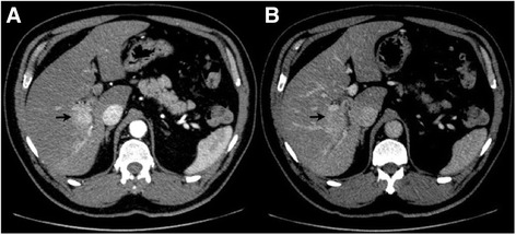
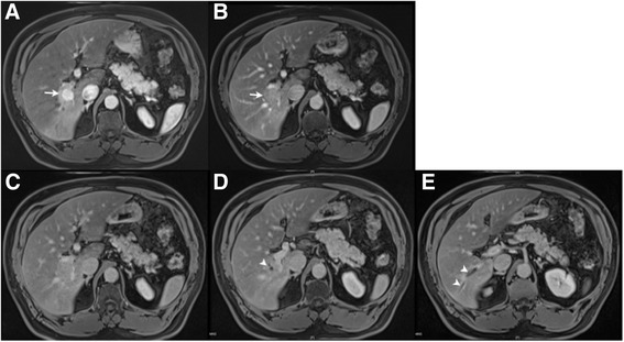
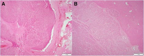
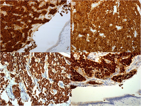
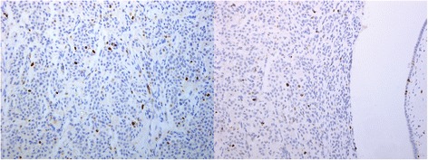
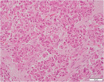
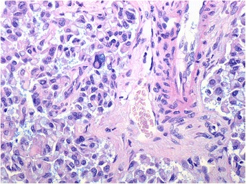
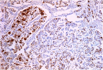
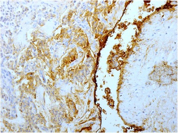
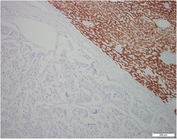
Case presentation: A 53-year old man presented with epigastric pain on a background of excessive alcohol consumption. Contrast-enhanced computed tomography imaging of the liver revealed a central enhancing mass located at the bifurcation of right anterior and posterior portal veins. Magnetic resonance imaging demonstrated intrahepatic biliary duct dilatation distal to the mass. The patient underwent a right lobe hepatectomy and excision of the extrahepatic biliary tree with formation of a hepaticojejunostomy. Histopathological finding of the specimen revealed an intraductal tumour with predominant neuroendocrine immunohistochemical phenotype and infiltration into nearby tissue. An element of glandular differentiation on immunohistochemistry confirmed the lesion as MANEC.
Conclusions: We present the first reported histopathological case of a MANEC arising from the intrahepatic bile ducts. This report aims to review what is known about primary neuroendocrine and mixed adeno-neuroendocrine carcinoma of the bile ducts, particularly in comparison to other types of biliary and hepatic tumours.
Figures
References
-
- Albores-Saavedra JSJ, Wittekind C, Sripa B, Menck HR, Soehandra N, Sriram PVJ. Tumours of the gallbladder and extrahepatic bile ducts. 2000.
-
- Albores-Saavedra J, HD, Klimstra DS. In: Rosai J SL, editor. Dysplasia, carcinoma-in-situ, and invasive carcinoma of the extrahepatic bile ducts. 3rd ed. Washington DC: Armed Forces Institute of Pathology; 2000.
-
- Komminoth P AR, Capella C, Klimstra DS, Kloppel G, Rindi G, Albores-Saavedra J. In: Bosman FT CF, Hruban RH, Theise ND, editor. Neuroendocrine neoplasms of the gallbladder and extrahepatic bile ducts. 4 ed. Lyon: IARC Press; 2010.
Publication types
MeSH terms
LinkOut - more resources
Full Text Sources
Other Literature Sources
Medical
