Review
. 2015:115:561-97.
doi: 10.1016/bs.ctdb.2015.09.003. Epub 2015 Oct 27.

Morphometrics, 3D Imaging, and Craniofacial Development

Affiliations
Review

Morphometrics, 3D Imaging, and Craniofacial Development

Benedikt Hallgrimsson et al. Curr Top Dev Biol. 2015.

Abstract

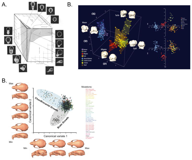
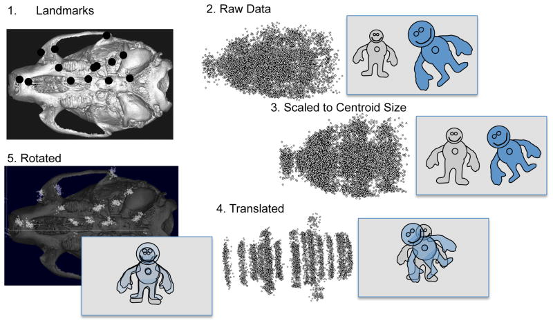
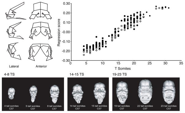
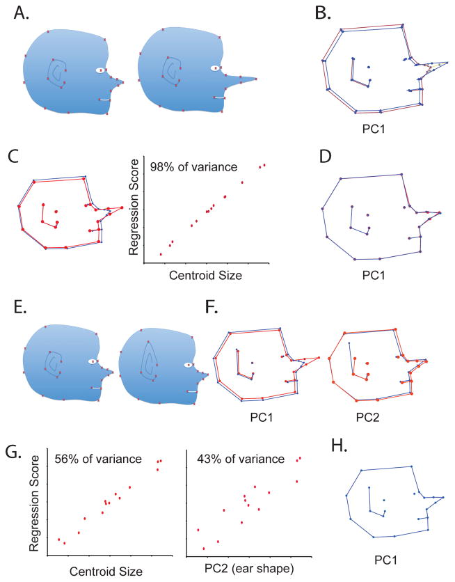
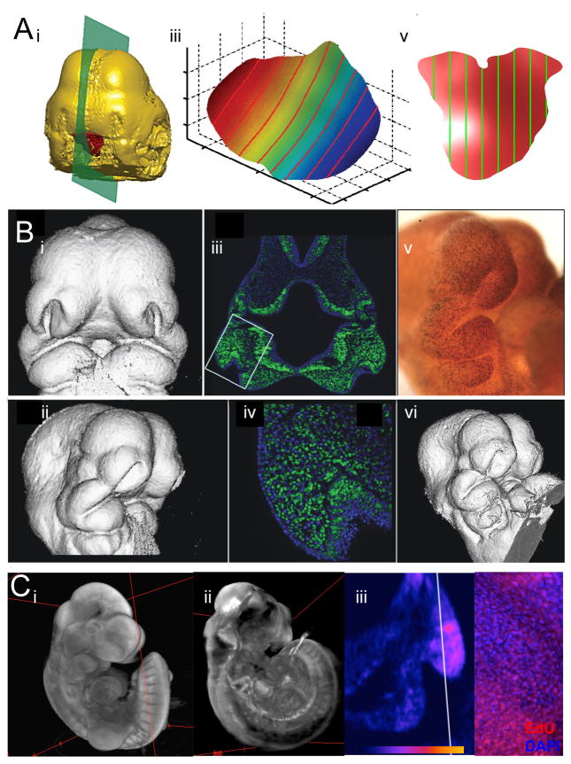
Recent studies have shown how volumetric imaging and morphometrics can add significantly to our understanding of morphogenesis, the developmental basis for variation, and the etiology of structural birth defects. On the other hand, the complex questions and diverse imaging data in developmental biology present morphometrics with more complex challenges than applications in virtually any other field. Meeting these challenges is necessary in order to understand the mechanistic basis for variation in complex morphologies. This chapter reviews the methods and theory that enable the application of modern landmark-based morphometrics to developmental biology and craniofacial development, in particular. We discuss the theoretical foundations of morphometrics as applied to development and review the basic approaches to the quantification of morphology. Focusing on geometric morphometrics, we discuss the principal statistical methods for quantifying and comparing morphological variation and covariation structure within and among groups. Finally, we discuss the future directions for morphometrics in developmental biology that will be required for approaches that enable quantitative integration across the genotype-phenotype map.

Keywords: Craniofacial; Imaging; MicroCT; Morphogenesis; Morphometrics; Mouse; Optical Projection Tomography; Phenomics.


Figures

Figure 1
Figure 1
A) Raup’s morphospace for shell coiling (Raup, 1966). B) Morphology for amniote embryos, including humans, is shown in a space constructed using Principal Components Analysis from 3D landmark data (Young et al. 2014). C) Analysis of mouse mutants with in a Canonical Variates Analysis based space. This plots shows craniofacial effects for mutations affecting chondrocanial growth and brain size forming two distinct common axes of covariation.
Figure 2
Figure 2
General Procrustes Superimposition. A shows a 2D view of a 3D adult mouse landmark set. B. shows the full scatter for the raw landmarks in this dataset. In this particular datasets, the two sides of each specimen are treated as separate individuals. B) The scatter for all individuals (left and right sides) after scaling the LM coordinates for centroid size. C) The scatter after translation so as to center the centroids of each individual. D). The distribution of Procrustes residuals after rotation and, where appropriate, reflection superimposed on a ventral view of a mouse.
Figure 3
Figure 3
Multivariate regression on Procrustes superimposed landmark data allows calculation of the shape changes that correspond to that regression. Here, tail somite stage is regressed on landmark coordinates that capture craniofacial shape variation during face formation. The wireframes show the deformations that correspond to the regression in frontal and lateral views while the scatter plot shows the regression scores plotted against tail somite stage.
Figure 4
Figure 4
Illustration of the Pinocchio effect. In A, a dataset is generated in which a profile of Pinocchio’s head varies only in that the nose gets longer. This only displaces the three landmarks on the nose. However, a PCA based on a Procrustes fit of those data shows small displacements of landmarks that were not actually displaced although the largest displacements involve the nose (B). However, the increase in nose length is perfectly correlated with centroid size and so regressing on CS completely removes both the nose length variation and its associated artifacts (C, D). In D, shape is standardized to the sample average and so shows an intermediate length nose. If we add an additional shape component, ear shape (E) that is uncorrelated with nose length, the Pinocchio effect is complicated slightly. Here, variation is spread across two PCs (F), one which captures nose length and another that captures ear length. These effects and all their associated changes in can be removed from the data as shown in G and H, either by regressing out centroid size and PC2 or PC1 and PC2. The artifactual shape changes, however, are preserved in the shape variation associated with these regressions.
Figure 5
Figure 5
The strongest (> 5% difference in means) significantly (α=0.05) different linear dimensions identified in a EDMA SHAPE comparison of skulls from 129 and C57 mouse strains, viewed from the lateral (top) and superior (bottom) aspects. Measurements were standardized by centroid size before analysis. Red lines are dimensions for which C57 is relatively longer than 129, while blue lines are relatively longer in 129 than C57.
Figure 6
Figure 6
Standard embryo craniofacial landmark set (Percival et al., 2014).
Figure 7
Figure 7
Right lateral view of the surface craniofacial morphology of E10.5, E11.5, and E12.5 mouse specimens in a standard orientation. As previously defined (Percival et al., 2014) the homologous location of a landmark between the maxillary and the lateral nasal prominences is shown. We consciously defined it so that it landmark such that it remains at the border between these cell populations rather than maintaining a standard geometric relationship with other cranial features like the eye.
Figure 8
Figure 8
Late fetal (Gonzalez et al. 2014), Neonatal (Boughner et al. 2008)), and adult mouse landmark sets used in recent studies by our group.
Figure 9
Figure 9
Principal Components Analysis in Geometric Morphometrics. A) Explanation of PCA (from Zeldicth, (2004)). The 2D plots illustrate PC1 and the projection of points (individuals) on to PC1 to create PC scores. The 3D plot shows how the scatter among three variables would correspond to principal components.
Figure 10
Figure 10
Allometry in adult mice and in mouse embryos (E1.0–12.5). A shows the allometric component of shape variation in the Parental strains and F1 crosses for the Collaborative Cross Mice. B and C show the results of multiple multivariate regression of shape on both tail somite stage (B) and size (C). B shows the component of shape variation that is related to tail somite stage while C shows the static allometry component that is perpendicular to the variation in C. A challenge in such analyses is the co-linearity among the effects of stage and size.
Figure 11
Figure 11
MicroCT scans of embryos fixed using different protocols shown at the same scale. Illustrating the effect of different fixation and scanning procedures on morphology. A) 4% formaldehyde + Bouin’s. B) 4% formaldehyde + 1% glutaraldehyde with Iothalamate meglumine for contrast. C) 4% formaldehyde + 5% glutaraldehyde plus contrast agent (Schmidt et al. 2010b).
Figure 12
Figure 12
Examples of Optical Projection Tomography Images. A) E10.5 mouse embryo stained with Sytox Green B) E12.5 mouse embryo stained with Sytox Green (blue) and Ser10 phosphohistone H3 (green). C) Hamburger Hamilton Stage 23 chick embryo showing Shh expression in the Frontonasal ectodermal zone (FEZ) highlighted in red.
Figure 13
Figure 13
A) Analysis of Shape of Gene Expression. i. OPT scan of whole mount in situ showing Shh expression (red). The green plane indicates the slices in i and ii. ii. Section of FEZ parallel to sagittal plane. iii. Surface spline extracted from raw FEZ. B) MicroCT renderings and cell proliferation data from the same specimens. i and ii) 3D reconstruction of μCT taken after processing but before sectioning. iii and iv) Hoescht 33342 staining to visualize cell nuclei (blue) with cells in S phase visualized using EdU + Alexa Fluor® 488 labeling (green) at 5X and 200X. v) Specimen processed wholemount for anti-PHH3 primary antibody to identify M-phase cells. vi) MicroCT rendering of the same specimen after treatment. C) Images derived from OPT imaging of EdU stained embryo counterstained with Sytox Green. B) Volume view of Sytox Green channel showing regional difference in cell density. C) Volume view of EdU channel showing regional difference in cell proliferation. D) Heat map of virtual section of lateral nasal prominence of (C) showing local proliferation differences. E) EdU incorporation as viewed in a traditional confocal section EdU stain (red), DAPI (blue).

References

    1. Alberch P. Developmental constraints in evolutionary processes. In: Bonner JT, editor. Development in Evolution. Berlin and New York: Springer-Verlag; 1982. pp. 313–332.
    1. Baghdadi L, Zamyadi M, Sled JG, Schneider JE, Bhattacharya S, Henkelman RM, Lerch JP. Semi-automatic segmentation of multiple mouse embryos in MR images. BMC Bioinformatics. 2011;12:237. - PMC - PubMed
    1. Bastir M, Rosas A. Hierarchical nature of morphological integration and modularity in the human posterior face. Am J Phys Anthropol. 2005;128(1):26–34. - PubMed
    1. Billington CJ, Jr, Schmidt B, Marcucio RS, Hallgrimsson B, Gopalakrishnan R, Petryk A. Impact of retinoic acid exposure on midfacial shape variation and manifestation of holoprosencephaly in Twsg1 mutant mice. Dis Model Mech. 2015;8(2):139–146. - PMC - PubMed
    1. Boehm B, Westerberg H, Lesnicar-Pucko G, Raja S, Rautschka M, Cotterell J, Swoger J, Sharpe J. The role of spatially controlled cell proliferation in limb bud morphogenesis. PLoS Biol. 2010;8(7):e1000420. - PMC - PubMed

Publication types

LinkOut - more resources