. 2016 Jun 14;37(23):1835-46.
doi: 10.1093/eurheartj/ehv579. Epub 2015 Nov 20.

Cardiac mesenchymal stromal cells are a source of adipocytes in arrhythmogenic cardiomyopathy

Affiliations

Cardiac mesenchymal stromal cells are a source of adipocytes in arrhythmogenic cardiomyopathy

E Sommariva et al. Eur Heart J. .

Abstract

Aim: Arrhythmogenic cardiomyopathy (ACM) is a genetic disorder mainly due to mutations in desmosomal genes, characterized by progressive fibro-adipose replacement of the myocardium, arrhythmias, and sudden death. It is still unclear which cell type is responsible for fibro-adipose substitution and which molecular mechanisms lead to this structural change. Cardiac mesenchymal stromal cells (C-MSC) are the most abundant cells in the heart, with propensity to differentiate into several cell types, including adipocytes, and their role in ACM is unknown. The aim of the present study was to investigate whether C-MSC contributed to excess adipocytes in patients with ACM.

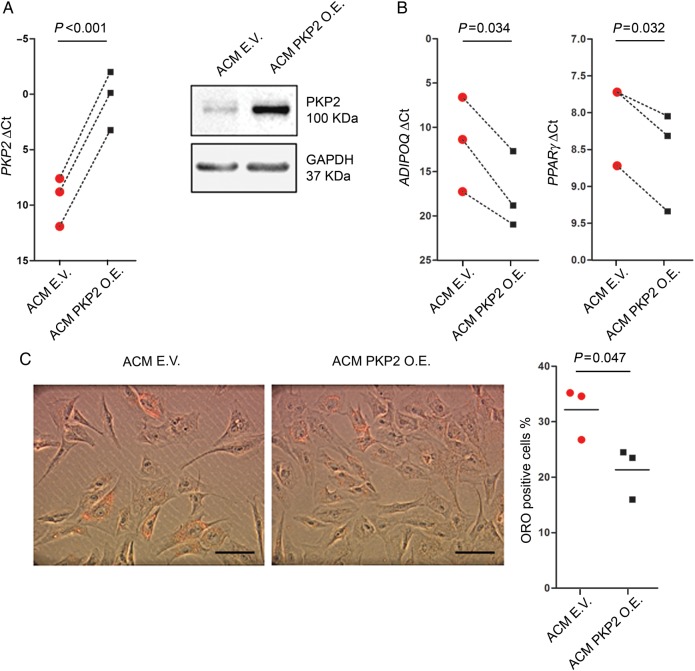
Methods and results: We found that, in ACM patients' explanted heart sections, cells actively differentiating into adipocytes are of mesenchymal origin. Therefore, we isolated C-MSC from endomyocardial biopsies of ACM and from not affected by arrhythmogenic cardiomyopathy (NON-ACM) (control) patients. We found that both ACM and control C-MSC express desmosomal genes, with ACM C-MSC showing lower expression of plakophilin (PKP2) protein vs.

Controls: Arrhythmogenic cardiomyopathy C-MSC cultured in adipogenic medium accumulated more lipid droplets than controls. Accordingly, the expression of adipogenic genes was higher in ACM vs. NON-ACM C-MSC, while expression of cell cycle and anti-adipogenic genes was lower. Both lipid accumulation and transcription reprogramming were dependent on PKP2 deficiency.

Conclusions: Cardiac mesenchymal stromal cells contribute to the adipogenic substitution observed in ACM patients' hearts. Moreover, C-MSC from ACM patients recapitulate the features of ACM adipogenesis, representing a novel, scalable, patient-specific in vitro tool for future mechanistic studies.

Keywords: Adipogenesis; Arrhythmogenic cardiomyopathy; Fibrofatty substitution; Mesenchymal stromal cells; Plakoglobin; Plakophilin2.


Figures

Figure 1
Figure 1
Characteristics of an arrhythmogenic cardiomyopathy patient (H1) who underwent heart transplant. (A) Twelve-lead electrocardiogram of a patient with an advanced form of arrhythmogenic cardiomyopathy, recorded 2 years before heart transplant, characterized by sinus rhythm, complete right bundle branch block, ST-T wave abnormalities in leads II, III, aVF, V1–V4 where an ε wave can also be identified. Frequent monomorphic premature ventricular beats originating from the inferior right ventricular wall are recorded. (B) Explanted heart of a late-stage arrhythmogenic cardiomyopathy patient, with biventricular involvement. Severe fibro-fatty infiltration is visible in the right ventricle, which is dilated with thin walls. A high-voltage implantable cardioverter defibrillator lead is visible in the right ventricular chamber. (C) Histological preparation of right ventricle tissue sample of the explanted heart (haematoxylin and eosin staining). The scale bar indicates 100 µm.
Figure 2
Figure 2
Cells undergoing adipogenic differentiation in arrhythmogenic cardiomyopathy hearts express the mesenchymal markers CD29 and CD105 and not the cardiomyocyte marker α-sarcomeric actin. Immunostaining of FFPE sections of explanted hearts from three arrhythmogenic cardiomyopathy patients. Nuclei are stained with Hoechst 33258. (A) PLIN1 antibody marks lipid drop membranes identifying pre-adipocytes and CD29 (upper panel), or CD105 (lower panel) as used as mesenchymal markers. The scale bar indicates 10 µm. (B) Transversal (upper panel) and longitudinal (lower panel) sections are immunostained with PLIN1 and α-sarcomeric actin for cardiomyocytes. The scale bar indicates 20 µm.
Figure 3
Figure 3
Representative diagnostic findings of an arrhythmogenic cardiomyopathy patient (B7) who underwent biopsy. (A) Twelve-lead electrocardiogram is characterized by sinus rhythm, normal conduction, negative T waves in V1–V3 leads. A ventricular premature beat originating from the right ventricular outflow tract is shown. (B) Cardiac magnetic resonance: four-chamber long axis (B, online videoA) and short-axis (B, online videoB) steady-state free procession sequences of left ventricle showed dilated rigth ventricle with multiple diastolic bulging in the subtricuspid region (arrows). The T1-weighted image (B′′) and post-contrast late gadolinium enhancement sequence (B′′′) show high-intensity signal suggesting fibro-adipose infiltration of the right ventricular-free wall (arrows). (C) Three-dimensional electroanatomical bipolar map of the right ventricular (endocardial aspect): normal signals are recorded over the entire right ventricular chamber (preserved voltage amplitude, >1.50 mV, purple), excepted from a portion of the anterior wall that is characterized by low-amplitude voltage (<1 mV, blue-yellow) with a small area of heterogeneous dense scar (<0.5 mV, red). (D) Endomyocardial biopsy bright field image. All biopsies used for cardiac mesenchymal stromal cells isolation were acquired in the area adjacent to the electro-anatomical scar and adipocytes are not evident at gross examination. (E) Histological preparation (haematoxylin and eosin staining) of a bioptic sample acquired in the pathological area, for diagnostic purposes. Confirmation of arrhythmogenic cardiomyopathy diagnosis is given by the evidence of large fibro-fatty substitution. (F) Histological preparation (Masson-trichrome staining) of a bioptic sample. Fibrotic tissue is evident in blue beside adipocytes (white round circles). All the scale bars indicate 100 µm.
Figure 4
Figure 4
Expression of desmosomal genes and proteins. ACM: samples from patients affected by arrhythmogenic cardiomyopathy; NON-ACM: samples from patients not affected by arrhythmogenic cardiomyopathy. (A) Desmosomal genes are expressed in ventricular mesenchymal stromal cells. Cardiac mesenchymal stromal cells RNA extracts either from patients not affected or affected by arrhythmogenic cardiomyopathy (n = 3 vs. n = 4) genes threshold cycles (Ct) with respect to the housekeeping gene GAPDH are examined. Total ventricle RNA extracts (n = 3) from patients not affected by arrhythmogenic cardiomyopathy are also reported as positive controls. (B) Desmosomal proteins are expressed in cardiac mesenchymal stromal cells, and PKP2 is expressed at a lower level in arrhythmogenic cardiomyopathy cardiac mesenchymal stromal cells. PKP2, PG, DSP1/2, DSG2, DSC2, and house-keeping gene GAPDH western blots of protein extracts of cardiac mesenchymal stromal cells from patients not affected by arrhythmogenic cardiomyopathy (n = 3) and affected (n = 4) are shown. Total ventricle lysates (n = 2) from patients not affected by arrhythmogenic cardiomyopathy are shown as positive controls. Densitometry, relative to the loading control GAPDH, is shown in the graphs, normalized for the expression of samples from patients not affected by arrhythmogenic cardiomyopathy.
Figure 5
Figure 5
Cardiac mesenchymal stromal cells obtained from arrhythmogenic cardiomyopathy patients (ACM), cultured in adipogenic medium, accumulate more lipid droplets than control cardiac mesenchymal stromal cells (NON-ACM). Representative images of cardiac mesenchymal stromal cells from bioptic samples of patients affected and not affected by arrhythmogenic cardiomyopathy, cultured for 72 h and 1 week in adipogenic medium, stained with Oil Red O (ORO; n = 6 vs. n = 4). The scale bar indicates 50 μm. Quantification of the luminance of the 255 red staining is reported on the right: intensity is expressed in arbitrary units.
Figure 6
Figure 6
Arrhythmogenic cardiomyopathy cardiac mesenchymal stromal cells in adipogenic medium show higher transcription of adipogenic genes and proteins and a lower transcription of anti-adipogenic and proliferation genes, compared to control cells. ACM: samples from patients affected by arrhythmogenic cardiomyopathy; NON-ACM: samples from patients not affected by arrhythmogenic cardiomyopathy. (A) Comparison of transcript abundance (genes threshold cycles [Ct] with respect to the house-keeping gene GAPDH) between RNA extract of cardiac mesenchymal stromal cells from patients not affected and affected by arrhythmogenic cardiomyopathy (n ≥ 4), after 72 h culture in adipogenic medium. (B) ADIPOQ and PLIN1 gene expression positively correlates with lipid accumulation (Oil Red O staining) in arrhythmogenic cardiomyopathy samples. (C) Comparison of PLIN1, PPARγ, and FABP4 protein abundance between protein extracts of cardiac mesenchymal stromal cells from patients not affected by arrhythmogenic cardiomyopathy (n = 3) and from arrhythmogenic cardiomyopathy patients (n = 5), after 72 h culture in adipogenic medium. Quantification of the mean protein abundance relative to GAPDH, and normalized for values of samples from patients not affected by arrhythmogenic cardiomyopathy, is shown in the graphs.
Figure 7
Figure 7
Arrhythmogenic cardiomyopathy cardiac mesenchymal stromal cells lipid accumulation is dependent on the expression of PKP2. Arrhythmogenic cardiomyopathy cardiac mesenchymal stromal cells (ACM; n = 3) were transfected with an empty vector (EV) or a vector overexpressing PKP2 (PKP2 OE). (A) Increased expression of PKP2 gene (left graph) and PKP2 protein (right blot) in the PKP2 OE cells compared with mock-transfected cells, 72 h after transfection. (B) Comparison of transcript abundance between arrhythmogenic cardiomyopathy cardiac mesenchymal stromal cells transfected either with the empty or the PKP2 expression vector, after 72 h culture in adipogenic medium. (C) Representative Oil Red O (ORO) staining images of arrhythmogenic cardiomyopathy cardiac mesenchymal stromal cells transfected either with the empty or the PKP2 expression vector, cultured for 72 h in adipogenic medium. The graph shows the percentage of cells containing at least one lipid droplet. The scale bar indicates 50 μm.
Figure 8
Figure 8
Cardiac mesenchymal stromal cells cultured in adipogenic medium show PLIN1 staining similar to differentiating cells in patients’ hearts. (A) Immunostaining for PLIN1 of cells from patients not affected by arrhythmogenic cardiomyopathy (NON-ACM) and from arrhythmogenic cardiomyopathy patients (ACM), cultured in adipogenic medium for 72 h. The scale bar indicates 10 μm. (B) Immunostaining for PLIN1 on FFPE sections of an arrhythmogenic cardiomyopathy (ACM) explanted heart. The scale bar indicates 20 μm. Nuclei are stained with Hoechst 33258.

Comment in

  • When the money is not in the bank.
    Jacoby D, Pantazis A. Jacoby D, et al. Eur Heart J. 2016 Jun 14;37(23):1847-9. doi: 10.1093/eurheartj/ehv645. Epub 2015 Dec 18. Eur Heart J. 2016. PMID: 26685139 Free PMC article. No abstract available.

References

    1. Asimaki A, Kleber AG, Saffitz JE. Pathogenesis of arrhythmogenic cardiomyopathy. Can J Cardiol 2015. 10.1016/j.cjca.2015.04.012. - DOI - PMC - PubMed
    1. Cerrone M, Noorman M, Lin X, Chkourko H, Liang FX, van der Nagel R, Hund T, Birchmeier W, Mohler P, van Veen TA, van Rijen HV, Delmar M. Sodium current deficit and arrhythmogenesis in a murine model of plakophilin-2 haploinsufficiency. Cardiovasc Res 2012;95:460–468. - PMC - PubMed
    1. Sato PY, Musa H, Coombs W, Guerrero-Serna G, Patino GA, Taffet SM, Isom LL, Delmar M. Loss of plakophilin-2 expression leads to decreased sodium current and slower conduction velocity in cultured cardiac myocytes. Circ Res 2009;105:523–526. - PMC - PubMed
    1. Oxford EM, Everitt M, Coombs W, Fox PR, Kraus M, Gelzer AR, Saffitz J, Taffet SM, Moise NS, Delmar M. Molecular composition of the intercalated disc in a spontaneous canine animal model of arrhythmogenic right ventricular dysplasia/cardiomyopathy. Heart Rhythm 2007;4:1196–1205. - PMC - PubMed
    1. Swope D, Cheng L, Gao E, Li J, Radice GL. Loss of cadherin-binding proteins beta-catenin and plakoglobin in the heart leads to gap junction remodeling and arrhythmogenesis. Mol Cell Biol 2012;32:1056–1067. - PMC - PubMed