. 2015 Nov 18;88(4):667-77.
doi: 10.1016/j.neuron.2015.10.038.

RAN Translation in Huntington Disease

Affiliations

RAN Translation in Huntington Disease

Monica Bañez-Coronel et al. Neuron. .

Abstract

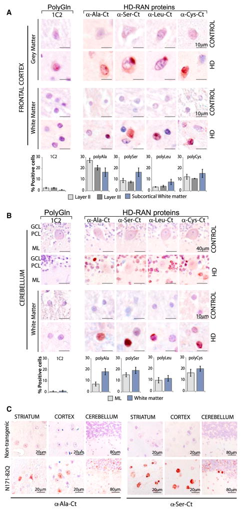
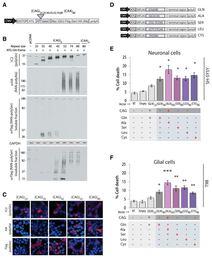
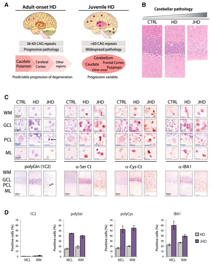
Huntington disease (HD) is caused by a CAG ⋅ CTG expansion in the huntingtin (HTT) gene. While most research has focused on the HTT polyGln-expansion protein, we demonstrate that four additional, novel, homopolymeric expansion proteins (polyAla, polySer, polyLeu, and polyCys) accumulate in HD human brains. These sense and antisense repeat-associated non-ATG (RAN) translation proteins accumulate most abundantly in brain regions with neuronal loss, microglial activation and apoptosis, including caudate/putamen, white matter, and, in juvenile-onset cases, also the cerebellum. RAN protein accumulation and aggregation are length dependent, and individual RAN proteins are toxic to neural cells independent of RNA effects. These data suggest RAN proteins contribute to HD and that therapeutic strategies targeting both sense and antisense genes may be required for efficacy in HD patients. This is the first demonstration that RAN proteins are expressed across an expansion located in an open reading frame and suggests RAN translation may also contribute to other polyglutamine diseases.


Conflict of interest statement

No other authors have conflicts.

Figures

Figure 1
Figure 1. RAN Translation in HD
(A) CAG and CAA HTT-exon1 minigenes with a 6×STOP codon cassette (two stops in each frame) upstream of HTT exon 1 and C-terminal epitope tags in each of the three reading frames. (B) Immunoblots of HEK293T cells show polyGln expression from all +ATG-constructs and RAN polyGln, polyAla and polySer proteins from (CAG)80 but not (CAG)23 or non-hairpin-forming (CAA)80 expansions. (C) Schematic diagram of putative HD-RAN proteins. C-terminal regions used to generate peptide for HD-RAN polyclonal antibodies are underlined. (D) IHC staining of human autopsy tissue shows positive staining (red) for sense (polyAla and polySer) and antisense (polyCys and polyLeu) RAN proteins in HD ([De]–[Dh]) but not HDL2 ([Di]–[Dl]), control ([Da]–[Dd]) or preimmune ([Dm]–[Dp]) controls. Red, positive staining; blue, nuclear counterstain. (E) Protein blots showing RAN proteins detected from the insoluble fraction of HD but not control frontal cortex lysates. See also Figures S1A and S1B. HMW, high molecular weight.
Figure 2
Figure 2. HD-RAN Proteins in Striatum
(A) Control and HD brain showing striatal sub-regions with summary of staining. (B) Quantification of IHC-positive cells for α-Gln, α-RAN, and IBA1 staining ± SEM in caudate nucleus. (C–I) IHC of striatal sub-sections from HD brains using (C) α-Gln antibody (1C2), ([D]–[G]) α-RAN antibodies, (H) α-IBA1 for microglia, and (I) α-active caspase-3 for cell death. (A)–(I): red, positive staining; blue, nuclear counterstain. (J) Double staining for RAN protein cocktail using mixture of all four anti-RAN antibodies (brown) plus anti-IBA1 antibody to label microglia with quantitation (± SEM) of single-, double-labeled, and IBA1 cells that are in close proximity to RAN-positive cells. (K) Double staining for RAN protein cocktail using mixture of all anti-RAN antibodies (brown) plus anti-caspase-3 antibody to label apoptotic cells with quantitation of the percent active Caspase-3-positive cells that are also positive for RAN proteins. See also Figure S2. Cau-WMB, caudate white matter bundles; Int-Cap, internal capsule; Put-WMB, putamen white matter bundles.
Figure 3
Figure 3. HD-RAN Proteins in Human Frontal Cortex and Cerebellum and HD Mice
(A and B) IHC staining of control and HD gray and white matter of (A) frontal cortex and (B) cerebellum using α-RAN and α-Gln (1C2) antibodies show punctate nuclear and cytoplasmic staining with α-polyAla, α-polySer, α-polyLeu, and α-polyCys. GCL, granule cell layer; PCL, Purkinje-cell layer; ML, molecular layer. Staining of the cortex and cerebellum in adult-onset HD cases is variable. IHC images and quantification of percent positive cells represent typical positive regions. (C) IHC staining of indicated brain regions in N171-82Q and control mice using the α-polyAla, α-polySer. Red, positive staining; blue, nuclear counterstain. See also Figures S3A–S3E.
Figure 4
Figure 4. Length-Dependent RAN Protein Expression, Aggregation, and Toxicity
(A) CAG and CAA HTT-exon1 minigenes. (B and C) (B) Immunoblots and (C) IF of HEK293T cells after transfection with indicated constructs. (D) Transfected minigenes (top) containing non-hairpin-forming alternative codons in the repeat region for polyGln90 (CAA), polySer90 (TCC-TCT), polyLeu90 (CTT-CTC), and polyCys90 (TGT) constructs. Because non-haipin forming codon substitutions encoding polyAla were not available, polyAla was expressed by a +ATG-GCA construct using a slightly longer repeat tract of 105 repeats. (E and F) LDH assays of SH-SY5Y and T98 cells expressing polyGln and individual HD-RAN proteins 42 hr post-transfection. Values equal percent of cell death ± SEM (n = 5; *p < 0.05, **p < 0.01, and ***p < 0.001). See also Figures S4A–S4F.
Figure 5
Figure 5. Increased RAN Protein Staining in Juvenile HD
(A) Schematic diagram summarizing features of adult-onset and juvenile HD pathology. (B) H&E staining in control, adult-onset, and juvenile-onset HD cases with cerebellar atrophy. (C) α-polyGln, α-polySer, α-polyCys and α-IBA1staining in cerebellar layers. (D) Quantitation of IHC-positive cells with 1C2 (polyGln) and α-polySer-Ct, α-polyCys-Ct, and α-IBA1 antibodies. WM, white matter; GCL, granule cell layer; PCL, Purkinje-cell layer; ML, molecular layer. Red, positive staining; blue, nuclear counterstain. Staining of cerebellum in adult-onset HD cases is variable. IHC images and quantification of percent positive cells represent typical positive regions. See also Figures S5A and S5B.

References

    1. Ash PE, Bieniek KF, Gendron TF, Caulfield T, Lin WL, Dejesus-Hernandez M, van Blitterswijk MM, Jansen-West K, Paul JW, 3rd, Rademakers R, et al. Unconventional translation of C9ORF72 GGGGCC expansion generates insoluble polypeptides specific to c9FTD/ALS. Neuron. 2013;77:639–646. - PMC - PubMed
    1. Bañez-Coronel M, Porta S, Kagerbauer B, Mateu-Huertas E, Pantano L, Ferrer I, Guzmán M, Estivill X, Martí E. A pathogenic mechanism in Huntington’s disease involves small CAG-repeated RNAs with neurotoxic activity. PLoS Genet. 2012;8:e1002481. - PMC - PubMed
    1. Bates G, Tabrizi SJ, Jones L, editors. Huntington’s Disease. 4. Oxford University Press; 2014.
    1. Björkqvist M, Wild EJ, Thiele J, Silvestroni A, Andre R, Lahiri N, Raibon E, Lee RV, Benn CL, Soulet D, et al. A novel pathogenic pathway of immune activation detectable before clinical onset in Huntington’s disease. J Exp Med. 2008;205:1869–1877. - PMC - PubMed
    1. Björkqvist M, Wild EJ, Tabrizi SJ. Harnessing immune alterations in neurodegenerative diseases. Neuron. 2009;64:21–24. - PubMed

Publication types

MeSH terms