Review
. 2015;50(6):532-49.
doi: 10.3109/10409238.2015.1091804. Epub 2015 Oct 13.

Torsins: not your typical AAA+ ATPases

Affiliations
Review

Torsins: not your typical AAA+ ATPases

April E Rose et al. Crit Rev Biochem Mol Biol. 2015.

Abstract

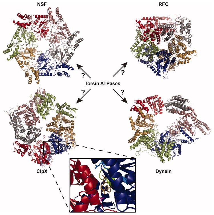
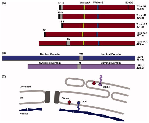
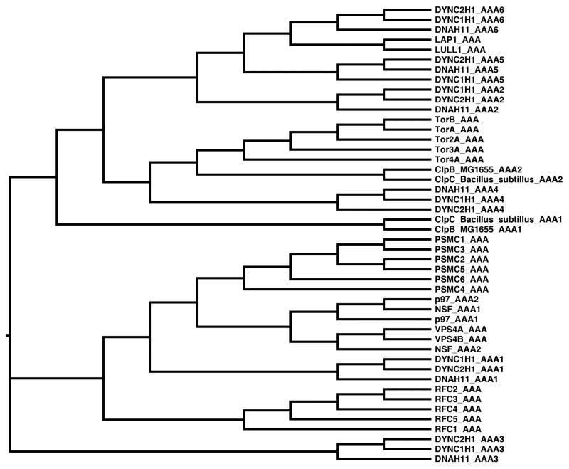
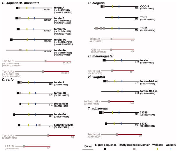
Torsin ATPases (Torsins) belong to the widespread AAA+ (ATPases associated with a variety of cellular activities) family of ATPases, which share structural similarity but have diverse cellular functions. Torsins are outliers in this family because they lack many characteristics of typical AAA+ proteins, and they are the only members of the AAA+ family located in the endoplasmic reticulum and contiguous perinuclear space. While it is clear that Torsins have essential roles in many, if not all metazoans, their precise cellular functions remain elusive. Studying Torsins has significant medical relevance since mutations in Torsins or Torsin-associated proteins result in a variety of congenital human disorders, the most frequent of which is early-onset torsion (DYT1) dystonia, a severe movement disorder. A better understanding of the Torsin system is needed to define the molecular etiology of these diseases, potentially enabling corrective therapy. Here, we provide a comprehensive overview of the Torsin system in metazoans, discuss functional clues obtained from various model systems and organisms and provide a phylogenetic and structural analysis of Torsins and their regulatory cofactors in relation to disease-causative mutations. Moreover, we review recent data that have led to a dramatically improved understanding of these machines at a molecular level, providing a foundation for investigating the molecular defects underlying the associated movement disorders. Lastly, we discuss our ideas on how recent progress may be utilized to inform future studies aimed at determining the cellular role(s) of these atypical molecular machines and their implications for dystonia treatment options.

Keywords: Early-onset torsion dystonia; LAP1; LULL1; endoplasmic reticulum; nuclear envelope; torsinA.


Conflict of interest statement

Declaration of Interest

The authors are funded by NIH (DP2 OD008624-01).

The authors report no declarations of interest.

Figures

FIGURE 1
FIGURE 1
Structural comparison of AAA+ ATPase assemblies. ClpX (PDB ID: 3HWS (Glynn et al. 2009)) and N-ethylmaleimide-sensitive factor (NSF) (PDB ID: 1D2N (Lenzen et al. 1998) are AAA+ ATPases that form homohexameric assemblies. Replication factor C (RFC) (PDB ID: 1SXJ (Bowman et al. 2004)) and dynein (4W8F (Bhabha et al. 2014)) are heteromeric AAA+ ATPases. RFC forms a heteropentameric assembly comprised of 5 different AAA subunits and dynein forms a heterohexameric assembly where all AAA domains are encoded in one continuous peptide. Individual AAA domains are depicted in different colors. Inset: Zoomed in image of the intersubunit interface of a ClpX dimer highlighting the Walker A site (green), Walker B site (light blue), and arginine finger (magenta) surrounding the nucleotide. Key residues K127, E185 (mutated to Q in the structure), and R307 are represented as sticks. ADP is shown in gold.
FIGURE 2
FIGURE 2
Domain structure of Torsin family ATPases. (A) Graphical comparison of the five members of the Torsin family of AAA+ ATPases depicting locations of various features in the primary sequence. SS: Signal Sequence; H: Hydrophobic Patch; Walker A: ATP binding motif; Walker B: ATP hydrolysis motif; E302: Glutamate deleted in the TorsinA disease mutant; TM: Predicted transmembrane segment. (B) Graphical comparison of cofactors LAP1 and LULL1 depicting locations of transmembrane segments and activator arginines. TM: transmembrane segment; R563/R449: activator arginines. (C) Cartoon depicting the subcellular localization of Torsins, LAP1, and LULL1. Torsin is anchored to the luminal face of the ER and NE membranes. LAP1 resides in the inner nuclear membrane and LULL1 in the ER membrane. Black rectangles: nuclear pore complex. Blue lines: nuclear lamina.
FIGURE 3
FIGURE 3
Phylogenetic comparison of AAA domains from various AAA ATPases. Phylogenetic tree built by MetaPIGA (Helaers & Milinkovitch 2010). Input sequences were individual AAA domains from each protein as determined by NCBI Conserved Domain Database (Marchler-Bauer et al. 2011), UniProt (Dyneins), or HHpred (LAP1 and LULL1) (Soding et al. 2005). All sequences used represent the human protein unless otherwise stated.
FIGURE 4
FIGURE 4
Evolutionary conservation of Torsin and LAP1/LULL1. Graphical representation of the primary sequence characteristics of Torsin (black) and LAP1/LULL1 (gray/red) homologs from various species. Red portions of cofactor homologs denotes domains predicted to have a AAA-fold.
FIGURE 5
FIGURE 5
Crystal structure of LAP1’s luminal domain and its potential impact on the Torsin ATPase oligomeric assembly. (A) Left Cartoon representation of a single NSF AAA domain (D2) from Cricetulus griseus (PDB ID: 1D2N (Lenzen et al. 1998)) showing the alpha/beta Rossman/RecA fold with the C-terminal four helix bundle. Right Cartoon representation of LAP1’s luminal domain from Homo sapiens (PDB ID:4TVS (Sosa et al. 2014)). LAP1’s luminal domain adopts a AAA-like fold without the C-terminal helical bundle. (B) Left TorsinA’s proposed homohexameric ring assembly. Each Torsin monomer’s nucleotide binding domain is depicted as a wedge where each monomer’s C-terminal helical bundle is depicted as a ball (red). Right Proposed heterohexameric ring assembly of TorsinA (red) with cofactor LAP1/LULL1 (blue). LAP1/LULL1 lack C-terminal helical bundles, which may prevent contact between adjacent subunits at the non-activating interfaces (arrows). (C) TorsinA (red ball) may form a homohexameric ring that, upon binding LAP1/LULL1 (blue ball), could cause the ATPase to adopt a split lock-washer assembly since LAP1/LULL1 lack the C-terminal helical bundle. Different nucleotide states could impact opening/closing of the split ring, possibly enabling a gating-mechanism to access the central pore of the ring. Membrane, gray dashed lines; LAP1/LULL1’s transmembrane domain, blue rectangle; LAP1/LULL1 nuclear/cytoplasmic domain, blue curved line.
FIGURE 6
FIGURE 6
Torsin activation by a strictly conserved arginine. (A) Surface representation of the heterodimeric model of LAP1 (PDB ID: 4TVS (Sosa et al. 2014)) and TorsinA (Phyre2 model (Brown et al. 2014)). The membrane is represented as dashed lines, membrane helices are represented as rectangular cylinders, and LAP1’s nuclear domain is shown as a curved line. (B) Zoomed in view of the active site showing the Walker A (K108) and Walker B (E171) residues of Torsin and the arginine (R563) provided by LAP1. AMPNP: gold (C) Sequence alignment (MUSCLE (Edgar 2004); colored by boxshade) of cofactor homologs diagrammed in Figure 4 showing the location of the strictly conserved arginine residue (arrow).
FIGURE 7
FIGURE 7
Potential biological functions of the Torsin/cofactor assembly. (A) Model of nuclear membrane fission mediated by LAP1, with subsequent disassembly and recycling of LAP1 oligomers by Torsin ATPase activity. (B) Model of Torsin and cofactor-mediated transmembrane signaling between the ER and cytoplasm (LULL1) or nucleoplasm (LAP1). (C) Model of cytoplasmic membrane tethering by LULL1’s nuclear domain that is disassembled by Torsin ATPase activity.
FIGURE 8
FIGURE 8
Physical locations of disease mutations in TorsinA and LAP1. Semi-transparent surface representation of the heterohexameric model of LAP1 (PDB ID: 4TVS (Sosa et al. 2014)) and TorsinA (Phyre2 model (Brown et al. 2014)) bound to AMPPNP using a least-squares superposition of alpha-carbons (Coot) onto hexameric p97 (PDB ID: 3CF2 (Davies et al. 2008)). LAP1 is depicted in light gray, TorsinA in dark gray, and disease causing mutations in blue. Mutations that do not map to the luminal domains of these proteins are highlighted with blue asterisks. The membrane is represented as gray dashed lines, membrane helices are represented as rectangular cylinders, and LAP1’s nuclear domain is shown as a curved line.

References

    1. AUGUSTIN S, GERDES F, LEE S, TSAI FT, LANGER T, TATSUTA T. An intersubunit signaling network coordinates ATP hydrolysis by m-AAA proteases. Mol Cell. 2009;35:574–85. - PMC - PubMed
    1. BABST M, WENDLAND B, ESTEPA EJ, EMR SD. The Vps4p AAA ATPase regulates membrane association of a Vps protein complex required for normal endosome function. EMBO J. 1998;17:2982–93. - PMC - PubMed
    1. BASHAM SE, ROSE LS. Mutations in ooc-5 and ooc-3 disrupt oocyte formation and the reestablishment of asymmetric PAR protein localization in two-cell Caenorhabditis elegans embryos. Dev Biol. 1999;215:253–63. - PubMed
    1. BASHAM SE, ROSE LS. The Caenorhabditis elegans polarity gene ooc-5 encodes a Torsin-related protein of the AAA ATPase superfamily. Development. 2001;128:4645–56. - PubMed
    1. BHABHA G, CHENG HC, ZHANG N, MOELLER A, LIAO M, SPEIR JA, CHENG Y, VALE RD. Allosteric communication in the dynein motor domain. Cell. 2014;159:857–68. - PMC - PubMed

Publication types