. 2015 Nov 2;25(21):2751-2762.
doi: 10.1016/j.cub.2015.09.025. Epub 2015 Oct 22.

Warts phosphorylates mud to promote pins-mediated mitotic spindle orientation in Drosophila, independent of Yorkie

Affiliations

Warts phosphorylates mud to promote pins-mediated mitotic spindle orientation in Drosophila, independent of Yorkie

Evan B Dewey et al. Curr Biol. .

Abstract

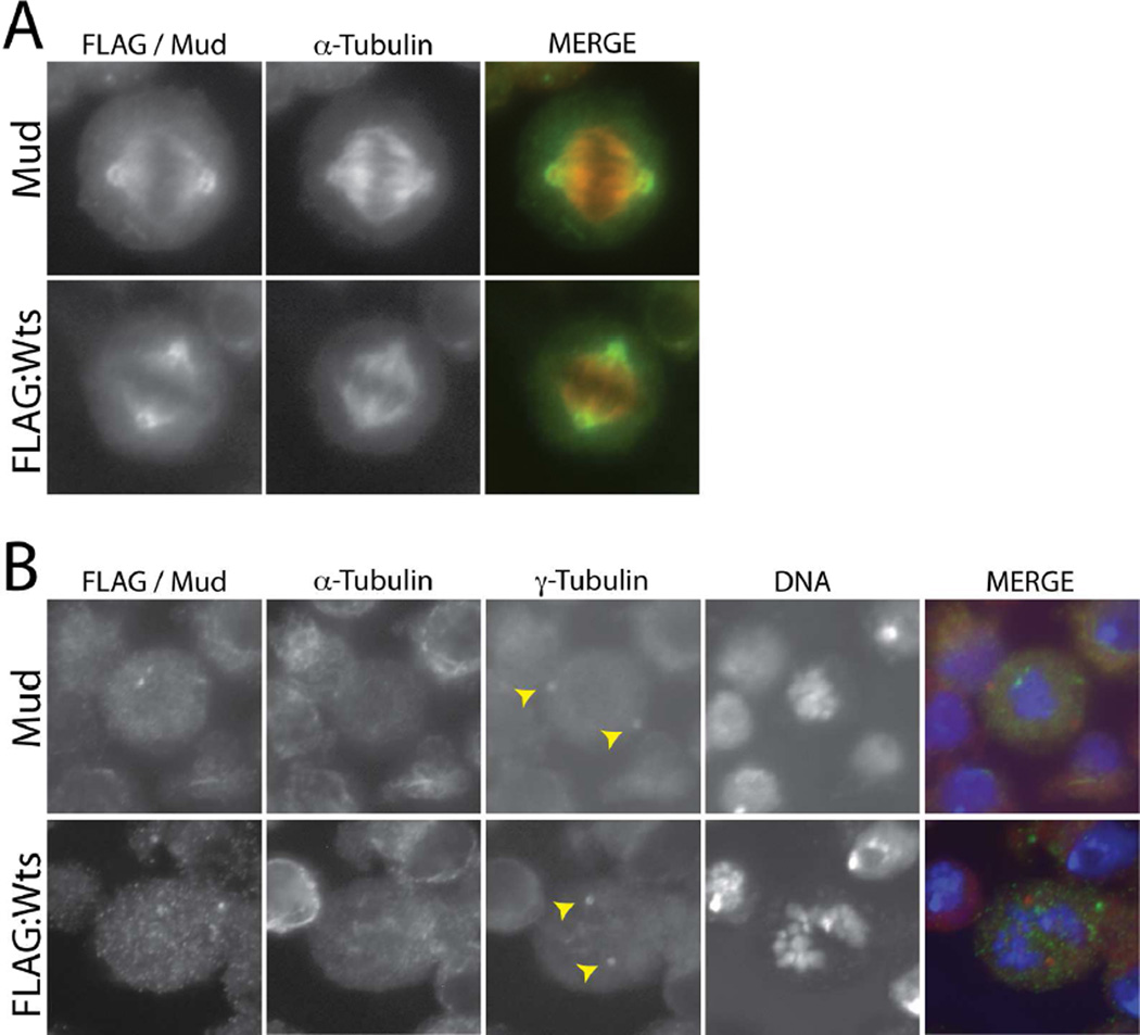
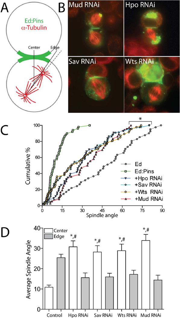
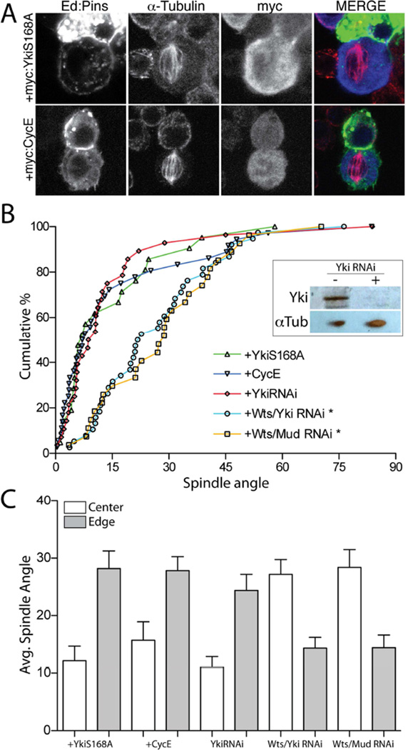
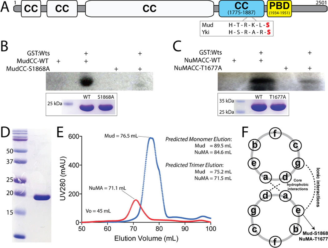
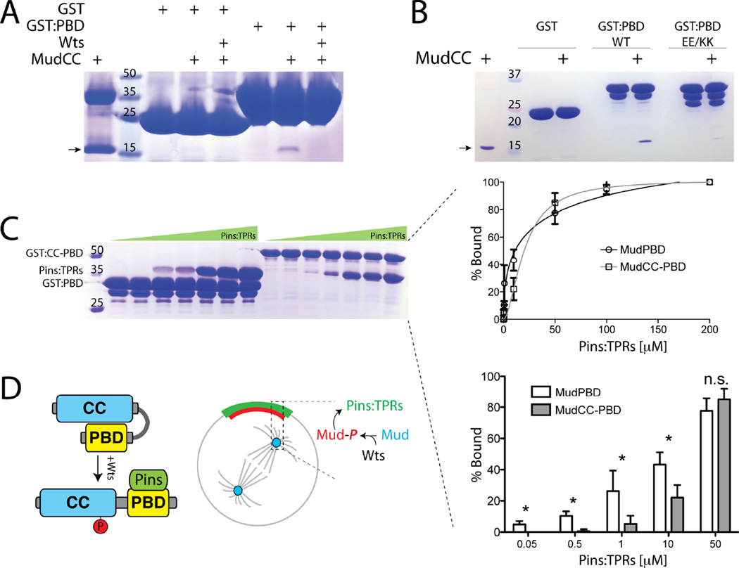
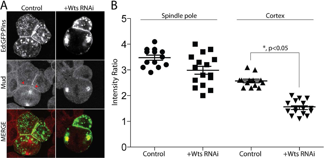
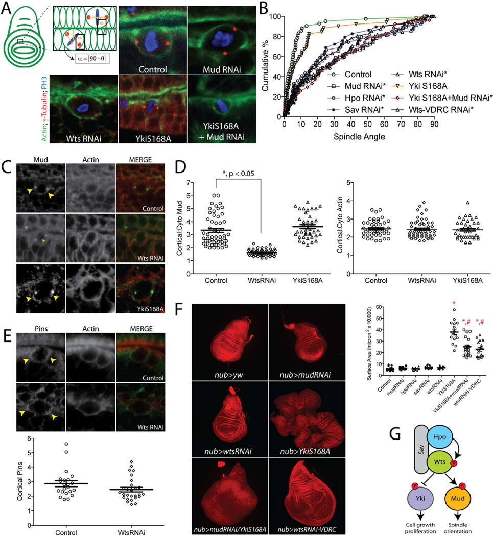
Multicellular animals have evolved conserved signaling pathways that translate cell polarity cues into mitotic spindle positioning to control the orientation of cell division within complex tissue structures. These oriented cell divisions are essential for the development of cell diversity and the maintenance of tissue homeostasis. Despite intense efforts, the molecular mechanisms that control spindle orientation remain incompletely defined. Here, we describe a role for the Hippo (Hpo) kinase complex in promoting Partner of Inscuteable (Pins)-mediated spindle orientation. Knockdown of Hpo, Salvador (Sav), or Warts (Wts) each result in a partial loss of spindle orientation, a phenotype previously described following loss of the Pins-binding protein Mushroom body defect (Mud). Similar to orthologs spanning yeast to mammals, Wts kinase localizes to mitotic spindle poles, a prominent site of Mud localization. Wts directly phosphorylates Mud in vitro within its C-terminal coiled-coil domain. This Mud coiled-coil domain directly binds the adjacent Pins-binding domain to dampen the Pins/Mud interaction, and Wts-mediated phosphorylation uncouples this intramolecular Mud interaction. Loss of Wts prevents cortical Pins/Mud association without affecting Mud accumulation at spindle poles, suggesting phosphorylation acts as a molecular switch to specifically activate cortical Mud function. Finally, loss of Wts in Drosophila imaginal disc epithelial cells results in diminished cortical Mud and defective planar spindle orientation. Our results provide new insights into the molecular basis for dynamic regulation of the cortical Pins/Mud spindle positioning complex and highlight a novel link with an essential, evolutionarily conserved cell proliferation pathway.


Figures

Figure 1
Figure 1. Warts localizes to mitotic spindle poles in mitotic S2 cells
(A) Cells were transfected with full-length Warts tagged with an N-terminal FLAG epitope sequence (FLAG:Wts) and stained with antibodies against FLAG and α-tubulin. To visualize endogenous Mud, untransfected cells were stained with an α-tubulin and Mud antibodies. (B) To depolymerize spindle microtubules, cells were treated with colchicine (12.5 µM) for 2 hours prior to fixation and antibody staining. Yellow arrowheads indicate both γ-tubulin-positive centrosomes, to which neither Wts nor Mud show significant localization. α-tubulin staining indicates successful depolymerization of spindle microtubules; note this channel is not shown in the merge panel.
Figure 2
Figure 2. The core Hippo kinase complex is required for robust Pins-mediated spindle orientation in polarized S2 cells
(A) Schematic overview of induced polarity assay. Cells are transfected with Pins fused to the truncated intracellular C-terminus of the adhesion protein Echnoid (Ed), which also contains GFP for visualization. Cell adhesion induces polarized Ed-based crescents, relative to which mitotic spindle orientation is measured. (B) S2 cells were transfected with Ed:GFP:Pins and subsequently treated with dsRNAi against indicated protein. Cells were fixed and stained with an α-tubulin antibody. In addition to the contact-induced cortical crescent, Ed:GFP:Pins often forms cytoplasmic puncta that can accumulate at peri-centrosomal regions. This localization can still occur in the presence of each RNAi treatment, suggesting that neither the Hippo complex nor Mud are required for this non-cortical localization. Also, previous studies have shown spindle orientation to be independent of the amount of puncta present [10]. (C) Spindle angles from at least 30 individual cells for each genotype are plotted as the cumulative percentage of cells at or below a given angle of spindle orientation. Each RNAi causes a phenotype intermediate between fully active Ed:Pins and the Ed alone negative control. Each RNAi condition was statistically different from both Ed and Ed:Pins but not one another (*, p < 0.05 ANOVA with Tukey’s post-hoc test). (D) Spindle angles were measured relative to the Ed:Pins crescent edge (grey filled bars) and center (open bars) for each condition. Fully active Ed:Pins (Control) positions spindles more closely to the center, whereas Hpo, Sav, and Wts RNAi preferentially orient spindles to the edge similar to Mud RNAi. *, p < 0.05 for ‘center’ versus ‘edge’ values within respective genotypes; #, p<0.05 for given RNAi ‘center’ compared to Control ‘center’, ANOVA followed by Tukey’s post-hoc test. The absence or presence of peri-centrosomal Ed:Pins puncta was similar across all conditions examined and did not show any significant correlation with spindle angle measurements, demonstrating they neither interfere with nor contribute to the spindle orientation process. See also Figure S1.
Figure 3
Figure 3. Hippo signaling acts independently of its canonical effector, Yorkie, to control S2 cell spindle orientation
(A) Cells were transfected with Ed:GFP:Pins together with either Yorkie-S168A (YkiS168A) or cyclin-E (CycE) as N-terminal c-myc tag fusion constructs. Cells were fixed and stained with antibodies against α-tubulin and the c-myc epitope. (B) Cumulative percentage plots for indicated genotypes. Neither CycE nor YkiS168A affect Ed:Pins function; RNAi against Yki was also without effect. Combined treatment of Wts and Yki RNAi or Wts and Mud RNAi resulted in spindle orientation similar to either Wts or Mud RNAi alone. Inset: western blot (20 µg total S2 cell lysate protein) indicating robust knockdown of Yki protein expression in cells treated with YkiRNAi. *, p < 0.05 compared to Ed:Pins, ANOVA followed by Tukey’s post-hoc test. (C) Spindle angles were measured relative to the Ed:Pins crescent edge (grey filled bars) and center (open bars) for each condition. In each genotype, edge and center measurements were statistically separated, p < 0.05, ANOVA followed by Tukey’s post-hoc test.
Figure 4
Figure 4. Warts directly phosphorylates Mud within its C-terminal coiled-coil domain
(A) The domain architecture of Mud consists primarily of coiled coil (CC) domains. The most C-terminal of these (MudCC; blue) contains a single consensus phosphorylation motif for Wts kinase that closely resembles that found in Yki. Immediately following this domain is the minimal Pins-binding domain (MudPBD; yellow). (B) MudCC, wild-type (MudCC-WT) or a single S1868A mutant (MudCC-S1868A), were incubated in the absence or presence of GST:Wts kinase together with [γ-32P]-ATP. Radioactive phosphate incorporation was assessed by autoradiography using Kodak BioMax-MS radioisotope film. Inset below: Coomassie stain of reaction inputs showing equal levels of Mud protein between phosphorylation conditions. (C) Identical experiments carried out with the NuMACC domain and the corresponding T1677A mutant. (D) Coomassie stained gel indicating homogenous purity of the MudCC protein purification. (E) Both MudCC and NuMACC domains elute from a size-exclusion column at volumes consistent with trimers. Predicted elution volumes were calculated from a standard curve performed on the HiLoad Superdex 200 column (GE Healthcare). (F) Schematic of coiled-coil heptad repeat residue interactions. Both S1868 in MudCC and T1677 in NuMACC are predicted to reside at ‘e’ positions, which participate in ionic interactions in an ideal coiled-coil. See also Figure S2.
Figure 5
Figure 5. Mud phosphorylation modulates its direct interaction with Pins
(A) MudCC was incubated in the absence or presence of Wts kinase to allow phosphorylation. Proteins were then incubated with GST or GST fused to MudPBD (GST:PBD), resolved by SDS-PAGE and coomassie stained to determine bound proteins. Wts inhibits the ability of GST:PBD to co-precipitate MudCC. The higher molecular weight band in lane 1 is an impurity that sometimes proved difficult to remove during MudCC purification. (B) MudCC was incubated with GST or GST:PBD, either as wild-type (WT) sequence or a E1939K/E1941K double mutant (EE/KK). The mutant PBD is severely impaired in its ability to bind MudCC. The arrow in panels A and B indicates MudCC(C) MudPBD alone or in tandem with the coiled-coil (MudCC-PBD) were fused to GST and incubated with increasing concentrations of the TPR domains of Pins (Pins:TPRs). Bands representing Pins:TPRs were quantified using densitometry analysis (ImageJ) and plotted as percent bound (right insets). Inclusion of the coiled-coil domain (i.e. MudCC-PBD) significantly reduced interaction with Pins:TPRs at concentrations up to 10 µM. (D) Model for Wts-mediated Mud regulation. Self-association between MudCC and MudPBD domains prevents Pins binding at low concentrations. Wts phosphorylation of MudCC allows for high affinity Pins binding at the cell cortex. Whether the cortical and spindle pole Mud pools are directly connected remains to be determined, as is the precise mechanism for cortical Mud translocation. Statistical analyses were performed using ANOVA with Tukey’s post-hoc test.
Figure 6
Figure 6. Warts kinase is dispensable for spindle pole Mud localization but necessary for its cortical association with Pins
(A) Cells were transfected with Ed:GFP:Pins and treated without (Control) or with RNAi against Warts (+Wts RNAi). Cells were fixed and stained for endogenous Mud. (B) The relative intensities of cortical and spindle pole localized Mud were calculated relative to cytoplasmic signal using ImageJ software. WtsRNAi reduces cortical Mud accumulation without affecting its localization to spindle poles. Statistical analysis was performed using ANOVA with Tukey’s post-hoc test.
Figure 7
Figure 7. Warts is required for cortical Mud localization and spindle orientation in wing imaginal disc epithelial cells
(A) Spindle orientation in individual epithelial cells of third instar larval imaginal wing discs were measured relative to actin rich folds. Representative images show spindle positioning in indicated genotypes. Discs were dissected and stained with antibodies against γ-tubulin (to mark centrosomes), phosphohistone-H3 (PH3), and phalloidin (to label actin). (B) Cumulative percentage plots for all collected measurements. Expression of YkiS168A does not significantly alter spindle orientation, whereas RNAi against Hpo, Sav, or Wts results in nearly identical loss of spindle orientation as MudRNAi expression. A second ‘Wts-VDRC’ RNAi causes statistically equal spindle orientation defects. Asterisks indicate conditions significantly different from control. (C) Expression of WtsRNAi, but not YkiS168A, causes loss of cortical Mud accumulation (yellow arrowhead indicates sharp cortical Mud signal; yellow asterisk indicates delocalized Mud signal). (D) Cortical Mud and actin intensity ratios relative to cytoplasmic signal were calculated for each condition. (E) Pins localizes to the cell cortex of dividing wing disc cells with preferential accumulation near spindle poles (yellow arrowheads). Quantifications are shown below images: in contrast to Mud, expression of WtsRNAi does not significantly affect cortical Pins localization. (F) Whole wing discs from L3 staged larvae were imaged at identical magnification for each genotype and maximum intensity projections of z-stacks were generated in Zen software (Carl Zeiss). Although neither MudRNAi nor Hpo, Sav, or WtsRNAi caused dramatic effects on disc size, expression of YkiS168A induced significant overgrowth. Animals expressing both MudRNAi and YkiS168A had a significantly reduced overgrowth compared to YkiS168A alone. Discs from the alternate Wts-VDRC animals were overgrown to a similar degree as double mutant discs. *, p < 0.05 compared to Control; #, p < 0.05 compared to YkiS168A, ANOVA with Tukey’s post-hoc test. (G) Model for dual Warts functionality in wing discs: an inhibitory phosphorylation of Yki restricts cell proliferation, whereas an activating phosphorylation of Mud promotes proper spindle orientation. See also Figures S3 and S4.

References

    1. Cabernard C, Doe CQ. Apical/basal spindle orientation is required for neuroblast homeostasis and neuronal differentiation in Drosophila. Developmental cell. 2009;17:134–141. - PubMed
    1. Lee CY, Andersen RO, Cabernard C, Manning L, Tran KD, Lanskey MJ, Bashirullah A, Doe CQ. Drosophila Aurora-A kinase inhibits neuroblast self-renewal by regulating aPKC/Numb cortical polarity and spindle orientation. Genes & development. 2006;20:3464–3474. - PMC - PubMed
    1. Lechler T, Fuchs E. Asymmetric cell divisions promote stratification and differentiation of mammalian skin. Nature. 2005;437:275–280. - PMC - PubMed
    1. Gonzalez C. Spindle orientation, asymmetric division and tumour suppression in Drosophila stem cells. Nature reviews. Genetics. 2007;8:462–472. - PubMed
    1. Bellaiche Y, Radovic A, Woods DF, Hough CD, Parmentier ML, O'Kane CJ, Bryant PJ, Schweisguth F. The Partner of Inscuteable/Discs-large complex is required to establish planar polarity during asymmetric cell division in Drosophila. Cell. 2001;106:355–366. - PubMed

Publication types

MeSH terms

LinkOut - more resources