Object and spatial memory after neonatal perirhinal lesions in monkeys
- PMID: 26593109
- PMCID: PMC4688056
- DOI: 10.1016/j.bbr.2015.11.010
Object and spatial memory after neonatal perirhinal lesions in monkeys
Abstract
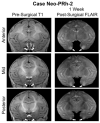
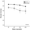
The contribution of the perirhinal cortex (PRh) to recognition memory is well characterized in adults, yet the same lesions have limited effect on recognition of spatial locations. Here, we assessed whether the same outcomes will follow when perirhinal lesions are performed in infancy. Monkeys with neonatal perirhinal (Neo-PRh) lesions and control animals were tested in three operant recognition tasks as they reached adulthood: Delayed Nonmatching-to-Sample (DNMS) and Object Memory Span (OMS), measuring object recognition, and Spatial Memory Span (SMS), measuring recognition of spatial locations. Although Neo-PRh lesions did not impact acquisition of the DNMS rule, they did impair performance when the delays were extended from 30s to 600s. In contrast, the same neonatal lesions had no impact on either the object or spatial memory span tasks, suggesting that the lesions impacted the maintenance of information across longer delays and not memory capacity. Finally, the magnitude of recognition memory impairment after the Neo-PRh lesions was similar to that previously observed after adult-onset perirhinal lesions, indicating minimal, or no, functional compensation after the early PRh lesions. Overall, the results indicate that the PRh is a cortical structure that is important for the normal development of mechanisms supporting object recognition memory. Its contribution may be relevant to the memory impairment observed with human cases of temporal lobe epilepsy without hippocampal sclerosis, but not to the memory impairment found in developmental amnesia cases.
Keywords: Developmental amnesia; Excitotoxic lesion; Medial temporal lobe epilepsy; Memory span; Recognition; Rhesus macaque.
Copyright © 2015 The Authors. Published by Elsevier B.V. All rights reserved.
Figures


Similar articles
-
Neonatal perirhinal cortex lesions impair monkeys' ability to modulate their emotional responses.Behav Neurosci. 2017 Oct;131(5):359-71. doi: 10.1037/bne0000208. Behav Neurosci. 2017. PMID: 28956946 Free PMC article.
-
The hippocampal/parahippocampal regions and recognition memory: insights from visual paired comparison versus object-delayed nonmatching in monkeys.J Neurosci. 2004 Feb 25;24(8):2013-26. doi: 10.1523/JNEUROSCI.3763-03.2004. J Neurosci. 2004. PMID: 14985444 Free PMC article.
-
Effects of combined and separate removals of rostral dorsal superior temporal sulcus cortex and perirhinal cortex on visual recognition memory in rhesus monkeys.J Neurophysiol. 2003 Oct;90(4):2419-27. doi: 10.1152/jn.00290.2003. Epub 2003 Jun 25. J Neurophysiol. 2003. PMID: 12826654
-
Recognition memory and the medial temporal lobe: from monkey research to human pathology.Rev Neurol (Paris). 2013 Jun-Jul;169(6-7):459-69. doi: 10.1016/j.neurol.2013.01.623. Epub 2013 Mar 6. Rev Neurol (Paris). 2013. PMID: 23473622 Review.
-
The role of the human medial temporal lobe in object recognition and object discrimination.Q J Exp Psychol B. 2005 Jul-Oct;58(3-4):326-39. doi: 10.1080/02724990444000177. Q J Exp Psychol B. 2005. PMID: 16194972 Review.
Cited by
-
Astrogliosis in juvenile non-human primates 2 years after infant anaesthesia exposure.Br J Anaesth. 2021 Sep;127(3):447-457. doi: 10.1016/j.bja.2021.04.034. Epub 2021 Jul 13. Br J Anaesth. 2021. PMID: 34266661 Free PMC article.
-
Neonatal perirhinal cortex lesions impair monkeys' ability to modulate their emotional responses.Behav Neurosci. 2017 Oct;131(5):359-71. doi: 10.1037/bne0000208. Behav Neurosci. 2017. PMID: 28956946 Free PMC article.
-
Neonatal Perirhinal Lesions in Rhesus Macaques Alter Performance on Working Memory Tasks with High Proactive Interference.Front Syst Neurosci. 2016 Jan 5;9:179. doi: 10.3389/fnsys.2015.00179. eCollection 2015. Front Syst Neurosci. 2016. PMID: 26778978 Free PMC article.
-
Monkeys do not show sex differences in toy preferences through their individual choices.Biol Sex Differ. 2023 Feb 3;14(1):3. doi: 10.1186/s13293-023-00489-9. Biol Sex Differ. 2023. PMID: 36737809 Free PMC article.
-
Impaired Cognitive Flexibility After Neonatal Perirhinal Lesions in Rhesus Macaques.Front Syst Neurosci. 2019 Jan 30;13:6. doi: 10.3389/fnsys.2019.00006. eCollection 2019. Front Syst Neurosci. 2019. PMID: 30760985 Free PMC article.
References
-
- Suzuki W, Naya Y. The perirhinal cortex. Annu Rev Neurosci. 2014;37:39–53. - PubMed
-
- Meunier M, Hadfield W, Bachevalier J, Murray EA. Effects of rhinal cortex lesions combined with hippocampectomy on visual recognition memory in rhesus monkeys. Journal of Neurophysiology. 1996;75:1190–1205. - PubMed
-
- Malkova L, Bachevalier J, Webster M, Mishkin M. Effects of neonatal inferior prefrontal and medial temporal lesions on learning the rule for delayed nonmatching-to-sample. Developmental Neuropsychology. 2000;18:399–421. - PubMed
Publication types
MeSH terms
Grants and funding
LinkOut - more resources
Full Text Sources
Other Literature Sources
Medical

