Review
. 2016 Jan;17(1):19-32.
doi: 10.1038/nrg.2015.3. Epub 2015 Nov 23.

RNA mis-splicing in disease

Affiliations
Review

RNA mis-splicing in disease

Marina M Scotti et al. Nat Rev Genet. 2016 Jan.

Abstract

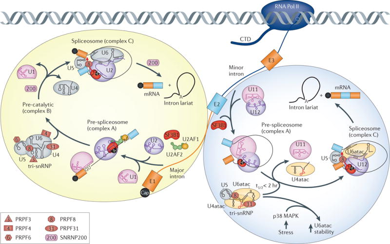
The human transcriptome is composed of a vast RNA population that undergoes further diversification by splicing. Detecting specific splice sites in this large sequence pool is the responsibility of the major and minor spliceosomes in collaboration with numerous splicing factors. This complexity makes splicing susceptible to sequence polymorphisms and deleterious mutations. Indeed, RNA mis-splicing underlies a growing number of human diseases with substantial societal consequences. Here, we provide an overview of RNA splicing mechanisms followed by a discussion of disease-associated errors, with an emphasis on recently described mutations that have provided new insights into splicing regulation. We also discuss emerging strategies for splicing-modulating therapy.


Conflict of interest statement

Competing interests statement

The authors declare no competing interests.

Figures

Figure 1
Figure 1. Mis-splicing of a single gene results in different diseases
Aberrant splicing of lamin A (LMNA) pre ‑mRNA is associated with multiple hereditary disorders. Normal exons are shown in blue, introns are shown as thick black lines, normal splicing is indicated by thin black lines, and disease-associated splicing is indicated in dotted lines or purple boxes (intron retention). a | Limb girdle muscular dystrophy type 1B (LGMD1B) is caused by a G>C 5′ splice site (5′ss) mutation that results in intron 9 retention, a premature termination codon (PTC) and nonsense‑mediated decay (NMD). c.1608 + 5 indicates that the mutations occurs 5 nucleotides into the intron that follows coding position (c) 1608. However, a lamin A/C protein truncated in intron 9 with a unique carboxy‑terminal sequence may also be produced. b | In familial partial lipodystrophy type 2 (FPLD2), a G>C transversion mutation occurs in the exon 8 5′ss, leading to intron 8 retention, NMD and potential translation of another truncated lamin A/C with a unique C‑terminal region. c | A common cause of Hutchinson–Gilford progeria syndrome (HGPS) is a C>T transition in exon 11, which activates a cryptic 5′ss and results in a 150 nucleotide deletion that is translated into the ageing‑associated protein progerin. d | For LMNA‑linked dilated cardiomyopathy (DCM), an alternative 3′ss is generated by an A>G mutation upstream of the normal exon 4 3′ ss so that nine additional nucleotides are inserted in-frame between exons 3 and 4, resulting in a 3-amino-acid insertion in the resultant protein.
Figure 2
Figure 2. Major and minor spliceosome mutations
The figure shows the splicing steps and core spliceosomal components of both the major (U2‑dependent) and minor (U12‑dependent) spliceosomes, including their interactions in the pre‑spliceosomal complex (complex A) and spliceosome (complex C). Pre‑mRNA processing factor 3 (PRPF3), PRPF4), PRPF6, PRPF8 and PRPF31 components of the U4/U6.U5 tri‑small nuclear ribonucleoprotein (tri‑snRNP) dysregulated in autosomal dominant retinitis pigmentosa (adRP) are shown. Also indicated is the SNRNP200 helicase, which is required at several dissociation steps in the spliceosomal cycle. Several PRPF components are common to both the U4/U6.U5 tri‑snRNP and the U4atac/U6atac.U5 tri‑snRNP complexes. Some mutations in the U4atac snRNA 5′ stem-loop found in microcephalic osteodysplastic primordial dwarfism type 1 (MOPD I) are highlighted in red. In addition, stress‑induced upregulation of p38 mitogen‑activated protein kinase (MAPK) leads to increased stability of U6atac (t1/2 <2 hours).
Figure 3
Figure 3. Co-transcriptional splicing factor recruitment and disease mutations
Models for splicing factor and precursor RNA mutations and disease-associated mis-splicing. a | Splicing factors recognize and bind to RNA polymerase II (RNA Pol II) transcripts in the nucleoplasm or directly at the carboxy‑terminal domain (CTD) of RNA Pol II. These factors may contain RNA‑binding motifs (such as RNA recognition motifs (RRMs) or zinc fingers (ZnFs)), as well as auxiliary domains composed of low complexity (LC) regions with prion‑like domains in heterogeneous nuclear ribonucleoprotein A1 (hnRNPA1), TDP‑43 and FUS (LC regions shown as green, yellow or red lines for hnRNPA1, TDP‑43 and FUS, respectively), or other regions that either mediate protein–protein interactions (in muscleblind‑like (MBNL)) or function as flexible linkers between RRMs (in hnRNPH). Splicing factors might bind to single‑stranded RNA (ssRNA) motifs or pre‑formed RNA structures (for example, G‑quadruplexes), resulting in the formation of dynamic RNA–RNP complexes that are continuously remodelled by RNA helicases and protein–protein interactions before nuclear export. b | Mutations (red star) in the LC regions of hnRNPA1, TDP‑43 and FUS could cause mis‑folding of RNA–RNP complexes and lead to abnormal splicing. c | For diseases caused by microsatellite expansions, splicing factors such as MBNL, which recognize a motif within the repeated sequence, are sequestered by the repeat expansion (ssRNA, top; RNA hairpin, bottom), leading to loss‑of‑function and mis‑splicing.
Figure 4
Figure 4. Therapeutic strategies
Examples of therapies based on antisense oligonucleotide (ASO) and small molecule approaches. a | Duchenne muscular dystrophy is often caused by chromosomal deletions (black triangle) that remove exons 48–50, resulting in a frameshift (blue rectangles, exons with intact codons; trapezoids, exons with incomplete codons) and loss of dystrophin protein. The red hexagon indicates the premature stop codon resulting from frameshifted exon 51. To prevent frameshifting, both phosphorodiamidate morpholino oligomer (PMO) and 2′OMePS (2′O‑methyl‑phosphorothioate) ASOs (black semicircle) block an exon 51 exonic splicing enhancer (ESE; green rectangle) and shift splicing to the in‑frame exon 52. b | In spinal muscular atrophy, survival of motor neuron 1 (SMN1), which produces the majority of SMN protein, is either deleted or inactivated by mutations, and the paralogous SMN2 expresses low levels of SMN due to a C>T transition (grey box) that suppresses exon 7 splicing. ASO‑10‑27 targets an intronic splicing silencer (ISS; red bar) and enhances exon 7 splicing to produce stable SMN protein. c | In myotonic dystrophy type 1, CUG expansion (CUGexp) RNA (red hairpin) binds muscleblind‑like (MBNL) proteins (green ovals) and causes mis‑splicing of MBNL RNA targets. Mutant MBNL–RNA complexes accumulate in the nucleus, and so ASO gapmers preferentially target mutant RNAs for degradation (dotted red line). Alternatively, small molecule compounds bind to mutant CUGexp RNA, displace MBNL and rescue abnormal splicing. DMPK, DM protein kinase.

References

    1. The ENCODE Project Consortium. An integrated encyclopedia of DNA elements in the human genome. Nature. 2012;489:57–74. - PMC - PubMed
    1. Kim MS, et al. A draft map of the human proteome. Nature. 2014;509:575–581. - PMC - PubMed
    1. Wilhelm M, et al. Mass-spectrometry-based draft of the human proteome. Nature. 2014;509:582–587. - PubMed
    1. Treutlein B, Gokce O, Quake SR, Sudhof TC. Cartography of neurexin alternative splicing mapped by single-molecule long-read mRNA sequencing. Proc Natl Acad Sci USA. 2014;111:E1291–E1299. Long-read sequencing of full-length neurexin mRNAs from pre-frontal cortex is performed to determine the extent of alternative splicing and provide evidence for thousands of neurexin isoforms. - PMC - PubMed
    1. Wang ET, et al. Alternative isoform regulation in human tissue transcriptomes. Nature. 2008;456:470–476. - PMC - PubMed

Publication types

LinkOut - more resources